
TenJet Procedure for Chronic Tendinopathy
Medically reviewed by Amit Sharma, MD. This page is educational and does not replace an individualized evaluation.
TenJet is an ultrasound-guided, minimally invasive tendon debridement procedure. Through a small access site, a physician uses a focused saline stream and suction to cut and evacuate selected abnormal tissue. TenJet is not PRP, a stem-cell treatment or a tendon-repair operation, and it is not automatically the next step whenever physical therapy or an injection has not helped.
The Short Answer
TenJet may be considered when chronic tendon pain has a well-defined clinical and ultrasound target, a suitable rehabilitation program has not provided enough improvement, and the amount and location of tendon damage can reasonably be treated without open repair.
TenJet and PRP solve different problems. TenJet removes selected tissue; PRP injects a blood-derived preparation and removes nothing. There is no universal rule that PRP must be tried first, and failed PRP alone does not establish TenJet candidacy.
Video: An overview of the TenJet procedure. Individual recommendations and recovery instructions vary by tendon and diagnosis.
What Is TenJet?
TenJet is a disposable instrument used for percutaneous tendon debridement. Under real-time ultrasound guidance, the physician places the instrument’s small cutting window at the selected target. A high-pressure saline stream cuts tissue within that window while a second channel creates suction and removes the cut material.
The manufacturer describes a dual-lumen, 12-gauge needle with a 1.65 mm cutting window and a non-thermal mechanism. Those technical characteristics explain how the instrument operates; they do not, by themselves, prove that TenJet produces better clinical outcomes than rehabilitation, PRP, another tenotomy method or surgery.
What TenJet Does
- Uses ultrasound to visualize the proposed target and nearby structures.
- Cuts and evacuates selected tissue through a small percutaneous access site.
- Avoids an open incision and does not leave a permanent implant.
- Is usually followed by protected activity and progressive tendon loading.
What TenJet Does Not Do
- It does not inject platelets, stem cells or medication.
- It does not reconnect a complete tendon rupture.
- It does not correct major joint instability or advanced arthritis.
- It does not guarantee that pain will resolve or that future surgery will be avoided.
- It has not been proven to prevent tendon rupture or future degeneration.
Tendinopathy, Tendinitis and Tendinosis: Why the Words Matter
Tendinopathy is the broad clinical term for persistent tendon pain and impaired function. Tendinitis is often used for a more acute inflammatory presentation. Tendinosis describes longer-standing degenerative or disorganized tendon changes. In practice, these processes may overlap, and an ultrasound or MRI abnormality does not prove that the imaged tendon is the source of pain.
The first-line treatment for many tendinopathies is a diagnosis-specific loading and rehabilitation program. Debridement should not be presented as required simply because imaging uses the word “tendinosis.”
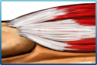
Healthy tendon
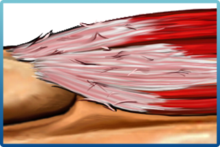
Tendinitis
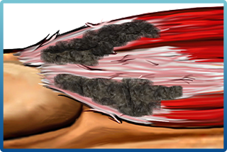
Tendinosis
Who May Be a Candidate for TenJet?
TenJet may be discussed for a carefully selected patient when the following findings fit together:
- Persistent, function-limiting tendon or fascia pain despite an adequate period of appropriate nonsurgical care.
- Symptoms and examination findings that localize to the proposed target.
- Ultrasound or other imaging that identifies a focal abnormality suitable for percutaneous debridement.
- No more likely untreated source of pain, such as nerve compression, referred pain, joint arthritis, instability or a different soft-tissue injury.
- No complete rupture or structural problem that requires repair rather than debridement.
- The ability and willingness to follow tendon-specific restrictions and rehabilitation.
- Realistic expectations about the limited comparative evidence and the possibility of incomplete relief.
When to Reconsider or Seek Surgical Evaluation
TenJet may be inappropriate when pain has not been localized, imaging findings are incidental, an infection is present, bleeding risk cannot be managed, or the patient has a complete rupture, a high-grade tear requiring repair, major weakness, significant retraction, joint instability or another condition better addressed surgically.
Which Tendons Can Be Treated?
TenJet is marketed for several tendon and fascia locations, including the common extensor or flexor tendon at the elbow, Achilles tendon, plantar fascia, patellar tendon, gluteal tendons, proximal hamstring and selected shoulder targets. The important limitation is that evidence from one body region cannot automatically be applied to another.
The most directly relevant published TenJet clinical study located for this review involved chronic elbow tendinopathy. That does not establish equal effectiveness for the plantar fascia, Achilles tendon, patellar tendon, gluteal or hamstring tendons, or rotator cuff. The diagnosis, degree of tearing, adjacent anatomy, rehabilitation requirements and alternative treatments differ at each site.
For shoulder pain, review whether symptoms arise from rotator-cuff tendinopathy, bursitis, calcific deposits, a partial tear, a full-thickness tear, arthritis, adhesive capsulitis or a cervical nerve problem before discussing debridement. Learn more about rotator-cuff tendinopathy.
How Is the TenJet Procedure Performed?
- Confirm the target. History, examination and imaging are reviewed to determine whether the visible abnormality matches the patient’s symptoms.
- Plan around nearby anatomy. Ultrasound is used to map the tendon, blood vessels, nerves and other structures.
- Prepare and anesthetize the area. The site is cleaned using sterile technique, and local anesthetic is administered. Sedation requirements vary.
- Position the instrument. The physician advances the device to the selected tissue under continuous ultrasound visualization.
- Debride the target. The saline jet cuts tissue in the instrument window while suction evacuates it.
- Dress the access site. A small dressing is applied. Bracing, a walking boot, crutches or other protection may be recommended depending on the treated location.
- Begin structured follow-up. Wound care, activity limits and progressive loading are tailored to the tendon and the extent of treatment.
The procedure itself may be brief, but procedure time should not be confused with biological recovery. Tendon adaptation and return to high-load work or sport generally take longer than skin healing.
TenJet vs. PRP: How Are They Different?
This is the core decision in Top 100 question #93. TenJet and platelet-rich plasma are not interchangeable, and neither is universally better.
| Decision factor | TenJet | PRP injection |
|---|---|---|
| Primary action | Mechanically cuts and evacuates selected tissue. | Injects a blood-derived platelet preparation; no tissue is removed. |
| Typical target | A focal chronic tendon or fascia abnormality that matches symptoms and can be reached safely. | A diagnosis-specific tendon, ligament or joint target for which a biologic injection is being considered. |
| Imaging | Real-time ultrasound is central to targeting and tissue removal. | Image guidance is commonly used, but the appropriate method depends on the target. |
| Tissue removal | Yes. | No. |
| Evidence | Direct TenJet evidence is limited; the published prospective elbow study was small and uncontrolled. | Evidence varies substantially by diagnosis and PRP preparation. Results from one tendon or joint should not be generalized to another. |
| Recovery | Requires access-site care, temporary protection and progressive reloading after debridement. | Often involves a smaller needle puncture, post-injection soreness and a target-specific loading plan. |
| Coverage | May be covered in some circumstances, but benefits and authorization must be verified. | Many orthopedic PRP injections are self-pay; plan-specific verification is still required. |
| Key limitation | Removing an imaging abnormality does not guarantee pain relief. | Symptom improvement does not prove that tissue was regenerated. |
Should PRP Be Tried Before TenJet?
Not automatically. No high-quality evidence establishes a universal PRP-before-TenJet sequence. For some diagnoses, rehabilitation remains the most important initial treatment and the evidence for PRP may be uncertain. For others, a clinician may discuss PRP before debridement because it does not remove tissue. The decision should be based on the specific diagnosis, tendon, degree of tearing, prior treatment, cost, coverage, recovery priorities and patient preference.
A failed PRP injection does not prove that abnormal tissue must be removed. The diagnosis, injection target, PRP preparation, rehabilitation and competing pain sources should be reassessed before escalating treatment.
Can PRP Be Used With or After TenJet?
A clinician may discuss PRP at a different stage of care, but high-quality evidence has not established that routinely adding PRP to TenJet improves outcomes over TenJet or rehabilitation alone. Same-session or staged combination treatment adds cost and may change recovery without a proven incremental benefit. If it is proposed, ask what diagnosis-specific evidence supports the combination.
For a broader overview, see our PRP patient guide, PRP recovery timeline and guide to why PRP sometimes does not work.
A Practical Treatment Pathway Before TenJet
- Confirm the pain generator. Do not treat an ultrasound or MRI finding without clinical concordance.
- Classify the structural problem. Distinguish load-related tendinopathy from acute tear, complete rupture, calcific disease, bursitis, arthritis, nerve pain and referred pain.
- Complete appropriate rehabilitation. Progressive loading is foundational for many tendinopathies, although the program must be adapted to the tendon and patient.
- Review modifiable factors. Training errors, smoking, diabetes, certain medications, nutrition and work demands may influence recovery.
- Discuss diagnosis-specific options. These may include continued rehabilitation, activity modification, bracing, medication, selected injections, shock-wave therapy, another tenotomy method or surgical consultation.
- Choose debridement only when the target and tradeoffs make sense. A minimally invasive procedure is still a procedure; the expected benefit should justify its risks, cost and recovery burden.
What Does the Evidence Actually Show?
A 2022 prospective multicenter study evaluated TenJet in 29 patients involving 32 elbows with chronic lateral or medial elbow tendinopathy. Participants had symptoms for more than three months, had not improved with nonsurgical care and had imaging consistent with the diagnosis. The investigators reported improved pain and function through 12 months.
The study also had important limitations: it was nonrandomized, had no comparison or sham group, enrolled a small number of patients and evaluated elbows rather than every tendon listed in marketing materials. Two patients later underwent open debridement. The absence of reported device-related complications in this small series does not establish zero risk.
These findings are encouraging enough to support an informed discussion for selected patients, but they do not prove that TenJet is superior to progressive rehabilitation, PRP, corticosteroid injection, another percutaneous tenotomy method or surgery. They also do not support a universal 80 to 90 percent success claim.
An additional prospective registry has been designed to study epicondylitis, gluteal tendinopathy and calcific shoulder tendinitis. Until results are available and independently assessed, enrollment or an ongoing registry should not be described as proof of effectiveness.
Do Not Combine Evidence From Different Devices
TenJet uses saline-jet hydrosurgery. Ultrasonic tenotomy devices use a different energy mechanism, and ordinary needle tenotomy uses neither system. Outcomes from one category should not be presented as TenJet outcomes unless the study actually used TenJet.
Recovery After TenJet
Recovery depends on the treated tendon, the amount of tissue removed, baseline strength, job demands and the rehabilitation plan. Same-day discharge does not mean immediate readiness for heavy lifting, running or sport.
| General phase | What may occur | Important qualification |
|---|---|---|
| First 48 to 72 hours | Access-site care, soreness, swelling and relative protection. Medication and icing instructions should come from the treating clinician. | Weight-bearing and motion rules differ for an elbow, shoulder, hip, knee, Achilles tendon or plantar fascia. |
| First two weeks | Light daily activity may be possible; a brace, boot, crutches or work restrictions may be prescribed. | Comfort with routine activity does not mean the tendon is ready for heavy loading. |
| Weeks two to six | Progressive range of motion and tendon-specific loading may begin or advance under guidance. | In the published elbow study, lifting was restricted to less than 10 pounds for six weeks. That was a study protocol, not a universal rule. |
| After six weeks | Strengthening and work or sport demands may progress if symptoms, examination and function permit. | Return to sport at eight to 12 weeks cannot be promised; some patients require longer and some need further treatment. |
Call the treating team promptly for fever, spreading redness, drainage, escalating swelling, uncontrolled pain, new numbness or weakness, color or temperature change in the limb, chest pain, shortness of breath or another concerning change.
Risks and Limitations
Potential risks include:
- Temporary pain flare, swelling, bruising or stiffness.
- Bleeding or hematoma.
- Infection or wound problems.
- Injury to nearby nerves, blood vessels or other structures.
- Unintended tendon or fascia injury.
- Incomplete relief, recurrent symptoms or worsening pain.
- Need for additional rehabilitation, injections, another procedure or surgery.
Risk is location-specific. For example, the adjacent neurovascular structures and the consequences of excessive loading differ between the elbow, shoulder, hip and Achilles region. A small published series cannot define uncommon complications reliably.
Alternatives to TenJet
Depending on the diagnosis and tendon, alternatives may include:
- Continued or redesigned progressive loading and physical therapy.
- Activity and ergonomic modification.
- Temporary bracing, orthotics or a walking boot when appropriate.
- Medication selected for the patient’s medical history and treatment goal.
- Extracorporeal shock-wave therapy for selected conditions.
- Diagnosis-specific injection options, including PRP or corticosteroid in limited circumstances.
- Percutaneous needle tenotomy or a different debridement technique.
- Surgical evaluation for a tear requiring repair, severe structural disease or persistent disability.
Neither PRP nor TenJet should be treated as a mandatory checkpoint before surgery. Conversely, surgery should not be recommended merely because a less-invasive option failed; the diagnosis and structural indication should be reassessed.
Insurance and Cost
Do not assume TenJet is covered. Coverage may depend on the insurer, diagnosis, duration and documentation of conservative treatment, medical policy, prior authorization, procedure code, network and site of service. Ask the office and insurer for a patient-specific estimate, including professional, facility, ultrasound, anesthesia and rehabilitation charges when applicable.
PRP is often self-pay for orthopedic indications. A coverage difference may affect the decision, but it should not substitute for determining which treatment, if any, fits the diagnosis.
Questions to Ask Before TenJet
- What is the exact diagnosis, and how do the examination and ultrasound findings agree?
- Is there a tear that requires repair rather than debridement?
- What rehabilitation program has been tried, and was it adequate for this tendon?
- What TenJet evidence applies to this body region?
- What alternatives are reasonable, including continued rehabilitation, PRP, shock-wave therapy or surgery?
- Why is TenJet preferred over another percutaneous tenotomy or debridement method?
- What are my lifting, walking, driving and work restrictions?
- What will the procedure and rehabilitation cost under my plan?
- What is the next step if symptoms do not improve?
Dr. Amit Sharma & our minimally invasive pain & spine team.
Frequently Asked Questions About TenJet
What is TenJet?
TenJet is an ultrasound-guided, minimally invasive debridement procedure. A small instrument uses a pressurized saline stream and suction to cut and evacuate selected abnormal tendon tissue through a small access site. It is not PRP, a stem-cell treatment or a tendon-repair operation.
Is TenJet the same as PRP?
No. TenJet mechanically removes selected tissue. PRP is an injection prepared from a patient’s blood and does not remove tissue. Their evidence, goals, costs and rehabilitation plans differ.
Should PRP always be tried before TenJet?
No. There is no universal PRP-before-TenJet sequence. The decision should depend on the diagnosis, tendon, imaging, prior rehabilitation, degree of tearing, patient preferences and the evidence for that specific condition. Failed PRP does not automatically make someone a TenJet candidate.
Can PRP be used with or after TenJet?
A clinician may discuss PRP at a different stage of care, but high-quality evidence has not established that routinely combining PRP with TenJet produces better outcomes than TenJet or rehabilitation alone. The timing, added cost and uncertain benefit should be discussed before treatment.
Which conditions may be considered for TenJet?
TenJet may be considered for selected chronic tendon or fascia disorders with a coherent clinical and ultrasound target after appropriate nonsurgical care. Published device-specific clinical evidence is most developed for chronic elbow tendinopathy; results cannot automatically be transferred to the Achilles, plantar fascia, patellar, gluteal, hamstring or rotator-cuff regions.
Is TenJet surgery?
TenJet is a minimally invasive percutaneous procedure that cuts and removes tissue through a small access site. It is less extensive than open tendon surgery, but it still deliberately alters tissue and requires sterile technique, informed consent and a structured recovery plan.
How long is recovery after TenJet?
Recovery depends on the tendon, extent of treatment, job and rehabilitation plan. Soreness and temporary protection are expected early, followed by progressive loading. In one elbow study, lifting was limited to less than 10 pounds for six weeks; that protocol should not be treated as a universal timeline for every tendon or patient.
How successful is TenJet?
A small prospective study reported improvement after treatment of 32 elbows, but it had no control group and two patients later underwent open debridement. This does not establish an 80 to 90 percent universal success rate, prove superiority to PRP or rehabilitation, or predict results for other tendons.
Is TenJet covered by insurance?
Coverage varies by insurer, diagnosis, plan, authorization requirements, coding and treatment setting. Coverage and out-of-pocket cost should be verified for the individual patient before the procedure; it should not be assumed from a general coverage statement.
What are the risks of TenJet?
Potential risks include pain flare, swelling, bruising, bleeding, infection, skin or wound problems, injury to nearby nerves or blood vessels, unintended tendon or fascia injury, incomplete relief, worsening symptoms and the need for additional treatment or surgery. Risk varies with the location and the patient’s health.
Selected Sources
- Barnes DE, Beckley JM, Smith J. Ultrasound-guided tenotomy via a hydrosurgery resection device improves symptoms of chronic elbow tendinopathy: a multicenter prospective study. Journal of Orthopaedic Experience & Innovation. 2022.
- HydroCision. TenJet technical overview. Manufacturer source used for device mechanics, not comparative effectiveness claims.
- ClinicalTrials.gov. Prospective evaluation of the TenJet system, NCT05352139.
- Cochrane. Autologous blood or PRP injection for lateral elbow pain. 2021.
- Millar NL, Silbernagel KG, Thorborg K, et al. Tendinopathy. Nature Reviews Disease Primers. 2021.
- Cleveland Clinic. Tendinopathy. Updated December 9, 2024.