

Percutaneous Discectomy: Procedure, Candidates & Recovery | Dr. Amit Sharma
Percutaneous discectomy is a category of minimally invasive lumbar disc procedures performed through a needle or small cannula under imaging guidance. A device removes or alters a limited amount of material from inside the disc. Reducing intradiscal volume may decrease the contribution of a carefully selected contained disc herniation.
This is not the same as removing a large free disc fragment under direct endoscopic or microscopic visualization. Percutaneous intradiscal decompression is most relevant when symptoms, examination and MRI findings identify the same contained protrusion and appropriate nonsurgical care has not provided enough relief.
Quick Answer
- What it is: limited treatment of nucleus material from inside a lumbar disc through a needle or small cannula.
- Best general fit: a symptomatic contained lumbar disc protrusion that corresponds with the clinical findings.
- What it is not: a broad decompression for major bone or ligament stenosis, or the usual method for retrieving a large extruded or sequestered fragment.
- Important distinction: mechanical, radiofrequency, plasma-assisted and other percutaneous systems are not identical and should not be treated as interchangeable.
- Most important step: deciding whether the disc finding is truly causing the symptoms and whether indirect intradiscal decompression can adequately treat it.
What Is Percutaneous Discectomy?
The word percutaneous means that the disc is reached through the skin rather than through a conventional surgical exposure. Under fluoroscopic or CT guidance, the physician advances a needle or small working cannula into the selected disc. A specialized device then removes, aspirates, ablates or otherwise modifies a limited quantity of nucleus material.
The proposed mechanical principle is volume reduction. A small decrease in nucleus volume may lower pressure within the disc and reduce the contribution of a contained protrusion. The compressed nerve is not necessarily viewed directly, and the protruding portion is not necessarily removed from beside the nerve.
Terminology can be confusing. Some older guidelines use percutaneous discectomy broadly enough to include endoscopic discectomy. On this page, the term refers more narrowly to non-endoscopic intradiscal decompression performed through a needle or small cannula. Full-endoscopic lumbar discectomy is discussed separately because it places a camera and working instruments near the herniation and nerve.
Disc protrusions are common on MRI, including in people without symptoms. A procedure should be considered only when the pain pattern, neurological examination and imaging identify the same clinically important disc.
Percutaneous Discectomy Is a Category, Not One Procedure
Different systems reach the disc through a similarly small access route but use different methods to treat nucleus material. Evidence, indications, technical details and insurance policies may differ by device.
Mechanical Disc Decompression
A mechanical system uses a cutting, rotating or aspiration mechanism to remove a measured or limited amount of nucleus material. The Dekompressor system is one example. Fluoroscopy or CT is used to guide the access device into the disc; a camera is not positioned beside the nerve.
Radiofrequency or Plasma-Assisted Decompression
Other platforms use radiofrequency, plasma or another energy source to ablate, coagulate or modify selected disc tissue. Some also permit limited manual removal of nucleus material. These methods have different device designs and should not be grouped together as though their clinical evidence were identical.
How Disc-FX Fits
Disc-FX is one specific percutaneous discectomy platform. It combines limited manual nucleus removal with bipolar radiofrequency ablation and coagulation of selected intradiscal tissue. It should not be used as a synonym for every percutaneous discectomy technique.
| Technique | How Disc Material Is Treated | Direct Camera View of the Nerve? | General Anatomic Concept |
|---|---|---|---|
| Mechanical percutaneous discectomy | Cuts, cores or aspirates a limited amount of nucleus material | No | Selected contained disc herniation |
| Radiofrequency or plasma-assisted decompression | Ablates, coagulates or modifies selected intradiscal tissue; technique varies | No | Selected contained disc herniation |
| Full-endoscopic discectomy | Directly visualizes and removes accessible compressive disc material | Yes | Contained, extruded or migrated herniation when accessible |
What Is a Contained Disc Herniation?
A lumbar disc has a softer central nucleus surrounded by a stronger outer ring called the annulus fibrosus. With a contained disc herniation, displaced nucleus material remains substantially held by the outer annular fibers and supporting structures. The disc may protrude, but there is no large separated fragment lying free within the spinal canal.
An extruded herniation extends through the outer disc boundary. A sequestered fragment has separated from the parent disc and may migrate. When a discrete fragment directly compresses a nerve, intradiscal volume reduction may not remove the structure causing the compression. Endoscopic or microscopic fragment removal may be more appropriate.
The words bulge, protrusion, extrusion and sequestration describe morphology. They do not, by themselves, prove which structure is causing pain or determine treatment. Symptoms, neurological findings, the location of the fragment, stenosis and stability all matter.
Who May Be a Candidate for Percutaneous Discectomy?
A possible candidate has persistent symptoms that correspond with a contained lumbar disc herniation, accessible anatomy and inadequate improvement after an appropriate course of nonsurgical care. The objective should be clear: limited intradiscal decompression must have a reasonable chance of treating the clinically important problem.
| Findings That May Support Consideration | Findings That May Favor Another Pathway |
|---|---|
| Contained lumbar disc protrusion | Large extruded, sequestered or migrated fragment |
| Symptoms and examination corresponding with the affected disc and nerve | MRI abnormality that does not match the clinical findings |
| Persistent radicular symptoms or carefully evaluated disc-related pain | Nonspecific axial back pain without a confirmed generator |
| Inadequate improvement with appropriate conservative treatment | Symptoms improving safely with continued conservative care |
| Sufficient disc height and an accessible posterolateral route | Advanced disc collapse or anatomy that prevents safe access |
| No clinically important instability or deformity | Significant spondylolisthesis, instability or deformity |
| No major bony or ligamentous compression requiring direct removal | Severe central, lateral-recess or foraminal stenosis |
| Elective situation without progressive neurological loss | Progressive weakness, cauda-equina symptoms, infection, fracture or tumor |
Why Symptoms and MRI Must Match
Percutaneous discectomy should not be offered merely because an MRI report uses the words disc bulge or herniation. The side and level of the imaging abnormality should plausibly explain the pain distribution, sensory change, weakness or reflex finding. Other causes of leg pain, including bony stenosis, hip disease, peripheral neuropathy and vascular claudication, may require different evaluation.
When a Limited Percutaneous Procedure Is Not the Next Step
New or progressive motor weakness, saddle-region numbness, loss of bowel or bladder control, or other signs of cauda-equina compression require urgent evaluation. A limited elective intradiscal procedure should not delay appropriate emergency or surgical assessment.
How Is Percutaneous Discectomy Performed?
The exact steps depend on the selected device and clinical plan. A typical non-endoscopic intradiscal procedure follows this general pathway:
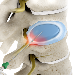
This procedure does not generally create the same direct operative view as full-endoscopic or microscopic discectomy. If a nerve is compressed by a free fragment, bone, ligament or severe foraminal narrowing, another approach may provide more complete treatment.
Percutaneous Discectomy Versus Other Treatments
| Treatment | Main Objective | Directly Sees the Nerve or Fragment? | General Clinical Fit |
|---|---|---|---|
| Epidural or selective nerve-root injection | Deliver medication near an inflamed nerve | No | Selected radicular pain when inflammation is an important component |
| Percutaneous intradiscal discectomy | Reduce or modify limited nucleus material from inside the disc | No | Selected symptomatic contained herniation |
| Full-endoscopic lumbar discectomy | Directly visualize and remove accessible compressive disc material | Yes | Contained, extruded or migrated herniation when endoscopically accessible |
| Microscopic discectomy | Direct surgical visualization and fragment removal | Yes | Disc fragment requiring direct removal when this access best matches the anatomy |
| Decompression with or without fusion | Remove broader bone or ligament compression and address instability when necessary | Yes | Extensive stenosis, instability, deformity or other complex pathology |
No procedure is automatically superior because its incision is smaller. The appropriate option is the least disruptive approach that can adequately treat the complete pain-generating anatomy.
Potential Benefits
For a carefully selected contained disc herniation, procedural characteristics may include:
- A needle or small-cannula access route
- Limited disruption of paraspinal muscle and posterior bone
- No spinal implant and no intended fusion
- Imaging-guided outpatient treatment in many cases
- Local anesthesia with appropriately selected sedation in many cases
- Removal or modification of only a limited amount of nucleus material
- Preservation of future treatment options if additional care becomes necessary
These are potential procedural advantages, not guarantees of pain relief, faster recovery or avoidance of future surgery. A small access route cannot overcome incorrect diagnosis, unsuitable disc morphology or untreated bony compression.
Recovery After Percutaneous Discectomy
Same-day discharge is common after an uncomplicated procedure. Mild access-site or disc-related soreness may occur. Walking generally begins early and increases gradually according to symptoms and individualized instructions.
- Driving: wait until sedating medication has been stopped and you can sit, turn, control the pedals and perform an emergency stop safely.
- Desk work: may resume relatively early in an uncomplicated recovery when pain is controlled and position changes are possible.
- Physical work: commonly requires a longer restriction period because lifting, bending, climbing, pushing and pulling place different demands on the spine.
- Exercise: should progress from walking to prescribed rehabilitation and later to higher-load activity after clearance.
There is no universal three-to-five-day return-to-normal rule. Recovery varies with the device, treated level, symptom duration, nerve health, neurological findings, medical history and job demands. Relief may be early or gradual because intradiscal volume reduction does not necessarily create immediate direct decompression beside the nerve.
Severe or escalating pain, new or worsening weakness, fever, wound drainage, saddle numbness, or new bowel or bladder dysfunction requires prompt assessment. Bowel or bladder dysfunction with saddle-region numbness or progressive weakness may require emergency evaluation.
Evidence and Important Limitations
The evidence for percutaneous discectomy varies substantially by technology. Mechanical, automated, laser, plasma-assisted, radiofrequency-assisted and full-endoscopic procedures should not be pooled conceptually as though they deliver the same treatment.
The North American Spine Society guideline states that automated percutaneous discectomy may be considered for lumbar disc herniation with radiculopathy, assigning a Grade C recommendation. The same guideline found insufficient evidence to determine equivalence with open discectomy and insufficient evidence for or against plasma disc decompression or nucleoplasty. The evidence reviewed was largely older and included small or observational studies.
A 2013 systematic review of automated percutaneous mechanical lumbar discectomy concluded that evidence was limited because of the lack of high-quality randomized trials. A 2025 systematic review of lumbar disc-herniation treatment guidelines found continuing disagreement among guidelines regarding several minimally invasive and percutaneous techniques.
These limitations do not mean that no selected patient can benefit. They do mean that broad claims such as a universal 90% success rate, lower complication rate than every surgical alternative, or guaranteed rapid recovery are not justified. Published averages cannot predict an individual result.
What Percutaneous Discectomy Cannot Reliably Treat
A limited intradiscal procedure may be inadequate when the dominant problem is:
- A large extruded, migrated or sequestered disc fragment
- Severe central, lateral-recess or foraminal bony stenosis
- Substantial ligamentum flavum hypertrophy
- Progressive motor weakness or another urgent neurological deficit
- Significant spondylolisthesis, instability or deformity
- Advanced disc collapse or severe multilevel degeneration
- Infection, fracture or tumor
- Symptoms caused by another nerve, joint, vascular or peripheral neurological disorder
- Nonspecific lower-back pain without a confirmed pain generator
If limited intradiscal decompression does not provide adequate relief, subsequent treatment may include an injection, endoscopic discectomy, microscopic discectomy, a broader decompression or, when a separate mechanical indication exists, stabilization or fusion.
Risks of Percutaneous Discectomy
Percutaneous discectomy is minimally invasive, but it is not risk-free. Potential risks include:
- Infection, including discitis
- Bleeding or hematoma
- Nerve irritation or injury
- Dural injury or spinal-fluid leak
- Vascular, visceral or other access-related injury
- Thermal injury with an energy-based system
- Temporary or persistent worsening of back or leg pain
- Inadequate decompression or failure to improve
- Recurrent or progressive disc herniation
- Continued disc degeneration
- Medication, contrast, sedation or anesthesia complications
- Need for another procedure or surgery
Risk depends on the device, access route, anatomy, medical history and operator experience. Individual informed consent should address the specific technique being proposed rather than treating all percutaneous systems as identical.
Alternatives to Percutaneous Discectomy
The most appropriate alternative depends on the diagnosis, neurological status and anatomy. Options may include:
- Continued observation when symptoms are improving and neurological function is stable
- Activity modification and physical therapy
- Diagnosis-appropriate medication
- Epidural steroid injection or selective nerve-root injection
- Disc-FX or another specifically selected percutaneous platform
- Full-endoscopic lumbar discectomy
- Microscopic discectomy
- Endoscopic lumbar decompression when bone or ligament materially contributes to compression
- Open or minimally invasive decompression for more extensive stenosis
- Fusion only when instability, deformity or another mechanical indication supports it
Insurance coverage varies by plan, diagnosis, device and medical-necessity policy. Authorization should be verified for the exact technique. Coverage does not prove that the procedure is the best anatomical match, and lack of coverage does not by itself answer the clinical question.
Related Disc and Sciatica Resources
- Disc-FX
- Endoscopic Lumbar Discectomy
- Endoscopic Spine Surgery
- Endoscopic Lumbar Decompression
- Epidural Steroid Injection
- Transforaminal Epidural Steroid Injection
- Herniated Disc
- Sciatica
Does Your MRI Show a Contained Disc Herniation?
A focused evaluation can determine whether intradiscal decompression, direct fragment removal, an injection or another treatment best matches the anatomy and symptoms.
Frequently Asked Questions About Percutaneous Discectomy
What is percutaneous discectomy?
Who is a candidate for percutaneous discectomy?
Is percutaneous discectomy the same as Disc-FX?
Is percutaneous discectomy the same as endoscopic discectomy?
Can percutaneous discectomy remove an extruded or sequestered fragment?
Does percutaneous discectomy treat spinal stenosis?
Will I need general anesthesia?
How long is recovery after percutaneous discectomy?
What are the risks of percutaneous discectomy?
Does insurance cover percutaneous discectomy?
References
- North American Spine Society. Diagnosis and Treatment of Lumbar Disc Herniation with Radiculopathy. Clinical guideline. 2012. Read source →
- Korean Spinal Neurosurgery Society. A Systematic Review of Treatment Guidelines for Lumbar Disc Herniation. Neurospine. 2025. Read source →
- Manchikanti L, et al. An updated review of automated percutaneous mechanical lumbar discectomy for the contained herniated lumbar disc. Pain Physician. 2013;16(2 Suppl):SE151-SE184. Read source →
- Lierz P, Alo KM, Felleiter P. Percutaneous lumbar discectomy using the Dekompressor system under CT-control. Pain Practice. 2009;9(3):216-220. Read source →
- Stryker. Dekompressor Disc Removal System. Manufacturer device information. Read source →



