

Basivertebral Nerve (BVN) Ablation for Vertebrogenic Back Pain: A Complete Patient Guide
Introduction
Chronic low back pain is one of the leading causes of disability worldwide, yet identifying its true source has often been challenging. While pain may arise from the intervertebral discs, facet joints, sacroiliac joints, muscles, or compressed spinal nerves, research over the past two decades has identified another important pain generator: the vertebral endplates.
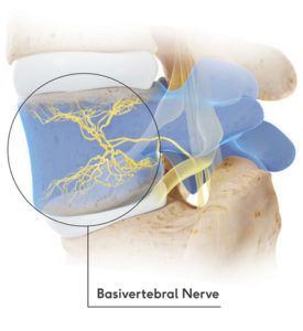 The vertebral endplates are thin layers of cartilage and bone located between each spinal disc and the adjacent vertebral body. When these endplates become damaged, inflammation can develop within the vertebral bone marrow, stimulating pain-sensitive nerve fibers carried by the basivertebral nerve (BVN). This condition is known as vertebrogenic pain.
Unlike traditional treatments that focus on the spinal discs or facet joints, basivertebral nerve ablation (BVN ablation) specifically targets the nerve responsible for transmitting pain signals from damaged vertebral endplates. For carefully selected patients with chronic vertebrogenic low back pain, this minimally invasive procedure may provide durable pain relief while improving function and quality of life.
This guide explains how vertebrogenic pain develops, the role of Modic changes on MRI, who may benefit from basivertebral nerve ablation, how the procedure is performed, expected recovery, long-term outcomes, and the differences between the Intracept® and OptAblate® systems.
Quick Answer
Basivertebral nerve ablation (BVN ablation) is a minimally invasive procedure used to treat chronic vertebrogenic low back pain caused by damaged vertebral endplates. During the procedure, a specialized probe is inserted into the affected vertebral body, where radiofrequency energy permanently interrupts the basivertebral nerve. Because this nerve carries pain signals from the inflamed endplates, carefully selected patients may experience significant and durable improvements in pain and function.
Clinical Perspective
Basivertebral nerve ablation is a diagnosis-driven procedure rather than a treatment for every patient with chronic low back pain. Success depends on identifying vertebrogenic pain through a careful combination of the patient’s history, physical examination, and MRI findings, particularly the presence of Modic type 1 or type 2 endplate changes. Appropriate patient selection remains one of the strongest predictors of a successful outcome.
Understanding Vertebrogenic Pain
For decades, chronic low back pain was commonly attributed to the intervertebral discs, facet joints, muscles, or compressed spinal nerves. While these structures frequently cause pain, researchers now recognize that the vertebral endplates can also be a significant source of chronic low back pain. This condition is known as vertebrogenic pain.
The vertebral endplates are thin layers of specialized bone and cartilage that separate each spinal disc from the vertebral body above and below it. Although they appear relatively small, they perform several essential functions, including distributing mechanical forces across the spine and allowing nutrients to pass between the vertebral body and the intervertebral disc.
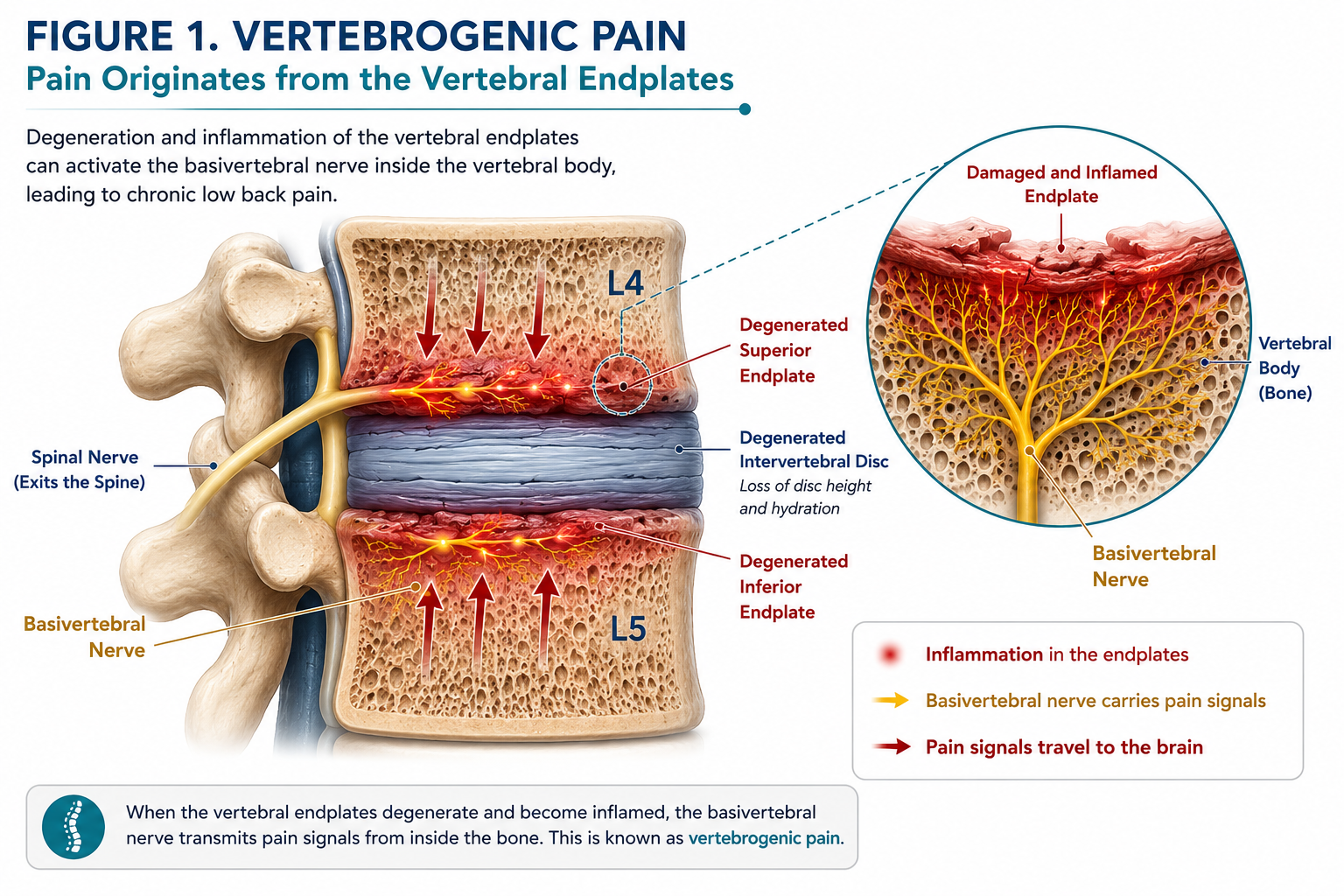
Degenerative disc disease may increase stress across the adjacent vertebral endplates. Damage to these endplates can produce inflammation within the vertebral marrow, activating the basivertebral nerve and generating chronic vertebrogenic low back pain.
Over time, repetitive loading, trauma, disc degeneration, or age-related changes can damage these endplates. When this occurs, inflammatory chemicals accumulate within the adjacent vertebral bone marrow and stimulate pain-sensitive nerve fibers carried by the basivertebral nerve. Rather than originating from the spinal disc itself, the pain originates within the vertebral body and its damaged endplates.
This represents an important shift in our understanding of chronic low back pain. Instead of assuming every patient suffers from “disc pain” or arthritis, physicians can now recognize a distinct group of patients whose symptoms arise primarily from damaged vertebral endplates. Identifying this source allows treatment to focus on the actual pain generator instead of surrounding structures that may not be responsible for the patient’s symptoms.
Today, vertebrogenic pain is recognized as a unique clinical diagnosis supported by the patient’s history, physical examination, and characteristic MRI findings. When these factors align, treatments directed specifically at the basivertebral nerve may provide meaningful and lasting relief for appropriately selected patients.
Why the Discovery of Vertebrogenic Pain Changed Spine Medicine
One of the greatest challenges in treating chronic low back pain has always been identifying its true source. Two patients may have nearly identical MRI scans yet experience completely different symptoms, while another patient may report disabling pain despite only modest degenerative changes.
For many years, physicians focused primarily on the intervertebral discs, facet joints, muscles, and spinal nerves as the major pain generators. Although these structures remain important, research over the past two decades has demonstrated that the vertebral endplates themselves can become painful structures capable of producing chronic axial low back pain.
The discovery that the basivertebral nerve carries pain signals from damaged vertebral endplates fundamentally changed the understanding of a subset of chronic low back pain. Rather than simply describing degenerative findings on MRI, physicians can now correlate specific imaging abnormalities, particularly Modic type 1 and type 2 changes, with a defined biological pain pathway.
This improved understanding has transformed treatment from being anatomy-driven to diagnosis-driven. Instead of repeatedly treating nearby structures that may not actually be responsible for the pain, physicians can identify patients whose symptoms originate inside the vertebral body itself and consider therapies specifically designed to interrupt this pain pathway.
Basivertebral nerve ablation represents one of the first procedures developed specifically to treat vertebrogenic pain. Its success reflects not only advances in technology, but also a better understanding of where chronic low back pain truly originates in carefully selected patients.
Understanding the Vertebral Endplates
The vertebral endplates are thin layers of cartilage and bone that separate each intervertebral disc from the vertebral body above and below it. Although they measure only a few millimeters in thickness, they are among the most important structures within the spinal motion segment.
Every spinal disc is bordered by two endplates. The superior endplate connects the disc to the vertebra above, while the inferior endplate connects it to the vertebra below. Together, these structures form the interface between the flexible intervertebral disc and the rigid vertebral body.
For many years, vertebral endplates received little attention because they were thought to serve only as structural support. Research has now shown that they perform several critical biological and mechanical functions that are essential for maintaining a healthy spine.
Functions of the Vertebral Endplates
Healthy vertebral endplates perform several important roles:
- Load Distribution.
They evenly distribute forces between the intervertebral disc and the vertebral body during standing, walking, bending, lifting, and twisting. - Nutrient Transport.
Because adult intervertebral discs have very little direct blood supply, oxygen and nutrients must diffuse through the vertebral endplates from the vertebral marrow to nourish the disc. - Mechanical Support.
The endplates provide a stable foundation that helps the disc maintain its normal height and resist excessive compression. - Barrier Function.
They help prevent disc material from herniating into the vertebral body while maintaining communication between the disc and adjacent bone.
A healthy vertebral endplate is remarkably strong yet remains sufficiently porous to allow nutrients to pass into the disc. This balance between strength and permeability is essential for preserving disc health throughout life.
When the Endplates Become Damaged
With aging, repetitive mechanical loading, spinal injury, smoking, obesity, or progressive degenerative disc disease, the vertebral endplates may begin to fail. Small microscopic cracks can develop within the cartilage and underlying bone, disrupting both their structural integrity and their ability to regulate nutrient exchange.
As these injuries accumulate, inflammatory chemicals released from the degenerating disc can enter the adjacent vertebral marrow. Instead of remaining confined to the disc, inflammation spreads into the vertebral body where pain-sensitive nerve fibers are abundant.
This process marks the transition from simple degenerative disc disease to vertebrogenic pain. Rather than being an incidental age-related finding, damaged vertebral endplates become an active source of chronic low back pain.
How Damaged Endplates Become Painful
Vertebrogenic pain usually develops gradually rather than after a single injury. In many patients, the process begins with age-related degeneration of the intervertebral disc. As the disc loses water content and elasticity, it becomes less capable of absorbing the forces generated during normal daily activities.
Instead of distributing pressure evenly, the degenerating disc transfers increasing mechanical stress directly to the adjacent vertebral endplates. Over months or years, these repetitive loads can produce microscopic fractures and structural damage within the endplate.
The injured endplate then triggers a local inflammatory response. Inflammatory cells, cytokines, and vascular changes develop within the adjacent vertebral marrow, creating an environment capable of stimulating pain-sensitive nerve fibers.
Unlike nerve pain caused by a herniated disc compressing a spinal nerve root, vertebrogenic pain originates inside the vertebral body itself. The pain signals are transmitted through branches of the basivertebral nerve that richly innervate the superior and inferior vertebral endplates.
The Biological Sequence
- Healthy disc begins to degenerate.
- Mechanical stress across the vertebral endplates increases.
- Microscopic endplate damage develops.
- Inflammation spreads into the adjacent vertebral marrow.
- The basivertebral nerve becomes activated.
- Persistent pain signals produce chronic vertebrogenic low back pain.
Understanding this biological sequence helps explain why many patients continue to experience disabling low back pain despite having no significant spinal stenosis, large disc herniation, or obvious nerve-root compression. Their primary pain generator may be the damaged vertebral endplates rather than the structures traditionally associated with back pain.
Key Concept
Degenerative disc disease is not synonymous with vertebrogenic pain. Many individuals have degenerative discs without symptoms. Vertebrogenic pain develops when degeneration produces biologically active damage within the vertebral endplates and adjacent bone marrow, leading to activation of the basivertebral nerve.
Modic Changes and Vertebrogenic Pain
Damage to the vertebral endplates cannot always be appreciated on routine X-rays or CT scans. Fortunately, magnetic resonance imaging (MRI) can reveal characteristic changes within the vertebral bone marrow that often accompany endplate injury. These findings are known as Modic changes, named after Dr. Michael Modic, the radiologist who first described them in 1988.
Modic changes are not diseases themselves. Rather, they are MRI findings that reflect biological changes occurring within the vertebral marrow immediately adjacent to damaged endplates. Depending on the stage of degeneration, these changes may represent active inflammation, fatty replacement of bone marrow, or chronic bony sclerosis.
When Modic changes occur in a patient with the appropriate symptoms and physical examination findings, they provide important evidence that the vertebral endplates may be the source of chronic low back pain.
Modic Type 1 Changes
Modic Type 1 changes represent an active inflammatory process within the vertebral marrow. They typically appear dark on T1-weighted MRI images and bright on T2-weighted images, reflecting increased water content caused by inflammation and bone marrow edema.
These changes are often associated with ongoing endplate injury and frequently correlate with more active or fluctuating pain. Because inflammation stimulates the basivertebral nerve, Type 1 changes are commonly seen in patients with vertebrogenic pain.
Modic Type 2 Changes
As degeneration progresses, the inflamed marrow may gradually be replaced by fatty tissue. This stage is known as Modic Type 2. On MRI, these changes appear bright on T1-weighted images and generally remain relatively bright on T2-weighted sequences.
Although Type 2 changes represent a more chronic stage of degeneration, they can still be associated with significant vertebrogenic pain. Many of the landmark clinical trials evaluating basivertebral nerve ablation included patients with both Type 1 and Type 2 Modic changes.
The MRI examples below illustrate the characteristic appearance of Modic Type 1 and Type 2 changes, the imaging patterns most commonly associated with vertebrogenic pain and evaluated when considering basivertebral nerve ablation.

Modic Type 1
Active inflammatory bone marrow edema
|

Modic Type 2
Fatty marrow replacement
|
Modic Type 3 Changes
The least common stage is Modic Type 3, which reflects dense bony sclerosis adjacent to the vertebral endplate. These areas appear dark on both T1- and T2-weighted MRI sequences because they contain little water or fat.
Unlike Types 1 and 2, Modic Type 3 changes have not been a major focus of clinical studies evaluating basivertebral nerve ablation and are generally not considered the typical imaging findings of vertebrogenic pain.
Why Modic Changes Matter
The presence of Modic changes does not automatically mean that a patient has vertebrogenic pain. Similar to degenerative disc disease, Modic changes may occasionally be seen in individuals with little or no back pain.
Likewise, not every patient with vertebrogenic pain demonstrates dramatic Modic changes. MRI findings must always be interpreted within the context of the patient’s symptoms, physical examination, and overall clinical presentation.
When these factors align, Modic Type 1 or Type 2 changes provide valuable imaging evidence that the vertebral endplates are likely contributing to a patient’s chronic low back pain and may help identify appropriate candidates for basivertebral nerve ablation.
Clinical Pearl
Modic changes should never be interpreted in isolation. A diagnosis of vertebrogenic pain requires agreement between the patient’s history, physical examination, MRI findings, and exclusion of other important pain generators such as facet joint arthritis, sacroiliac joint dysfunction, spinal stenosis, fracture, infection, tumor, or symptomatic nerve-root compression.
What Does Vertebrogenic Pain Feel Like?
Vertebrogenic pain typically presents as a deep, aching pain located in the center of the lower back. Unlike sciatica, which follows the course of a spinal nerve into the buttock or leg, vertebrogenic pain is primarily axial, meaning it remains centered over the lumbar spine. Patients often describe the pain as constant, difficult to localize with one finger, and severe enough to interfere with daily activities.
The pain usually develops gradually over months or years as degenerative changes affect the vertebral endplates. Some patients recall a specific injury that initiated their symptoms, while others notice that the pain slowly worsened without a clear inciting event.
Common Symptoms
- Deep aching pain in the middle of the lower back.
- Pain that is worse with prolonged sitting or standing.
- Difficulty bending forward or returning to an upright position.
- Pain that increases with lifting, twisting, or repetitive bending.
- Morning stiffness that gradually improves with gentle movement.
- Pain that interferes with work, exercise, and routine daily activities.
Pain Distribution
Although vertebrogenic pain is usually confined to the lower back, some patients experience aching into the buttocks or upper thighs. Unlike lumbar radiculopathy, the discomfort generally does not travel below the knee and is not accompanied by significant numbness, tingling, or muscle weakness.
Many patients notice that sitting for prolonged periods, driving, bending forward, or repeatedly lifting objects significantly worsens their symptoms. Because these activities increase stress across the vertebral endplates, they may provoke pain originating from the damaged vertebral body.
Symptoms Alone Cannot Make the Diagnosis
While this pattern of pain is characteristic of vertebrogenic pain, symptoms alone cannot determine the source of chronic low back pain. Similar complaints may occur with degenerative disc disease, facet joint arthritis, sacroiliac joint dysfunction, muscle strain, spinal stenosis, or other spinal disorders.
For this reason, physicians combine the patient’s history with the physical examination, MRI findings, and exclusion of other pain generators before concluding that the vertebral endplates are responsible for the pain.
Clinical Perspective
Many patients with vertebrogenic pain have been told for years that they simply have “degenerative disc disease.” While disc degeneration is common with aging, chronic pain often develops because of inflammation and injury involving the adjacent vertebral endplates rather than the disc alone.
How Is Vertebrogenic Pain Diagnosed?
Vertebrogenic pain is diagnosed by combining the patient’s symptoms, physical examination, MRI findings, and exclusion of other possible causes of chronic low back pain. There is no single blood test, physical examination maneuver, or imaging finding that can establish the diagnosis by itself.
The diagnosis becomes more convincing when a patient has a typical pattern of axial low back pain, has not improved with appropriate conservative treatment, and has Modic Type 1 or Type 2 changes adjacent to one or more lumbar vertebral endplates.
1. Clinical History
The evaluation begins with a detailed history of the patient’s pain. The physician will ask when the symptoms began, where the pain is located, which activities worsen it, whether the pain travels into the legs, and how it affects work, sleep, exercise, and daily function.
Features that may suggest vertebrogenic pain include:
- Chronic pain centered in the lower back.
- Pain lasting six months or longer.
- Pain worsened by sitting, bending, lifting, or prolonged standing.
- Difficulty returning to an upright position after bending forward.
- Limited or temporary relief from physical therapy, medication, or injections.
- No dominant pattern of leg pain, numbness, tingling, or weakness.
These features are supportive, but they are not unique to vertebrogenic pain. Similar symptoms can arise from discs, facet joints, sacroiliac joints, muscles, hips, or compressed spinal nerves.
2. Physical Examination
The physical examination is used both to identify findings consistent with vertebrogenic pain and to search for competing pain generators. The physician may assess lumbar range of motion, neurologic function, gait, hip mobility, sacroiliac joint tenderness, facet loading, and signs of nerve-root irritation.
Patients with vertebrogenic pain may have pain with forward bending, sitting, or movements that load the anterior spinal column. However, there is no single examination maneuver that confirms the diagnosis.
A normal neurologic examination may support an axial pain diagnosis, particularly when there is no evidence of significant nerve-root compression. However, patients may have more than one spinal condition at the same time, so the examination must be interpreted carefully.
3. MRI Evaluation
MRI is the most important imaging study when evaluating suspected vertebrogenic pain because it can show the intervertebral discs, vertebral endplates, bone marrow, spinal nerves, and surrounding soft tissues.
The physician looks for Modic Type 1 or Type 2 changes adjacent to the vertebral endplates, most commonly between the L3 and S1 levels. These changes may support an endplate-related pain source when they occur at a level that matches the patient’s symptoms.
MRI is also used to identify other abnormalities that may be responsible for the pain, including:
- Large disc herniation.
- Significant spinal or foraminal stenosis.
- Nerve-root compression.
- Compression fracture.
- Infection.
- Tumor.
- Severe facet joint disease.
- Spondylolisthesis or spinal instability.
4. Excluding Other Pain Generators
One of the most important parts of the evaluation is determining whether another condition better explains the patient’s symptoms. Chronic low back pain is often multifactorial, and MRI abnormalities are common even in individuals without pain.
A physician may need to distinguish vertebrogenic pain from:
- Discogenic pain arising from the intervertebral disc.
- Facet-mediated pain.
- Sacroiliac joint pain.
- Lumbar radiculopathy or sciatica.
- Spinal stenosis.
- Hip arthritis or other hip disorders.
- Myofascial pain.
- Vertebral compression fracture.
In some patients, diagnostic injections or additional imaging may be used to evaluate competing pain sources. These tests do not directly diagnose vertebrogenic pain, but they may help determine whether another structure is the dominant pain generator.
5. Clinical Correlation
The final diagnosis is based on correlation. The physician must determine whether the patient’s pain pattern, examination findings, MRI abnormalities, treatment history, and overall clinical presentation tell a consistent story.
A patient should not be diagnosed with vertebrogenic pain simply because the MRI report mentions Modic changes. Likewise, the absence of dramatic pain during a single examination maneuver does not automatically exclude the condition.
Why MRI Alone Is Not Enough
Modic changes are an important piece of evidence, but they must match the patient’s symptoms and clinical examination. Successful basivertebral nerve ablation depends less on simply finding an abnormal MRI and more on correctly identifying the vertebral endplates as the patient’s dominant pain source.
Who Is a Candidate for Basivertebral Nerve Ablation?
Basivertebral nerve ablation is intended for carefully selected patients with chronic vertebrogenic low back pain. The procedure is most successful when the diagnosis has been established through a comprehensive evaluation rather than based on MRI findings alone.
Most patients considered for basivertebral nerve ablation have experienced persistent low back pain for months or years despite appropriate non-surgical treatment. Their symptoms significantly interfere with work, recreation, exercise, or daily activities.
You May Be a Candidate If You Have:
- Chronic low back pain lasting at least six months.
- Pain primarily located in the lower back rather than the legs.
- Failure to improve after conservative treatment such as physical therapy, medications, activity modification, or spinal injections.
- MRI demonstrating Modic Type 1 or Type 2 changes involving one or more lumbar vertebral endplates.
- Clinical evaluation consistent with vertebrogenic pain.
You May Not Be an Ideal Candidate If:
- Your primary complaint is leg pain caused by nerve compression.
- Your symptoms are better explained by spinal stenosis, facet arthritis, sacroiliac joint dysfunction, hip disease, or another condition.
- Your MRI does not correlate with your symptoms.
- Your pain is related to infection, fracture, tumor, or significant spinal instability.
The goal is not simply to identify abnormalities on MRI, but to determine whether the vertebral endplates are the dominant source of your pain. Careful patient selection is one of the strongest predictors of successful treatment.
When Conservative Treatment Isn’t Enough
Most patients with chronic low back pain begin with conservative treatment. These therapies often improve symptoms and remain the first-line approach for many spinal conditions. Unfortunately, some patients continue to experience disabling pain despite months of appropriate care.
Conservative treatment may include physical therapy, home exercise programs, anti-inflammatory medications, activity modification, weight management, chiropractic care, massage therapy, spinal injections, or other non-operative treatments.
Patients with vertebrogenic pain frequently experience temporary improvement with these therapies but continue to have recurrent pain because the underlying pain generator within the vertebral endplates remains active.
When conservative treatment fails and the clinical evaluation supports vertebrogenic pain, basivertebral nerve ablation may become an appropriate treatment option.
Important Note
Basivertebral nerve ablation is not considered a first-line treatment for routine back pain. It is generally reserved for patients whose symptoms persist despite appropriate conservative management and whose evaluation supports vertebrogenic pain as the primary pain source.
How Basivertebral Nerve Ablation Works
Basivertebral nerve ablation is a minimally invasive procedure that interrupts pain signals traveling from damaged vertebral endplates to the brain. Rather than repairing the disc or removing bone, the treatment targets the sensory nerve responsible for transmitting vertebrogenic pain.
During the procedure, a specialized probe is advanced into the vertebral body using fluoroscopic (X-ray) guidance. Once correctly positioned, controlled radiofrequency energy creates a small lesion around the basivertebral nerve.
The goal is to prevent the nerve from continuing to transmit chronic pain signals generated by inflamed vertebral endplates while preserving the overall structural integrity of the spine.
Because the procedure addresses the pain pathway rather than the degenerative changes themselves, patients do not receive immediate relief during the procedure. Instead, improvement typically occurs gradually as the treated nerve stops transmitting pain signals.
How Is Basivertebral Nerve Ablation Performed?
Basivertebral nerve ablation is a minimally invasive outpatient procedure performed under fluoroscopic (X-ray) guidance. Although the exact technique varies slightly depending on the physician and technology used, the overall goal is to accurately place a specialized probe within the vertebral body and interrupt pain transmission through the basivertebral nerve.
Before the Procedure
Before treatment, your physician will review your MRI, medical history, medications, and imaging studies to confirm that vertebrogenic pain remains the most likely source of your symptoms. Most patients receive intravenous sedation or light anesthesia to maximize comfort during the procedure.
During the Procedure
- The skin is cleansed using sterile technique.
- Fluoroscopy (live X-ray) is used to identify the appropriate vertebral level.
- A small introducer cannula is advanced through the pedicle into the vertebral body.
- A specialized radiofrequency probe is positioned at the location of the basivertebral nerve.
- Controlled radiofrequency energy creates a precise thermal lesion that interrupts pain transmission.
- The probe is removed, and a small bandage is applied.
No spinal fusion, hardware, screws, rods, or implants are required. The procedure is performed through a small skin incision and typically involves minimal blood loss.
Recovery After Basivertebral Nerve Ablation
Most patients return home the same day. Mild soreness around the needle entry site is common for several days and usually improves with time. Because the procedure interrupts pain transmission rather than removing damaged tissue, improvement is typically gradual rather than immediate.
First Week
- Walking is encouraged.
- Light daily activities may be resumed as tolerated.
- Avoid heavy lifting and strenuous exercise unless instructed otherwise.
- Mild soreness at the treatment site is expected.
First Month
Many patients begin noticing gradual improvement during the first several weeks, although the exact timeline varies. Some individuals experience earlier relief, while others continue to improve over several months.
Three Months and Beyond
Clinical studies have demonstrated continued improvement in pain, physical function, and quality of life over the first three to twelve months following treatment. Many patients report increased activity levels and decreased reliance on pain medication.
Recovery Tip
Although many patients feel better within weeks, the full benefit of basivertebral nerve ablation may continue to develop over several months as pain signaling decreases and normal activity gradually resumes.
Risks and Potential Complications
Basivertebral nerve ablation has demonstrated a favorable safety profile in published clinical studies. As with any medical procedure, however, potential risks and complications should be discussed before treatment.
Fortunately, serious complications are uncommon when the procedure is performed by experienced physicians using appropriate imaging guidance and careful patient selection.
Potential Risks Include:
- Temporary soreness at the needle insertion site.
- Bruising or localized bleeding.
- Infection.
- Temporary increase in back pain during the early recovery period.
- Rare injury to nearby nerves or surrounding structures.
- Failure to achieve meaningful pain relief.
Because no spinal implants are inserted and the structural stability of the vertebra is preserved, recovery is generally faster than with many traditional spinal operations.
Your physician will review your medical history, imaging studies, medications, and individual risk factors before recommending treatment.
Clinical Evidence and Success Rates
Basivertebral nerve ablation is one of the few minimally invasive spine procedures supported by multiple prospective clinical trials specifically designed to treat vertebrogenic pain. Unlike treatments directed at generalized low back pain, these studies enrolled carefully selected patients whose symptoms, MRI findings, and clinical evaluations were consistent with pain originating from damaged vertebral endplates.
Over the past decade, clinical research has consistently demonstrated meaningful improvements in pain, physical function, quality of life, and patient satisfaction following basivertebral nerve ablation. Importantly, these improvements have remained durable for years after treatment.
The SMART Trial
The SMART trial was the first multicenter, randomized, sham-controlled study evaluating basivertebral nerve ablation. Patients who underwent treatment experienced significantly greater improvements in pain and function than those receiving the sham procedure. Long-term follow-up demonstrated that these benefits were maintained for at least five years in the majority of treated patients.
The INTRACEPT Trial
The INTRACEPT trial compared basivertebral nerve ablation with standard non-surgical care. Patients treated with basivertebral nerve ablation experienced substantially greater improvements in pain, disability, and quality of life than those who continued conservative treatment alone. These findings further strengthened the evidence supporting vertebrogenic pain as a distinct clinical condition with a targeted treatment option.
Long-Term Outcomes
Long-term follow-up combining patients from multiple prospective studies has shown that improvements in pain and function remain durable for at least five years after treatment. Many patients reported returning to higher activity levels, reducing their use of pain medications, and requiring fewer spinal injections or additional procedures during follow-up. Serious device-related complications remained uncommon. [Citation]
What These Studies Mean for Patients
Clinical studies do not suggest that basivertebral nerve ablation is appropriate for every patient with chronic low back pain. Instead, they demonstrate that carefully selected patients with vertebrogenic pain and Modic Type 1 or Type 2 changes can experience meaningful and lasting improvement when the vertebral endplates are correctly identified as the primary pain source.
Key Takeaway from the Research
The strongest predictor of success is appropriate patient selection. Basivertebral nerve ablation is not designed to treat every form of chronic low back pain. It is intended for patients whose symptoms, examination, and MRI findings consistently indicate vertebrogenic pain arising from damaged vertebral endplates.
Intracept® vs. OptAblate®: What’s the Difference?
Patients researching basivertebral nerve ablation frequently encounter two names: Intracept® and OptAblate®. While these names represent different commercially available systems, both are designed to accomplish the same fundamental goal: interrupting pain signals carried by the basivertebral nerve in carefully selected patients with vertebrogenic pain.
Rather than thinking of these as different procedures, it is more accurate to think of them as different technologies used to perform the same therapeutic concept. Both systems utilize radiofrequency energy to create a controlled lesion around the basivertebral nerve within the vertebral body.
What Matters Most?
Although patients often focus on device names, published research consistently suggests that the most important determinants of success are:
- Accurate diagnosis of vertebrogenic pain.
- Appropriate patient selection.
- Correlation of symptoms with MRI findings.
- Careful procedural technique.
- Physician experience treating spinal disorders.
Choosing the right patient for treatment is generally far more important than selecting one commercially available system over another.
Should Patients Be Concerned About Which System Is Used?
Most patients should focus less on the brand name of the technology and more on whether their physician has determined that vertebrogenic pain is truly responsible for their symptoms. An experienced spine specialist will recommend treatment only after excluding other potential causes of chronic low back pain and confirming that the clinical presentation matches the imaging findings.
For patients interested in a more detailed discussion of these technologies, including similarities, technical differences, and currently available evidence, please see our comprehensive article → Intracept® vs. OptAblate®
Frequently Asked Questions About Basivertebral Nerve Ablation
How long does basivertebral nerve ablation take?
The procedure typically takes between 60 and 90 minutes, depending on the number of vertebral levels being treated. Most patients return home the same day after a brief recovery period.
Will I be awake during the procedure?
Most patients receive intravenous sedation or anesthesia so they remain comfortable throughout the procedure. Your physician will discuss the most appropriate anesthesia plan based on your medical history and the planned treatment.
Is basivertebral nerve ablation painful?
Patients generally tolerate the procedure well. Mild soreness around the needle insertion site is common for several days afterward, but significant procedural pain is uncommon because sedation or anesthesia is typically used.
When will I notice improvement?
Some patients begin noticing improvement within several weeks, while others experience gradual relief over two to three months. Because the procedure interrupts pain transmission rather than removing damaged tissue, recovery is usually progressive rather than immediate.
How long do the results last?
Published clinical studies have demonstrated durable improvements in pain and function for at least five years in appropriately selected patients. Individual results vary depending on overall spinal health and other underlying conditions.
Can the basivertebral nerve grow back?
Current evidence suggests that pain relief can remain durable for many years after treatment. While nerves possess some capacity for healing, clinical studies have shown sustained improvement in many patients without routine repeat treatment.
Can both sides be treated?
The basivertebral nerve lies within the vertebral body rather than on the right or left side of the spine. Treatment is directed toward the affected vertebral level, allowing both sides of the endplate to be addressed through the same targeted approach.
Can multiple vertebrae be treated during one procedure?
Yes. When clinically appropriate, multiple vertebral levels may be treated during the same procedure. The decision depends on your MRI findings, symptoms, and your physician’s evaluation.
Will I need physical therapy afterward?
Many patients benefit from gradually increasing activity and participating in a structured rehabilitation program once pain improves. Physical therapy may help restore flexibility, improve core strength, and maximize long-term function.
Will I still have degenerative disc disease after the procedure?
Yes. Basivertebral nerve ablation does not reverse degenerative disc disease or restore damaged discs. Instead, it reduces pain by interrupting nerve signals arising from injured vertebral endplates.
Can I still have the procedure if I have a disc bulge?
Many patients have disc bulges that are unrelated to their primary pain. Your physician will determine whether the disc abnormality or the vertebral endplates are more likely responsible for your symptoms.
Can I have basivertebral nerve ablation after lumbar fusion?
Some patients continue to experience vertebrogenic pain following lumbar fusion surgery. Depending on the location of the fusion and MRI findings, basivertebral nerve ablation may still be considered. Learn more about BVN ablation after lumbar fusion.
Will insurance cover basivertebral nerve ablation?
Insurance coverage varies among Medicare and commercial insurance plans. Coverage depends on medical necessity, imaging findings, and individual payer policies. Our office can help determine your specific benefits before scheduling treatment.
What happens if I am not a candidate?
Not every patient with chronic low back pain has vertebrogenic pain. If your evaluation suggests another pain generator, your physician will discuss alternative treatment options that are better suited to your specific diagnosis.
Key Takeaways
- Vertebrogenic pain is a distinct cause of chronic low back pain that originates from damaged vertebral endplates rather than the intervertebral disc alone.
- The basivertebral nerve carries pain signals from the vertebral endplates to the brain.
- Modic Type 1 and Type 2 changes on MRI may indicate vertebral endplate injury when they correlate with the patient’s symptoms and examination.
- Basivertebral nerve ablation is designed to interrupt these pain signals using targeted radiofrequency energy.
- Successful treatment depends on accurate diagnosis, careful patient selection, and exclusion of other causes of chronic low back pain.
- Clinical studies have demonstrated durable improvements in pain and function for appropriately selected patients.
- If conservative treatment has failed and your symptoms are consistent with vertebrogenic pain, consultation with an experienced spine specialist may help determine whether basivertebral nerve ablation is an appropriate treatment option.
Conclusion
Chronic low back pain is one of the most common reasons patients seek medical care, yet identifying its true source can be challenging. The recognition of vertebrogenic pain has transformed our understanding of chronic axial low back pain by identifying the vertebral endplates and basivertebral nerve as important pain generators in carefully selected patients.
Basivertebral nerve ablation offers a minimally invasive treatment option for patients whose symptoms, physical examination, and MRI findings consistently indicate vertebrogenic pain. While the procedure is not appropriate for every individual with back pain, clinical studies have demonstrated meaningful and durable improvement in pain and function when the diagnosis is accurate and patient selection is appropriate.
If you have persistent low back pain despite conservative treatment and have been told you have Modic changes or degenerative disc disease, a comprehensive evaluation by an experienced spine specialist can help determine whether vertebrogenic pain is contributing to your symptoms and whether basivertebral nerve ablation may be an appropriate treatment option.
Schedule a Consultation
Every patient with chronic low back pain deserves an accurate diagnosis before treatment is recommended. If you have persistent axial low back pain despite conservative care, our practice offers comprehensive evaluations to determine whether vertebrogenic pain or another spinal condition is responsible for your symptoms.
Treatment recommendations are individualized and may include physical therapy, medications, spinal injections, minimally invasive procedures, or surgery when appropriate. Our goal is to identify the underlying pain generator and develop the treatment plan that best fits your specific condition.
Dr. Amit Sharma & our minimally invasive pain & spine team.



