
Vertebrogenic Pain: 9 Critical Facts and Lasting Relief
Vertebrogenic pain is an increasingly recognized and clearly defined cause of chronic axial low back pain. Unlike other pain generators such as discs, facet joints, or sacroiliac joints, vertebrogenic pain originates specifically from the vertebral endplates and is transmitted via the basivertebral nerve (BVN). With advancements in spinal imaging and targeted therapies like basivertebral nerve ablation, including the Intracept Procedure and OptaBlate, vertebrogenic pain is no longer a vague diagnosis but a treatable condition grounded in evidence. Before proceeding with this article, let us review the key difference between vertebral body-related pain, vertebrogenic pain and discogenic pain.
Important: Vertebrogenic pain is the diagnosis. Basivertebral nerve ablation (BVN ablation) is the treatment category. Intracept and OptaBlate are two procedure platforms used to target the same basivertebral nerve pain pathway.
Vertebral Body vs. Vertebrogenic Vs. Discogenic
Although they may sound similar, vertebrogenic pain and vertebral body-related pain are not the same. Understanding their differences is essential for accurate diagnosis and treatment planning.
🔹 Vertebral Body-Related Pain
This term refers to any pain that originates from the vertebral body, the thick, load-bearing part of the spine. It encompasses a wide range of causes including:
- Osteoporotic compression fractures
- Tumors or metastatic lesions
- Infections such as vertebral osteomyelitis
- Traumatic injuries
- Degenerative structural changes
It is a broad anatomical descriptor and can involve mechanical, nociceptive, or inflammatory pain sources within the vertebral body.
🔹 Vertebrogenic Pain
Vertebrogenic pain is a more specific clinical diagnosis that refers to chronic low back pain originating from endplate damage and inflammation, mediated by the basivertebral nerve (BVN).
Key features include:
- Presence of Modic type 1 or 2 changes on MRI
- Inflammation at the vertebral endplates
- Not caused by disc herniation, facet joints, or sacroiliac joints
This condition is often treated with basivertebral nerve ablation, which includes technologies such as the Intracept Procedure and OptaBlate, both of which involve ablation of the BVN.
🔹 Discogenic Pain
Discogenic pain arises from the intervertebral disc itself, particularly the outer annulus fibrosus, which is innervated by the sinuvertebral nerve. This type of pain is often triggered by:
- Annular fissures or internal disc disruption
- High-intensity zones (HIZ) on MRI
- Intradiscal pressure (e.g., sitting, forward flexion)
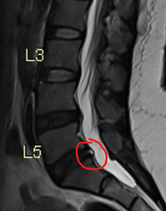
HIZ |
 |
Unlike vertebrogenic pain, discogenic pain may be confirmed by provocative discography and is typically addressed using regenerative therapies (e.g., PRP, stem cells) or intradiscal electrothermal therapy (IDET), or biacuplasty.
🔹 Key Anatomical Differences
| Feature | Discogenic Pain | Vertebrogenic Pain |
|---|---|---|
| Source | Annulus fibrosus | Vertebral endplates |
| Nerve Pathway | Sinuvertebral nerve | Basivertebral nerve (BVN) |
| Typical Imaging | High-intensity zone, annular tear | Modic type 1 or 2 changes |
| Symptoms | Axial pain worsened by sitting, flexion | Deep midline pain, prolonged standing, activity |
| Treatment | Regenerative (PRP, stem cells), IDET | BVN ablation using platforms such as Intracept or OptaBlate |
✅ Clarifying Statement
💡 Clinical Pearl: While both discogenic and vertebrogenic pain stem from degenerative spinal changes, they are distinct entities with different anatomical origins, neural pathways, and treatment strategies. Accurate differentiation is essential for optimal care.
Category clarification: Patients often search specifically for Intracept, but the broader treatment category is basivertebral nerve ablation. Newer systems such as OptaBlate are also designed to treat the same vertebrogenic pain pathway.
Stages of Degenerative Disc Disease and Endplate Involvement
Degenerative Disc Disease (DDD) is a progressive condition in which intervertebral discs lose hydration, elasticity, and structural integrity over time. This degeneration places increased stress on adjacent vertebral endplates, often contributing to vertebrogenic pain.
Common Stages of DDD Progression:
- Stage 1 – Early Disc Dehydration: Nucleus pulposus begins to lose water content. MRI shows mildly decreased T2 signal. Patients may have little or no symptoms.
- Stage 2 – Disc Narrowing and Annular Fissures: Disc height starts to reduce. Small annular tears may develop. Endplates show signs of early stress and inflammation.
- Stage 3 – Advanced Degeneration: Disc collapses. There is significant loss of height and signal. Endplates appear sclerotic or eroded. This stage is commonly associated with Modic changes and vertebrogenic pain.
- Stage 4 – Segmental Instability and Facet Arthropathy: Severe disc collapse causes abnormal motion and facet joint overload. May lead to stenosis and nerve root involvement.
Modified Pfirrmann Grading Scale (Grades 1–8)
The Modified Pfirrmann Scale is a widely used MRI grading system for lumbar disc degeneration. It assesses disc hydration, signal intensity, structure, and disc height. A modified classification was proposed in 2007 by Griffith et al. to better categorize degenerative discs in older patients where most discs are Pfirrmann grade III or IV. It has, however, not been as widely adopted.
| Grade | T2 Signal Intensity | Annular Fiber Junction | Disc Height |
|---|---|---|---|
| Grade 1 | Uniformly hyperintense, equal to CSF | Distinct junction between inner and outer annular fibers posteriorly | Normal |
| Grade 2 | Hyperintense (between presacral fat and CSF), ± hypointense intranuclear cleft | Distinct junction | Normal |
| Grade 3 | Hyperintense (less than presacral fat) | Distinct junction | Normal |
| Grade 4 | Mildly hyperintense (slightly more than outer annular fibers) | Indistinct junction | Normal |
| Grade 5 | Hypointense (equal to outer annular fibers) | Indistinct junction | Normal |
| Grade 6 | Hypointense | Indistinct junction | ~30% reduction |
| Grade 7 | Hypointense | Indistinct junction | 30–60% reduction |
| Grade 8 | Hypointense | Indistinct junction | >60% reduction |

Integration into Clinical Decision-Making
Recognizing whether a patient’s axial pain is discogenic or vertebrogenic helps guide the therapeutic approach:
- Stages 2–5 Pfirrmann: More favorable for regenerative intradiscal therapy
- Stages 6–8 Pfirrmann: Often associated with Modic changes and vertebrogenic pain → BVN ablation is often more appropriate, including options such as Intracept or OptaBlate
Self-Assessment Scoring Tool
This simple point-based system helps non-specialists gauge whether their low back pain may be discogenic or vertebrogenic in origin. While not diagnostic, it can encourage appropriate next steps:
- 1 point: Persistent midline low back pain (axial)
- 1 point: Absence of radicular pain or sciatica extending past the knee (learn more)
- 1 point: Pain duration >3 months
- 1 point: Pain worsens while sitting
- 1 point: Pain worsens during forward flexion
- 1 point: Pain improves with standing
- 1 point: MRI shows Modic changes (Type 1 or 2)
- 1 point: MRI shows high-intensity zones or annular tears
- 1 point: Facet joint syndrome ruled out via diagnostic medial branch block
- 1 point: SI joint dysfunction ruled out via diagnostic SI joint injection
Interpretation:
- 0–4 points: Low probability — conservative care may be sufficient
- 5–7 points: Moderate probability — likely structural pain generator; clinical imaging and expert evaluation recommended
- 8–10 points: High probability — strong candidate for advanced diagnostic workup, including targeted image-guided injections
Note: This tool is an educational guide and not a substitute for formal clinical diagnosis. Accurate evaluation requires high-quality spine MRI, physical examination, and expert correlation using validated diagnostic techniques.
📞 Ready to Take the Next Step?
If your symptoms align with the scoring criteria above, or if you’re seeking expert evaluation for persistent back pain, schedule a consultation with Dr. Amit Sharma.
Treatment Strategy Overview
The most important aspect of effective treatment lies in arriving at a high score in the self-assessment tool. A high score increases confidence that the pain generator is truly internal to the disc or vertebral body, allowing clinicians to avoid unnecessary treatments or surgeries that don’t address the root cause.
Conservative Care
- Core strengthening programs and posture retraining
- Chiropractic adjustments (with focus on spinal mobility and mechanics)
- Physical therapy involving stabilization and flexibility training
- Intermittent mechanical traction (limited evidence for short-term relief)
Evolution of Interventional Spine Care
- Epidural Steroid Injections: Work better when disc has herniated into the spinal canal and is compressing a spinal nerve root causing sciatica. The success rate of epidural steroids for discogenic or vertebrogenic low back pain is 50% at best, and is short lasting. Transforaminal injections (instead of interlaminar approach) tend to be better in this clinical scenario. Discuss the choice of technique and medication with your doctor.
- IDET (Intradiscal Electrothermal Therapy): Applies controlled thermal energy to seal annular tears.
- Biacuplasty: Uses bipolar radiofrequency to target painful discs without open surgery.
- Disc-FX System (off-label for annular fissures): Combines mechanical and thermal nucleus modulation through a minimally invasive access.
Regenerative Medicine Options
These treatments focus on biologic repair and inflammation reduction using orthobiologic materials:
- Platelet-rich plasma (PRP)
- Bone marrow aspirate concentrate (BMAC)
- Stem Cells (Wharton’s Jelly or amniotic-derived) injectable
They have been extensively used intradiscal, epidural and even as a “segmental approach” for wider coverage to include secondary pain generators. Always discuss the target structures and the rationale with your interventional provider.
Disclaimer: Regenerative therapies for discogenic back pain are considered investigational and are not currently FDA-approved. While early studies and clinical experience are promising, individual outcomes may vary. Patients should discuss risks, benefits, and alternatives with a qualified spine physician.
Basivertebral Nerve (BVN) Ablation
This procedure targets the basivertebral nerve, which carries pain signals from damaged vertebral endplates. By ablating this nerve within the vertebral body, pain transmission is disrupted, providing sustained relief for patients with vertebrogenic back pain.
- Known As: BVN ablation, including the FDA-cleared Intracept Procedure and newer platforms such as OptaBlate
- Indications: Axial low back pain with Modic type 1 or 2 changes
- Setting: Minimally invasive outpatient procedure under fluoroscopic guidance
- Benefits: Durable pain relief, improved function, opioid reduction
Clinical Insight: BVN ablation is a breakthrough in targeting vertebrogenic pain, but requires careful patient selection, appropriate imaging, and exclusion of other pain generators.
Technology note: While many patients recognize the term Intracept, physicians increasingly discuss vertebrogenic pain in terms of the broader BVN ablation category. OptaBlate is another procedure platform designed to treat the same basivertebral nerve target in appropriately selected patients.
Spinal Fusion Surgery
Spinal fusion is often considered the final step in the treatment ladder for chronic axial back pain, especially when conservative and interventional options fail. It involves permanently joining two or more vertebrae using screws and rods/plates to eliminate bone motion.
- Indications: Severe instability, gross mechanical collapse, or failure of all other modalities
- Outcomes: Variable success rates (~50-60%) depending on indication, imaging findings, and patient selection
- Risks: Adjacent segment disease, non-union, infection, persistent pain, and reduced spinal mobility
Clinical Insight: Fusion should be reserved for select cases with strong radiologic and clinical correlation. Most patients with vertebrogenic or discogenic pain can avoid fusion through earlier identification and targeted therapies.
Patient Perspective
“After years of failed injections and physical therapy, I finally found relief through the Intracept procedure. My pain is no longer controlling my life.”
— Patient treated with BVN ablation, shared with permission
Additional Note on Vertebrogenic Pain Treatment Options
As understanding of vertebrogenic pain has evolved, so has the treatment landscape. Historically, many patients with chronic midline low back pain were grouped under broad labels such as degenerative disc disease or nonspecific axial back pain. However, the recognition of vertebral endplate damage, Modic changes, and basivertebral nerve involvement has made it possible to target this pain generator more precisely. This is why basivertebral nerve ablation has become such an important concept in modern interventional spine care.
Today, patients may encounter different names while researching treatment. Some will search for the Intracept Procedure, while others may hear about OptaBlate. These are not competing diagnoses. Rather, they are procedure platforms within the same broader therapeutic category of BVN ablation. Both aim to treat pain carried by the basivertebral nerve in patients with vertebrogenic pain. This distinction matters because it helps patients understand that the diagnosis comes first, and the selected platform comes second.
For that reason, anyone with chronic low back pain, Modic changes on MRI, and failure of conservative care should not simply ask, “Do I need Intracept?” A better question is, “Do I have vertebrogenic pain, and am I a candidate for BVN ablation?” Once that diagnosis is established, the discussion can naturally include technologies such as Intracept or OptaBlate based on physician assessment, anatomy, procedural planning, and clinical judgment.
Frequently Asked Questions (FAQ)
What is vertebrogenic pain?
How is vertebrogenic pain different from discogenic pain?
Who is a candidate for the Intracept Procedure?
Is OptaBlate also used for vertebrogenic pain?
How long do the results of BVN ablation last?
Can regenerative therapies help with vertebrogenic pain?
Key References
- Fischgrund JS, et al. Intraosseous basivertebral nerve ablation for the treatment of chronic low back pain: a prospective randomized double-blind sham-controlled multi-center study. Eur Spine J. 2018.
- Truumees E, et al. Basivertebral nerve ablation for the treatment of chronic low back pain associated with Modic changes: a review. Pain Med. 2021.
- Bogduk N. The anatomical basis for spinal pain syndromes. Pain Med. 2013.
- Kallewaard JW, et al. 2020 guidelines for lumbar discogenic pain. Pain Pract. 2020.
- Becker S, et al. Correlation between Modic changes and pain severity: a prospective MRI-based study. Spine J. 2008.


