
Radiofrequency Ablation (RFA) or Rhizotomy
Introduction
Welcome to our information page about radiofrequency ablation (RFA), neurotomy, or rhizotomy. This article will focus on the basics of this technique and the painful conditions it can be used to treat.
Dr. Sharma is one of the few physicians on Long Island who is currently offering all types of radiofrequency ablation options; from conventional continuous high heat RFA (for neck, mid-back, and low back pain) using the most advanced techniques (NIMBUS), water-cooled moderate temperature RFA (Coolief for shoulder, hip, knee, sacroiliac joint-related pain, intracept for vertebrogenic low back pain), low temperature RFA for delicate structures (pulsed RFA for chronic radiculopathy), to cryoablation (occipital neuralgia) and chemical neurolysis (intercostal nerves for chest pain related to shingles).
Dr. Sharma also performs neurotomy for several painful nerve conditions like occipital headaches, neuralgia paresthetica, intercostal neuralgia, and chemoablation for cancer-related pain. He is the only physician on Long Island currently offering the most advanced option of pulsed radiofrequency ablation of the dorsal root ganglion for patients who cannot (or do not want to) undergo decompressive back surgery for “pinched nerve-related sciatica“. He is also performing advanced radiofrequency ablation techniques like intracept (basivertebral nerve ablation).
Understand Basic Neuroanatomy
Your brain works as a supercomputer. To run your body efficiently, it sends and receives signals from most organs and tissues (skin, muscles) through small neuronal-wires, which are called “nerves” in medical terms. A short lesson on the basics of human neuroanatomy is important to understand this topic fully.
The central nervous system (CNS), consisting of the brain and spinal cord, serves as the control center of the body. Peripheral nerves act as communication lines, transmitting signals between the CNS and the rest of the body. Damage to nerves can result in various pain conditions, making treatments like radiofrequency ablation critical for pain management.
According to the Cleveland Clinic (Cleveland Clinic), radiofrequency ablation is a safe and effective treatment for managing chronic pain, particularly in areas such as the spine, hip, knee, and shoulder.
The Concept Behind Neuronal Destruction
Pain is a sensory and emotional experience. Past experiences of pain help you avoid injuries. When you do get injured, pain helps you avoid further damage to the involved area. In some conditions, pain becomes a nuisance and needs to be blocked. Pain can be blocked, or reduced, by:
- Curing the underlying condition
- Suppressing the central nervous system from receiving painful signals (using drugs)
- Destroying the nerves that carry painful signals to the spinal cord and brain
In a perfect world, the only part of central or peripheral nervous system that we ever want to destroy are the small nerves that carry pain signals to the brain. The idea of destroying pain carrying nerves has always fascinated scientists and doctors. It can be achieved using a chemical agents (neurolysis), freezing cold temperatures (cryoablation) or heat (radiofrequency ablation or rhizotomy).
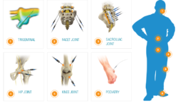 Radiofrequency ablation has been used to destroy pain carrying nerve endings for many medical conditions. In pain management world, it has historically been used to deaden the pain carrying nerve endings of spinal facet joints (medical branches). Recently this technology has been sophisticated to destroy pain carrying nerve fibers of:
- Vertebral bone (Intarcept)
- Degenerated disc (IDET, Transdiscal Coolief)
- Sacroiliac joint (L5 dorsal ramus and sacral lateral branches)
- Hip (articular branches of the obturator and femoral nerves) and
- Knee joint (genicular nerves)
- Shoulder joint (articular branches of the axillary, suprascapular and subscapular nerves)
According to Mayo Clinic (Mayo Clinic), radiofrequency ablation works by sending radiofrequency energy through a needle to heat and destroy nerve tissue that transmits pain signals. This technique has been proven effective for managing various chronic pain conditions.
Radiofrequency Ablation Procedure
During a radiofrequency ablation procedure, the patient lies face down. The procedure is outpatient, performed under local anesthesia with intravenous sedation. It typically takes approximately 20-30 minutes. After numbing the site, a small needle is inserted through the skin to target the specific nerve causing pain.
Fluoroscopy, an imaging technique, is used to guide the needle precisely to the targeted nerve. Once in place, radiofrequency energy generates heat, creating a lesion that disrupts the nerve’s ability to transmit pain signals.
According to the American Society of Regional Anesthesia and Pain Medicine (ASRA), this procedure is especially effective for pain originating from facet joints, sacroiliac joints, and other specific structures.


After The Radiofrequency Rhizotomy
Patients typically resume normal activities the day after undergoing radiofrequency ablation. Some soreness, numbness, or weakness in the targeted region is common for a few weeks. However, pain relief generally begins within 2-3 weeks and can last anywhere from several months to over a year.
According to the John Hopkins Medicine (Johns Hopkins Medicine), patient recovery is usually rapid with minimal side effects, making radiofrequency ablation a preferred choice for managing chronic pain.
Risks of Radiofrequency Rhizotomy
Although radiofrequency ablation is considered a safe procedure, some risks exist. Potential complications include:
- Infection at the injection site
- Bleeding
- Nerve damage
- Temporary pain increase
- Allergic reaction to medications used during the procedure
Consulting a highly skilled pain specialist like Dr. Sharma ensures that the risk of complications remains minimal.


