
Baastrup’s Syndrome: 5 Critical Facts You Must Know
Baastrup’s syndrome is a frequently misunderstood cause of low back pain. On imaging, radiologists often describe “kissing spines” or “Baastrup’s disease” when adjacent lumbar spinous processes are enlarged and touch each other. But those radiologic findings alone do not mean the patient truly has painful Baastrup’s syndrome.
In many people, these changes are simply part of age-related degeneration and are completely painless. In others, the same anatomy can cause focal midline pain that worsens with extension and improves with flexion. This page explains how to distinguish a harmless radiology finding from a true clinical syndrome, how Baastrup’s syndrome is diagnosed, and which minimally invasive treatments can help.
For clinicians and patients who want to read more, useful background is available in open-access reviews such as the pictorial essay on Baastrup’s disease in the medical literature and radiology resources like Radiopaedia’s Baastrup disease overview and other peer-reviewed summaries.
1. Baastrup’s Sign vs. Baastrup’s Disease vs. Baastrup’s Syndrome
The terminology around Baastrup can be confusing, so it is important to separate three different concepts:
 |
|
Benvenuto, P., Attarian, A. Kissing spines: an underdiagnosed cause of low back pain.Intern Emerg Med 13, 955–956 (2018). https://doi.org/10.1007/s11739-018-1848-4.
|
In short: Baastrup’s sign and Baastrup’s disease describe what the spine looks like.
Baastrup’s syndrome describes what the patient feels.
2. What Causes Baastrup Changes in the Spinal Column?
Baastrup-related findings are considered part of the family of spinal column conditions. They involve the posterior elements of the lumbar spine rather than the discs themselves. Over time, several factors contribute:
- Degenerative changes and disc height loss – as discs collapse, the spinous processes come closer together in extension.
- Repetitive flexion–extension loading – chronic strain on the interspinous and supraspinous ligaments can lead to thickening, inflammation, and eventual bony remodeling.
- Hyperlordosis – increased lumbar curvature brings the spinous processes into repeated contact.
- Global degenerative disease – Baastrup changes often coexist with facet arthrosis, disc degeneration, and sometimes spondylolisthesis.
Over time, the tissue between the spinous processes can develop inflammation and even interspinous bursae. In some patients, this process becomes painful and evolves into true Baastrup’s syndrome; in others, it remains a silent byproduct of age-related wear and tear.
3. Symptoms of Baastrup’s Syndrome: When “Kissing Spines” Actually Hurt
Not everyone with Baastrup changes on MRI or X-ray has Baastrup’s syndrome. The clinical picture matters. Typical symptom patterns include:
- Focal midline low back pain – usually in the lower lumbar region (often L3–L5), felt directly over the spinous processes.
- Worse with extension – standing upright, arching back, or lying flat on the stomach can reproduce or intensify pain.
- Improved with flexion – bending forward, sitting in a flexed position, or curling slightly often reduces symptoms.
- Localized tenderness – pressing on the involved spinous processes reproduces the familiar pain.
- Minimal or no leg symptoms – unlike classic spinal stenosis or disc herniation, leg numbness or true radiculopathy may be absent or mild.
Because other spinal column conditions—such as facet arthropathy, spinal stenosis, and myofascial pain—can mimic this pattern, careful examination is essential. True Baastrup’s syndrome is suspected when the pain is sharply localized to the interspinous region and correlates with imaging at the same level.
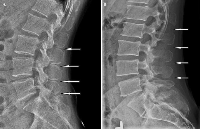
4. Imaging Findings and Radiology Pitfalls
On imaging, Baastrup changes are often striking. Common features include:
- Enlarged, flattened, or sclerotic spinous processes in the lumbar spine.
- Narrowed interspinous space with “kissing” of adjacent spinous processes, especially in extension views.
- Interspinous bursitis or fluid on MRI in some patients.
- Associated degenerative disc disease, facet arthrosis, and sometimes mild spondylolisthesis.
However, one of the key dilemmas with Baastrup’s syndrome is that radiologic findings are common while clinically significant pain is much less common. Several studies have shown that Baastrup changes increase with age and can be found in many people who have no back pain at all.
Radiology pitfalls include:
- Labeling every case of “kissing spines” as the primary pain source.
- Overlooking other major causes of pain such as spinal stenosis, sacroiliac joint dysfunction, or vertebral compression fractures.
- Failing to correlate the exact level of tenderness on exam with the level identified on imaging.
For clinicians, it is crucial to treat the patient, not just the picture. Baastrup’s syndrome should only be diagnosed when clinical findings, imaging, and—ideally—diagnostic injections all agree.
5. How Is Baastrup’s Syndrome Diagnosed?
A confident diagnosis of Baastrup’s syndrome usually requires three components:
1. Clinical Evaluation
- Detailed history focused on pain location, aggravating and relieving factors, prior injuries, and previous treatments.
- Physical examination assessing posture, lumbar lordosis, range of motion, and specific maneuvers in extension and flexion.
- Direct palpation of the lumbar spinous processes to identify focal tenderness over the suspected level.
2. Imaging Correlation
- X-rays – particularly lateral and flexion–extension views showing approximation of spinous processes.
- MRI – helpful for demonstrating interspinous bursitis, marrow edema, or cysts, and for assessing discs and facets.
- CT – can show bony sclerosis and remodeling when detailed bone evaluation is needed.
3. Diagnostic Interspinous Injection
The most convincing way to confirm Baastrup’s syndrome is often a targeted diagnostic injection:
- Under fluoroscopic or ultrasound guidance, a small amount of local anesthetic is injected into the painful interspinous space.
- Sometimes a small dose of steroid is added if both diagnosis and treatment are desired.
- Significant, short-term relief of the patient’s typical pain strongly supports the diagnosis.
If the injection does not change the pain, it is important to re-evaluate for other spinal column conditions such as facet joint pain, spinal stenosis, sacroiliac joint dysfunction, or vertebrogenic pain.
6. Treatment Options for Baastrup’s Syndrome
Once Baastrup’s syndrome is confirmed as a true pain generator, treatment is usually stepwise and minimally invasive where possible.
Conservative Management
- Activity modification – avoiding prolonged extension or positions that repeatedly load the interspinous region.
- Physical therapy – focused on core strengthening, hip mobility, postural training, and strategies to reduce excessive lumbar lordosis.
- Medication management – anti-inflammatories, analgesics, and other non-opioid strategies when appropriate.
Image-Guided Injections
- Interspinous injections – anesthetic and steroid delivered into the symptomatic interspinous space can calm inflammation and provide meaningful relief.
- Facet joint or medial branch blocks – if facet arthropathy coexists and contributes to pain.
- Epidural injections – if there is associated spinal stenosis or nerve-related leg pain.
Radiofrequency and Other Interventions
In select patients, radiofrequency techniques may be considered to target the interspinous region or adjacent pain pathways when diagnostic blocks have been clearly positive. This can extend the duration of relief compared to a single injection.
Surgical Options
Surgery is reserved for highly selected cases of Baastrup’s syndrome that have:
- Persistent, disabling pain despite comprehensive conservative and interventional therapies.
- Clear correlation between symptoms, imaging, and diagnostic injections.
Surgical options may include partial resection of the offending spinous process or wider decompression when there is coexisting spinal stenosis. As with other spinal column conditions, the best outcomes are achieved when the symptomatic level is correctly identified and other pain generators are not overlooked.
7. When Baastrup’s Findings Are Not the Problem
Perhaps the most important clinical fact is that many patients with “kissing spines” on imaging do not have Baastrup’s syndrome. In these cases, the Baastrup changes are incidental, and the true pain source might be:
- Degenerative disc disease or annular tears.
- Facet joint arthropathy at the same or adjacent levels.
- Spinal stenosis with neurogenic claudication.
- Sacroiliac joint dysfunction or hip pathology.
- Myofascial pain in the paraspinal muscles.
Treating the wrong target—such as operating on the spinous processes when stenosis or disc pathology is the main issue—can lead to persistent symptoms and frustration. This is why a structured approach to diagnosis is so important for Baastrup’s syndrome and other spinal column conditions.
Dr. Amit Sharma & our minimally invasive pain & spine team.
If your imaging report mentions “kissing spines,” “Baastrup’s disease,” or Baastrup’s sign—and you still do not have clear answers—consider a focused evaluation. Clarifying whether you truly have Baastrup’s syndrome or another spinal column condition is the first step toward precise, minimally invasive treatment tailored to you.


