Disc Bulge: 7 Valuable Insights for Fast, Lasting Relief
If your MRI report mentions a disc bulge or protrusion, it’s natural to wonder how serious it is—and whether it explains your back or neck pain. Many patients are told that a “bulging disc” is either nothing to worry about or, on the other extreme, that it will inevitably lead to surgery. The truth lies somewhere in between.
A disc bulge is one of the most common findings on spinal imaging. Sometimes it is completely painless. Other times, a bulging disc can irritate nerves, narrow the spinal canal, and cause symptoms that are just as intense as a herniated disc.
In this guide, we break down 7 powerful facts about disc bulges—what they are, how they differ from herniations and annular tears, when they become painful, and which non-surgical and minimally invasive treatments can help. Our goal is to replace fear and confusion with clarity and a plan.
1. What Is a Disc Bulge?
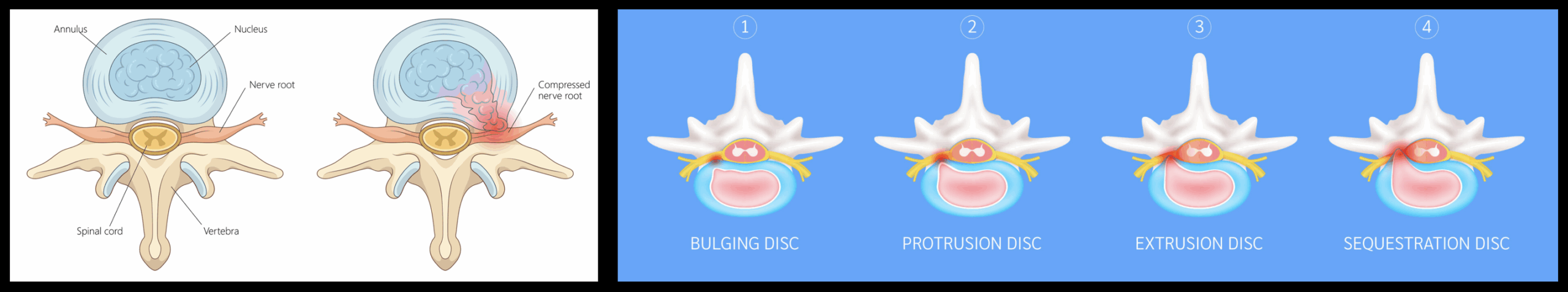
The human spine is made up of stacked bones called vertebrae, separated by intervertebral discs that act as shock absorbers. Each disc has:
- Annulus fibrosus – a tough outer ring of fibrous cartilage.
- Nucleus pulposus – a soft, gel-like center that distributes load and allows flexibility.
A disc bulge occurs when the disc extends beyond its normal boundary around most or all of its circumference, but the outer annulus remains reasonably intact. You can think of it like a tire that has flattened and pushed out slightly on all sides, without a complete blowout.
On MRI, a disc bulge may:
- Extend a few millimeters beyond the edges of the vertebral bodies.
- Push into the spinal canal or nerve exit holes (foramina).
- Coexist with other degenerative changes such as facet arthritis or ligament thickening.
Importantly, a disc bulge is a shape description, not a pain diagnosis. It only matters clinically if it compresses or irritates nearby nerves or structures that match your symptoms.
2. Disc Bulge vs Herniated Disc vs Annular Tear
Patients often hear several terms at once—disc bulge, herniated disc, annular tear—and understandably get confused. These are related but distinct problems.
Disc Bulge
- Broad, often symmetric extension of the disc beyond its normal margin.
- Annulus is stretched and degenerated but not necessarily fully torn.
- May or may not touch the spinal cord or nerve roots.
Disc Herniation
- More focal displacement of disc material (nucleus) through a weakened or torn area of the annulus.
- Can be a protrusion, extrusion, or even sequestration (free fragment).
- Often has a higher chance of directly compressing a nerve root.
Annular Tear
- Actual disruption in the annulus fibrosus.
- May be painful even without a large bulge or herniation.
- Can coexist with both disc bulges and herniations.
You can read more about these related disc problems on our dedicated pages:
3. Which Hurts More: Disc Bulge or Herniated Disc?
One of the most common questions we hear is: “Is a disc bulge better than a herniated disc?” The honest answer: it depends.
Pain is driven less by the label and more by:
- Location – central canal, lateral recess, foraminal, or far lateral.
- Size and shape – how much the bulge or herniation narrows the canal or foramina.
- Structures compromised – spinal cord, nerve roots, dorsal root ganglion, or only the disc itself.
- Inflammatory response – chemical irritation from disc material can hurt even without major mechanical compression.
Some patients have a large disc bulge that looks impressive on MRI but causes no symptoms. Others have a small focal herniation in a tight foraminal space that produces severe sciatica or arm pain.
The key message: a disc bulge is not automatically “mild,” and a herniation is not automatically catastrophic. Both can be:
- Completely asymptomatic, or
- Just as painful and disabling as each other—depending on size, location, and nerve involvement.
This is why we never treat the MRI alone. We correlate what we see on imaging with your actual symptoms and neurologic exam to determine whether the disc bulge is truly the pain generator.
4. How Common Are Disc Bulges—and Are They Always Serious?
Disc bulges are extremely common, especially with age. In fact, large population studies show that many adults with no back pain at all have disc bulges or herniations on MRI. One widely cited review in the American Journal of Neuroradiology and related research in journals like the New England Journal of Medicine have demonstrated that disc “abnormalities” are common incidental findings.
This means:
- Seeing the words “disc bulge” on your MRI does not automatically mean something is wrong.
- Even moderate or multilevel bulges may be benign if they do not match your symptoms.
- Some disc bulges are simply part of the natural aging process of the spine.
Our job is to determine if your disc bulge is:
- Clinically relevant – clearly matching your pain pattern, exam findings, and nerve distribution.
- Or incidental – present on imaging but not responsible for your symptoms.
This distinction protects you from both under-treatment and over-treatment, including unnecessary injections or surgery.
5. Symptoms of a Symptomatic Disc Bulge
When a disc bulge is truly causing problems, symptoms often resemble a herniated disc:
- Localized neck or back pain that may worsen with bending, loading, or prolonged sitting.
- Sciatica – sharp, shooting pain radiating down the leg, often from a foraminal or paracentral lumbar bulge.
- Cervical radiculopathy – arm pain, numbness, or tingling from a bulge in the neck.
- Numbness or tingling in a specific nerve distribution.
- Weakness in the muscles controlled by the affected nerve.
- Pain with coughing, sneezing, or straining if the bulge increases epidural pressure.
Bulges in different regions may behave differently:
- Lumbar disc bulge – low back pain, sciatica, or neurogenic claudication if combined with stenosis.
- Cervical disc bulge – neck pain, arm symptoms, or in rare cases spinal cord compression (myelopathy).
- Thoracic disc bulge – mid-back pain or band-like chest/abdominal pain, though symptomatic thoracic bulges are less common.
6. How We Diagnose a Disc Bulge (Beyond the MRI Report)
At SpinePain Solutions, we take a structured approach to evaluating a disc bulge:
Step 1: Detailed History
- Onset and pattern of pain.
- Activities that worsen or relieve symptoms.
- Work demands, hobbies, and prior injuries.
- Red flag symptoms such as significant weakness or bowel/bladder changes.
Step 2: Focused Physical and Neurologic Exam
- Spine range of motion and pain provocation testing.
- Strength, reflexes, and sensation in arms or legs.
- Special tests such as Straight Leg Raise, Spurling’s test, or neural tension testing.
Step 3: Imaging Review—Not Just the Radiology Summary
- We personally review your MRI or CT images—not just the written report.
- We look at the exact level, side, and location of the disc bulge.
- We correlate findings with your specific symptoms and exam (for example, an L4–L5 central bulge vs an L5–S1 foraminal bulge).
Step 4: Advanced Diagnostics When Needed
In selected complex cases, we may consider additional testing such as EMG (electromyography) through trusted neurologists with subspecialty training. However, we only use these tests when they will genuinely change management—not as a reflexive step for every disc bulge.
Unsure If Your Disc Bulge Is Really the Problem?
Many patients are told that a disc bulge explains all their pain—only to find out later that the real issue was somewhere else. We offer careful, second-opinion evaluations that focus on clinical relevance, not just the MRI picture.
7. Disc Bulge Treatment Options (Without Rushing to Surgery)
A diagnosis of a disc bulge does not mean you are headed straight to the operating room. Most patients improve with a combination of conservative care, interventional procedures, and, when appropriate, advanced minimally invasive techniques.
Conservative Management
- Targeted physical therapy – to improve core strength, posture, hip mobility, and mechanics that reduce disc stress.
- Activity modification – avoiding extreme flexion/extension or heavy lifting during flare-ups while staying gently active.
- Medications – short-term anti-inflammatories, muscle relaxants, or nerve pain agents used judiciously.
- Collaborative chiropractic care – when appropriate, we partner with experienced chiropractors to coordinate safe and effective manual therapies.
Precision Interventional Treatments
- Transforaminal Epidural Steroid Injections (TF ESI) – delivering medication to the front of the epidural space, where a disc bulge or herniation typically compresses the nerve.
- Selective nerve root blocks – both diagnostic and therapeutic for radiating arm or leg pain.
- Epidural injections with directed catheters (e.g., Versa-Kath) – allowing more precise treatment around complex multilevel bulges, including at challenging cervical levels.
Advanced Options for Persistent Radicular Pain
- Pulsed radiofrequency (PRF) of the dorsal root ganglion (DRG) – to modulate chronic nerve pain without destroying the nerve.
- Neuromodulation (spinal cord or DRG stimulation) – in carefully selected patients with severe, longstanding symptoms.
Regenerative and Disc-Focused Procedures
For some patients with discogenic pain or early degeneration, we may discuss emerging options such as PRP or other biologic therapies in collaboration with our regenerative medicine division, Bloom Infusions & Wellness. These treatments are considered investigational and are not FDA-approved for disc bulges or herniations, but they may play a role in select cases.
Disclaimer: Regenerative medicine therapies are considered investigational and are not FDA-approved for treating disc bulges or herniated discs. Individual results vary and all options should be discussed thoroughly.
When Minimally Invasive Surgery Is Considered
True surgical indications for a disc bulge are similar to those for a herniated disc:
- Severe or progressive neurologic deficits (significant weakness, myelopathy).
- Intractable pain that does not respond to thorough non-surgical care.
- Emergencies such as cauda equina syndrome.
When surgery is truly necessary, minimally invasive techniques—such as full endoscopic discectomy or targeted decompression—can often address the bulge while preserving stabilizing structures. We collaborate with high-quality surgeons who share a patient-first, evidence-based philosophy and do not operate based on MRI findings alone.
When to See a Spine Specialist for a Disc Bulge
You should seek evaluation for a disc bulge if:
- Pain lasts longer than 2–3 weeks despite rest and simple measures.
- You have radiating symptoms into an arm or leg.
- You notice new numbness, tingling, or weakness.
- Pain interferes with work, sleep, or daily activities.
- You experience any loss of bowel or bladder control (this is an emergency).
At SpinePain Solutions, we provide detailed evaluations and individualized care plans across our Long Island locations in Commack, Bay Shore, Bethpage, and Huntington, NY.
Dr. Amit Sharma & our minimally invasive pain & spine team.
If your MRI report mentions a disc bulge and you are unsure what it really means for your health, we are here to help. A careful, minimally invasive evaluation can clarify whether the bulge is truly the problem and guide you toward the safest, most effective treatment options.


