

ION Minimally Invasive Facet Fusion: Candidacy, Evidence and Recovery
ION minimally invasive facet fusion uses facet screw fixation and bone graft to support posterior spinal fusion in selected surgical cases. It is not simply a longer-lasting version of radiofrequency ablation, and it is not the automatic next step after successful diagnostic blocks.
The decision comes before the device: identify the condition being treated, determine whether fusion is justified, and then select an appropriate fixation strategy. A small incision or low-profile implant does not establish candidacy or guarantee a better result.
Medial branch blocks test the response to temporarily anesthetizing the facet nerve supply. They are commonly used to help select patients for medial branch RFA. They do not show whether a spinal segment is unstable or whether fusion will succeed. Fusion requires a separate clinical and structural assessment.
For broader surgical options, read our minimally invasive spinal fusion guide. This page focuses on facet-fusion selection and how ION fits into that decision.
What Is ION Minimally Invasive Facet Fusion?
The facet joints are paired joints at the back of the spine. They help guide movement and contribute to stability. A facet fixation procedure places screws across the selected joint pathway; bone preparation and graft are used to support fusion over time. Initial fixation and completed biological fusion are not the same thing.
ION is a device family, not one identical operation for every patient. The exact implant, treated level, bone preparation, graft material and accompanying procedures must be specified. Facet fixation can be part of a larger fusion plan rather than a replacement for every pedicle screw, interbody or decompression procedure.
FDA-Cleared, Not FDA-Approved
The original ION Facet Screw System received 510(k) clearance in 2022; ION 3D received clearance in 2024. Their documentation describes posterior surgical indications at C2-S1, including spondylolisthesis, spondylolysis, symptomatic failed fusion or pseudoarthrosis, and defined degenerative conditions including facet disease with instability. The devices are intended for use with bone graft. A listed spinal range does not make every patient or level suitable. Original ION clearance; ION 3D clearance.
The appropriate term is FDA-cleared. Clearance is not a demonstration that this technique is superior to RFA or conventional fusion, and it does not establish insurance coverage. The instructions for the actual proposed model must be checked; different ION variants should not be treated as interchangeable.
When Is Facet Fusion Considered After Successful Diagnostic Blocks?
Fusion may be considered when the symptoms, imaging and overall surgical assessment support an appropriate structural indication and the expected benefit justifies the risks. Persistent pain alone, facet arthritis on an MRI, or relief after a block does not independently establish that indication.
| What a medial branch block can support | What it does not establish |
|---|---|
| The patient’s familiar pain responds to temporary anesthesia of the targeted nerve supply | Spinal instability or a need to immobilize a segment |
| A prognostic assessment for medial branch RFA in an appropriate clinical setting | Reliable prediction of facet-fusion benefit |
| One part of evaluating a suspected facet contribution | Proof that no disc, SI-joint, hip or nerve source also contributes |
The multispecialty lumbar-facet guidelines address blocks as prognostic tests for RFA, not stand-alone surgical clearance. Read our medial branch block guide for injection interpretation and documentation.
Record the usual pain, activity response and timing rather than relying only on a recalled percentage. Sedation, medication changes, low activity and anesthetic spread can affect interpretation. Block thresholds for RFA should not be imported as a universal facet-fusion rule.
Findings That Support Further Surgical Assessment
| Potential supporting findings | Reasons to evaluate or treat further first |
|---|---|
| Persistent, meaningful functional limitation despite appropriate care | Improving symptoms or mild pain without major functional limitation |
| A structural diagnosis that matches the proposed operation and level | Incidental degeneration without a coherent surgical target |
| Clinical and imaging findings supporting an appropriate fixation plan | Positive blocks alone or discordant symptoms and imaging |
| Competing lumbar, SI-joint, hip and neurological causes evaluated | Predominant untreated radiculopathy or another likely pain generator |
| Bone quality and general medical risk assessed | Active infection, impaired healing or unacceptable anesthesia risk |
| Ability to follow restrictions and realistic goals | Inadequate home support or an expectation of guaranteed complete relief |
Standing radiographs, MRI or CT, and selected dynamic radiographs may be useful depending on the question. Imaging must be interpreted alongside symptoms and examination; the presence of arthritis is not proof that it causes the patient’s pain. The level of instability or deformity may require a more comprehensive surgical plan rather than limited facet fixation.
A Practical Decision Pathway
- Localize the problem: review symptoms, examination and competing disc, nerve-root, SI-joint and hip sources.
- Interpret the block for its purpose: determine whether it supports a facet contribution and RFA selection.
- Consider lower-risk care: rehabilitation, medication optimization and appropriately selected RFA may be suitable.
- Reassess persistent pain: failed or short-lived RFA does not itself demonstrate instability.
- Assess the structural surgical indication: specify the level, pathology and why fusion rather than another treatment is justified.
- Select the actual operation: decide whether ION fixation, another construct, decompression or combined surgery fits the anatomy and goals.
A poor RFA result should prompt review of diagnosis, target, technique and other pain sources. When a structural surgical problem is already established, RFA is not a mandatory prerequisite to every operation. The two treatment pathways should not be collapsed into one automatic sequence.
Facet RFA Versus Facet Fusion
| Decision factor | Medial branch RFA | Facet fixation and fusion |
|---|---|---|
| Primary goal | Reduce pain transmission from selected facet nerve supply | Provide fixation and support bone fusion for an appropriate surgical indication |
| Permanent implant | No | Yes |
| Instability | Does not mechanically stabilize the segment | Can provide fixation within an appropriate surgical construct |
| Selection | Clinical assessment and suitable prognostic blocks | Separate structural, anatomical and surgical risk assessment |
| Healing | Does not require bone fusion | Requires internal healing and development of fusion |
| Loss of benefit | May require reassessment; repeat treatment can be considered in selected cases | Requires reassessment of healing, implants and competing pain sources |
For the nonsurgical pathway, see radiofrequency ablation. Do not infer a head-to-head ION advantage from studies of RFA, other facet devices or conventional lumbar fusion.
How the Procedure Is Performed
The plan should identify the exact device model, spinal level or levels, approach and whether fixation accompanies decompression or another fusion technique. Anesthesia and the setting depend on the operation and patient risk; an outpatient setting does not automatically mean a low-risk procedure.
- Planning: clinical findings and imaging are reviewed with bone quality, previous surgery and medical risk.
- Positioning and access: the patient is positioned for the planned posterior approach and imaging guidance.
- Bone preparation and graft: the appropriate surfaces are prepared and graft is placed according to the technique and device instructions.
- Fixation: the selected facet screws are placed and their positions assessed with the planned imaging.
- Additional treatment: decompression or other fixation is performed if required by the agreed surgical plan.
- Closure and follow-up: wound care, medications, restrictions and follow-up are specified before discharge.
Procedure length, incision size, anesthesia requirements and same-day discharge vary. They should not be advertised as fixed advantages for every ION case.

Procedure illustration: the actual access, level and fixation strategy depend on the selected device and surgical plan. An illustration of a smaller approach is not comparative evidence of lower infection, anesthesia or revision risk.
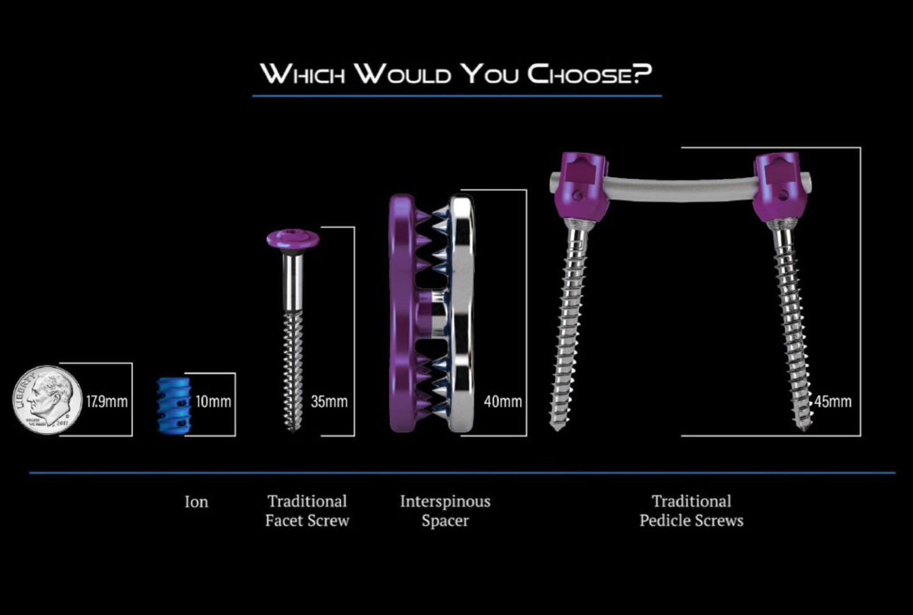
Illustrative device material from the existing page. Implant configurations and clinical applications vary; this image is not a patient outcome comparison.
ION Procedure Demonstration
Open the original ION demonstration video (MOV) if the player does not work in your browser. Demonstration material illustrates a technique; it does not establish candidacy or comparative benefit.
Evidence and Important Limitations
| Evidence source | What it addresses | What it does not establish |
|---|---|---|
| FDA 510(k) documents | Specified indications, device design and substantial equivalence; nonclinical performance testing | A clinical success rate or superiority over other operations |
| 2022 facet-stabilization systematic review | Published experience with multiple fixation techniques and surgical contexts | An ION-specific recommendation based on positive blocks alone |
| Manufacturer surface and histology material | Design concepts and preclinical findings | Faster human fusion, less postoperative pain or fewer revisions |
| ION observational study registration | A planned framework for assessing outcomes | Completed comparative results or a patient benefit guarantee |
Evidence for the exact stand-alone, percutaneous facet-fusion application after successful blocks should not be equated with evidence for conventional fusion or for screws used as supplemental fixation. The 2022 systematic review considers multiple techniques; its findings are not interchangeable across devices, levels and combined procedures.
A registered study is not itself proof of effectiveness. The ION Facet Screw System Ambispective Evaluation is described as observational; assess the published methods and results for the exact application before using an outcome statistic.
Nanotex, Bone Graft and Biologic Adjuncts
The manufacturer describes Nanotex surface characteristics intended to support bone integration, but specifically cautions that animal findings may not represent clinical performance. Fluid attraction or animal bone ingrowth is not proof that patients fuse faster or experience less pain. Manufacturer design and preclinical information.
Discuss the actual graft separately from the implant. Autograft, allograft and any other proposed material have different purposes, evidence and risks. Do not assume that PRP, bone marrow aspirate or a marketed cellular product improves ION outcomes because it can be placed near an implant. The patient should understand the specific product, clinical rationale, regulatory status and any additional cost.
Alternatives and Conditions ION Does Not Directly Treat
- Rehabilitation: progressive conditioning, strength, mobility and function-focused care.
- Medication management: individualized symptom control with attention to risk.
- Medial branch RFA: a potential option for appropriately selected facet-mediated pain without an independent need for fixation.
- Decompression: may be needed when stenosis or disc disease compresses neural structures.
- Other fusion strategies: may be required for anatomy, instability, deformity or a construct needing more extensive support.
- Treatment of another source: SI-joint, hip, disc or neurological disorders may need a different approach.
- Continued nonsurgical care: reasonable when expected surgical benefit does not justify risk.
Facet fixation does not directly remove a herniated disc or open a narrowed spinal canal. It may accompany decompression when fixation is also appropriate, but should not replace necessary treatment of nerve compression. Likewise, basivertebral nerve ablation and facet fixation address different clinical questions; combining them requires a separately supported indication for each treatment.
Facet-joint debulking and FacetFuse are different procedures or technologies. Their suitability and evidence should be reviewed rather than presented as equivalent alternatives for every patient.
Recovery After ION Facet Fusion
Recovery depends on the level, number of treated segments, approach, accompanying procedures and the patient’s health. A limited fixation operation and fixation combined with decompression or interbody fusion should not share an identical recovery promise.
| Recovery stage | General priorities |
|---|---|
| Immediately after the operation | Monitor pain, neurological status and safe mobility; protect the wound; clarify discharge and medication instructions |
| Early wound-healing period | Take permitted short walks, attend wound follow-up and observe the written lifting, bending and twisting limits |
| Ongoing internal healing | Progress activity and rehabilitation only as directed; reassess symptoms and healing at planned visits |
| Return to work and exercise | Base clearance on the actual operation, functional recovery, commuting and physical demands rather than a universal date |
Bone fusion can take months even when the incision feels better. This is general spinal-fusion counseling, not a measured ION-specific timeline. Follow the operating clinician’s instructions. AAOS spinal-fusion recovery guidance.
Walking, Showering, Driving and Work
- Walking: increase permitted activity gradually; use assistance if balance or leg symptoms require it.
- Wound care: follow the actual dressing and closure instructions. Showering and soaking are separate decisions.
- Driving: wait for clearance, stop impairing medication and ensure safe entry, exit, turning and emergency braking.
- Work: sedentary duties may need a different plan from lifting, climbing, patient transfers or machinery.
- Exercise and therapy: respect bone-healing precautions; early aggressive loading is not justified by a small incision.
- Medication: ask the team about blood thinners, anti-inflammatory drugs and any other changes. Do not stop prescribed medication on your own.
Smoking, diabetes, impaired bone health, poor conditioning and additional procedures can complicate healing. Pain improvement does not prove that fusion is complete, and persistent soreness does not by itself establish failure.
Risks and Warning Signs
Potential complications include:
- Infection and wound complications
- Bleeding or hematoma
- Nerve injury; risk to other neural structures depends on the spinal level and operation
- Implant malposition, loosening, migration or breakage
- Bone injury or fracture
- Nonunion or pseudoarthrosis
- Persistent, recurrent or worsening pain
- Symptoms involving adjacent levels or another pain source
- Anesthesia complications
- Need for further fixation, revision or removal
Contact the treating team promptly for fever, drainage, spreading redness, wound opening, increasing swelling or severe escalating pain. New major weakness, saddle numbness, loss of bowel or bladder control, chest pain or shortness of breath require urgent medical evaluation.
Fusion is intended to reduce motion at the treated region. It does not guarantee complete relief, eliminate every source of spinal pain or ensure that future surgery will never be needed.
Insurance and Preoperative Planning
FDA clearance is not automatic insurance approval. Coverage depends on the diagnosis, operation, device and technique, documentation, treated levels and current plan policy. Obtain an authorization review and a written estimate of patient costs before proceeding; prior authorization is not an unconditional guarantee of payment.
Before choosing ION, ask:
- What structural diagnosis justifies fusion, rather than treatment of facet pain alone?
- Which exact ION model and spinal levels are planned?
- What does my block response support, and what does it not answer?
- Why is this fixation strategy appropriate for my anatomy and bone quality?
- Is the proposed fixation stand-alone or part of a combined operation?
- Do I also need decompression, and which symptoms might remain?
- What independent clinical evidence applies to this exact application?
- What graft or biologic material will be used, and why?
- What are my written activity, wound-care and work instructions?
- What happens if fusion does not develop or pain persists?
- What are the authorization requirements and expected personal costs?
ION Facet Fusion Frequently Asked Questions
Do successful medial branch blocks mean I should have facet fusion?
No. Successful medial branch blocks support the inference that the facet nerve supply contributes to pain and can help select patients for medial branch RFA. They do not establish spinal instability or predict fusion success. Facet fusion requires a separate clinical, imaging and surgical risk assessment.
What is ION minimally invasive facet fusion?
ION is a facet screw system used with bone graft to provide posterior spinal fixation and support fusion in selected surgical cases. The implant configuration, treated levels and whether it is used alone or with other procedures depend on the specific device labeling, anatomy and surgical plan.
Is ION FDA-approved?
The appropriate term is FDA-cleared. FDA 510(k) records describe the ION Facet Screw System and ION 3D for specified posterior spinal surgical indications. Clearance does not prove superiority over RFA or other fusion techniques, guarantee successful fusion or establish insurance coverage.
Should I try RFA before facet fusion?
For selected facet-mediated pain without an appropriate structural fusion indication, medial branch RFA may be a less invasive option after suitable prognostic blocks. RFA is not a mandatory step before every structural spine operation, and failed RFA does not automatically establish a need for fusion.
Can facet fusion treat sciatica or spinal stenosis?
Facet fixation does not directly remove a herniated disc or decompress a narrowed spinal canal. When nerve compression is a major problem, decompression or another surgical strategy may be needed. Facet fixation can be part of a combined operation, but it should not be presented as a substitute for required decompression.
How long is recovery after ION facet fusion?
Recovery depends on the treated levels, implant technique, any accompanying decompression or fusion, health conditions and job demands. Incisional discomfort may improve before internal bone healing is complete. Walking, lifting, bending, twisting, physical therapy, driving and return to work should follow the operating clinician’s written instructions rather than a universal few-day recovery promise.
Does a smaller implant guarantee less pain or fewer complications?
No. Implant size and a minimally invasive approach describe design and access, not guaranteed outcomes. Complications and recovery also depend on the indication, anatomy, number of treated levels, accompanying procedures, bone quality and surgical technique.
Do Nanotex, bone marrow aspirate or PRP guarantee successful fusion?
No. Surface-design, laboratory and animal findings do not establish a guaranteed patient benefit. Bone graft and any additional biologic material must be discussed separately, including the exact product, evidence, regulatory status, risks and cost. PRP or bone marrow aspirate should not be described as proven to improve ION fusion outcomes without appropriate clinical evidence.
Is ION facet fusion covered by insurance?
Coverage is not automatic. It depends on the insurer, proposed operation, diagnosis, treated levels, device and technique, documentation and applicable policy. Request a review of authorization requirements and estimated out-of-pocket costs before proceeding.
What if pain continues after facet fusion?
Persistent pain requires reassessment of healing, implant position, possible nonunion or infection, and competing disc, nerve, SI-joint or hip sources. Further imaging, rehabilitation, medication adjustment or revision evaluation may be appropriate depending on the cause. Persistent pain alone does not identify which next treatment is needed.
Selected References
- Cohen SP, et al. Consensus practice guidelines on interventions for lumbar facet joint pain from a multispecialty, international working group. 2020. Read source →
- U.S. Food and Drug Administration. ION Facet Screw System, K211855. Cleared February 25, 2022. Read source →
- U.S. Food and Drug Administration. ION 3D, K240086. Cleared February 8, 2024. Read source →
- Musso S, et al. Lumbar facet joint stabilization for symptomatic spinal degenerative disease: a systematic review of the literature. 2022. Read source →
- American Academy of Orthopaedic Surgeons. Spinal Fusion. General surgical and recovery guidance, not ION-specific outcome evidence. Read source →
- SurGenTec. ION 3D product information. Manufacturer design and preclinical information, not independent comparative clinical evidence. Read source →
- ClinicalTrials.gov. The ION Facet Screw System Ambispective Evaluation (FixatION), NCT06720948. Study registration is not a completed efficacy result. Read source →
Dr. Amit Sharma & our minimally invasive pain & spine team.



