
Endoscopic Lumbar Discectomy: Procedure & Recovery
“My MRI shows a herniated disc. Can it be removed endoscopically, and will that fix my back pain?”
Endoscopic lumbar discectomy is designed primarily to relieve a compressed lumbar nerve responsible for sciatica or radiculopathy. It is not a general treatment for every disc bulge or every form of axial lower-back pain.
Endoscopic lumbar discectomy is a minimally invasive procedure that removes selected lumbar disc material compressing a nerve root through a small camera-guided working portal. The physician approaches the herniation through a transforaminal or interlaminar corridor and views the nerve, disc fragment, and surrounding anatomy on a monitor.
The objective is focused: remove enough of the offending fragment to free the nerve while preserving as much normal disc, bone, ligament, and muscle as reasonably possible. The operation usually does not require an implant or fusion when the treated segment is stable.
The Quick Answer
- The best-established indication is leg-dominant sciatica or lumbar radiculopathy caused by a matching disc herniation.
- An MRI abnormality alone does not establish candidacy.
- Transforaminal and interlaminar approaches reach the disc through different anatomic corridors.
- Selected cases may use local anesthetic with monitored sedation; others require general anesthesia.
- A randomized trial found transforaminal endoscopic discectomy noninferior to open microdiscectomy for leg-pain reduction.
- Endoscopic access may improve early recovery, but it does not eliminate recurrence, residual symptoms, nerve injury, or revision surgery.
- Large central compression, instability, deformity, severe bony stenosis, or anatomy that limits the endoscopic corridor may require another approach.
What Is Endoscopic Lumbar Discectomy?
A lumbar disc consists of a fibrous outer ring and a softer inner nucleus. A fissure in the outer ring can allow disc material to protrude or extrude. When that material compresses or inflames a lumbar nerve root, patients may develop sciatica, numbness, tingling, or weakness.
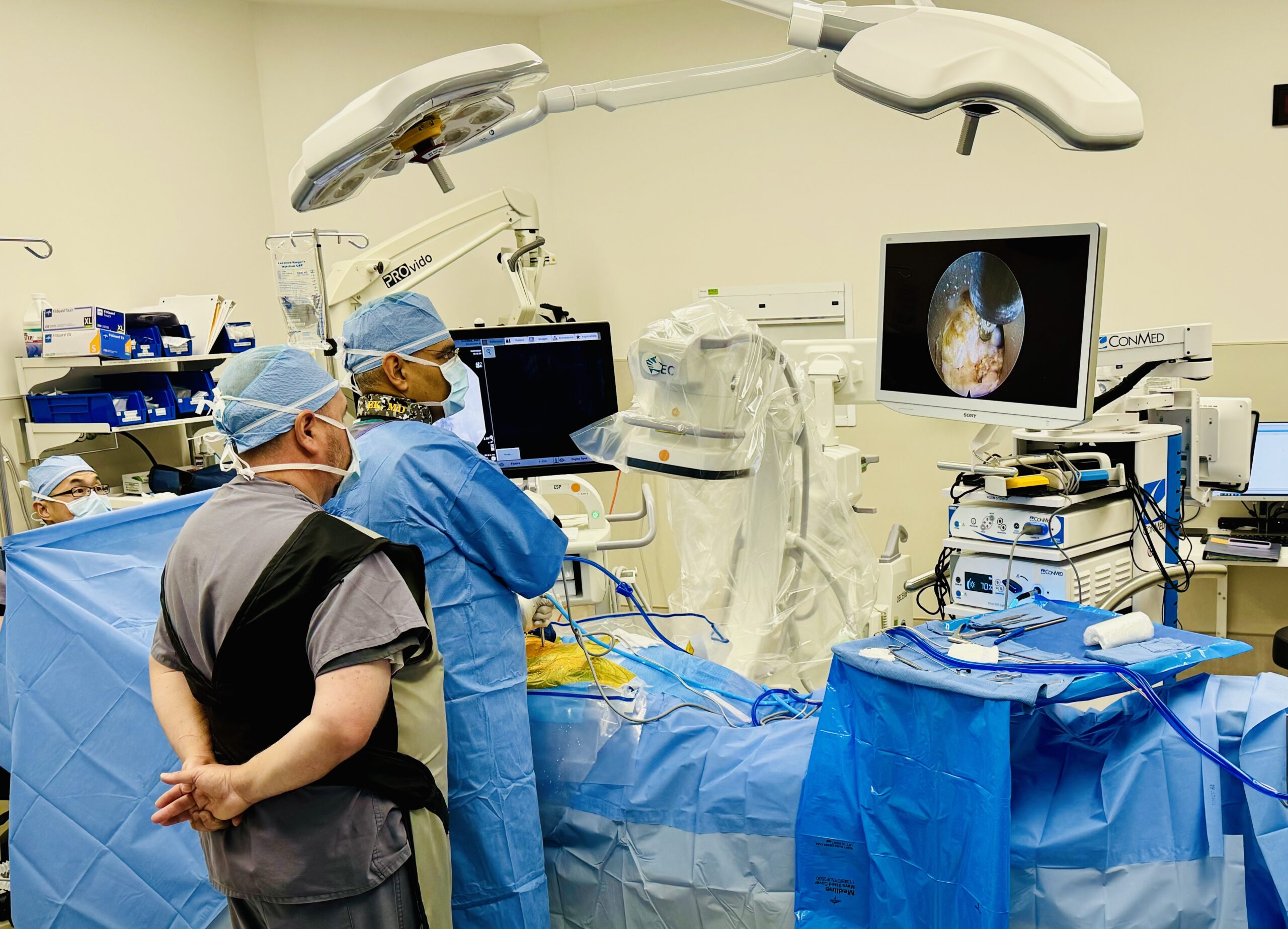
During endoscopic lumbar discectomy, a working-channel endoscope is positioned near the herniation under fluoroscopic guidance. Small instruments remove the fragment while the nerve and surrounding tissues are viewed directly. The procedure may also include a limited foraminoplasty or removal of small amounts of ligament or bone when necessary to reach and release the nerve safely.
The entire disc is not removed. The goal is to remove the material responsible for nerve compression while preserving the remaining disc and spinal anatomy.
Leg-dominant radicular pain caused by a matching disc fragment is more predictable than nonspecific axial back pain associated only with disc degeneration or a broad bulge.
Which Symptoms Is the Procedure Intended to Treat?
Endoscopic lumbar discectomy may be considered for symptoms caused by a compressed lumbar nerve, including:
- Sharp, burning, electric, or shooting pain from the buttock into the leg
- Pain traveling below the knee in a recognizable nerve-root pattern
- Numbness or tingling corresponding to the compressed nerve
- Weakness caused by nerve-root dysfunction
- Persistent radicular symptoms after appropriate nonsurgical treatment
The spinal level and affected nerve are not always numbered the same way. For example, an L4-5 paracentral disc herniation commonly compresses the traversing L5 nerve root. The examination and symptom distribution help determine whether the MRI finding is clinically meaningful.
Who May Be a Candidate for Endoscopic Lumbar Discectomy?
A patient may be considered when:
- Leg-dominant sciatica or radiculopathy limits function or quality of life
- MRI or CT demonstrates a lumbar disc herniation at the correct level and location
- Symptoms, examination findings, and imaging correspond
- Appropriate treatment such as activity modification, medication, physical therapy, or an epidural injection has not provided sufficient improvement
- The fragment can be reached and removed adequately through an endoscopic corridor
- There is no dominant instability, deformity, infection, fracture, tumor, or other condition requiring a different operation
Not every patient must complete the same duration or sequence of conservative treatment. Progressive motor weakness or a cauda-equina pattern may require more urgent evaluation. Conversely, many disc herniations improve without surgery, so tolerable symptoms without neurological progression may continue to be managed nonsurgically.
MRI Findings That Require Clinical Correlation
Disc protrusions, extrusions, migrated fragments, foraminal herniations, and recurrent herniations can sometimes be treated endoscopically. The decisive issue is not the label alone. Fragment location, migration, calcification, canal dimensions, iliac-crest anatomy, facet overgrowth, prior surgery, and the affected nerve determine the most practical corridor.
Transforaminal vs Interlaminar Endoscopic Discectomy
Both approaches use direct endoscopic visualization, but they reach the herniation through different pathways.
| Feature | Transforaminal Endoscopic Lumbar Discectomy | Interlaminar Endoscopic Lumbar Discectomy |
|---|---|---|
| Access route | Posterolateral route through or near the neural foramen | Posterior route through the interlaminar window |
| Often considered for | Selected foraminal, extraforaminal, and intracanal herniations; upper lumbar levels | Selected central, paracentral, or migrated fragments; commonly useful at L5-S1 |
| Anatomic considerations | Foraminal size, exiting nerve, high iliac crest, and fragment migration | Interlaminar-window size, dura, traversing nerve, and fragment position |
| Anesthesia | May use local anesthetic with monitored sedation or general anesthesia | Often performed under general anesthesia, although practice varies |
| Characteristic concern | Exiting-nerve or dorsal-root-ganglion irritation; limited access to some migrated fragments | Dural or traversing-nerve manipulation; access depends on window anatomy |
Neither route is inherently superior. The best corridor is the one that reaches the actual fragment safely and permits adequate decompression with the least necessary disruption.
How the Procedure Is Performed
The exact workflow varies, but commonly includes:
- Preoperative planning: MRI and examination identify the disc level, fragment location, affected nerve, and preferred approach.
- Positioning and anesthesia: the patient is positioned according to the access route and receives individualized anesthesia.
- Level confirmation: fluoroscopy confirms the correct spinal level and trajectory.
- Working-channel placement: dilators and a cannula create a narrow path to the disc or canal.
- Endoscopic visualization: irrigation and the camera provide a magnified view of the disc fragment, nerve, and surrounding tissues.
- Fragment removal: small instruments remove the offending disc material. Limited bone or ligament may be removed when necessary for access or decompression.
- Endpoint confirmation: the physician confirms that the nerve is adequately released before removing the instruments and closing the portal.
Procedure duration varies with the level, fragment location, migration, calcification, prior surgery, required bone work, and anesthesia. A fixed promise such as “45 minutes” is not appropriate for every case.
Endoscopic Discectomy vs Microdiscectomy
Both procedures aim to free a lumbar nerve by removing herniated disc material. The main difference is the access and visualization strategy.
| Feature | Endoscopic Lumbar Discectomy | Conventional Microdiscectomy |
|---|---|---|
| Visualization | Camera at the target through a working channel | Microscope through a small posterior exposure |
| Access | Transforaminal or interlaminar portal | Posterior midline or paramedian exposure with muscle retraction |
| Anesthesia | Selected cases under monitored sedation; others under general anesthesia | Usually general anesthesia |
| Early recovery | May have less early incisional pain and faster mobilization | Often outpatient with a relatively rapid recovery as well |
| Clinical outcome | Effective in appropriately selected patients | Established and effective for matching lumbar disc herniation |
| Important limitation | Narrow corridor, approach limitations, and learning curve | More posterior tissue exposure, but broader familiar access |
A multicenter randomized noninferiority trial involving 613 patients found transforaminal endoscopic discectomy noninferior to open microdiscectomy for reduction of sciatica-related leg pain. Some secondary outcomes favored the endoscopic group, but the differences were small and may not have been clinically meaningful.
More recent meta-analyses generally report similar pain and functional improvement, with possible advantages in blood loss, hospital stay, and early recovery. However, findings concerning recurrence and reoperation are not uniform. A 2025 meta-analysis reported a higher reoperation rate in the endoscopic cohort. It is therefore inaccurate to promise that endoscopic discectomy has a lower recurrence or revision rate than microdiscectomy.
Recovery After Endoscopic Lumbar Discectomy
Most procedures are outpatient, and walking usually begins after recovery from anesthesia. Recovery should be based on the actual operation and nerve condition rather than an advertised timetable.
| General Period | What Patients May Expect |
|---|---|
| Day of surgery | Walking and same-day discharge are common after appropriate observation. A responsible adult may be required for transportation. |
| First several days | Incisional soreness, back tightness, and fluctuating leg symptoms may occur. Short walks are commonly encouraged. |
| Approximately one to two weeks | Some patients resume desk-based work and light activity when pain control, mobility, and medication use permit. |
| Following weeks | Lifting, bending, twisting, exercise, and physical work are advanced according to healing and physician instructions. |
Leg pain caused by mechanical nerve compression may improve quickly, but numbness, tingling, or weakness can take weeks or months to recover. Long-standing neurological loss may be incomplete. Temporary postoperative nerve inflammation can also cause fluctuating or burning symptoms before improvement settles.
How Long Can Pain Last After Endoscopic Discectomy?
The answer depends on what type of pain remains.
- Incisional or muscular soreness: commonly improves over days to a few weeks.
- Residual nerve pain: may fluctuate while an inflamed or chronically compressed nerve recovers.
- Numbness or weakness: often improves more slowly than pain and may not resolve completely.
- Persistent axial back pain: may reflect disc degeneration, facet pain, muscle pain, instability, or another source not corrected by removing a nerve-compressing fragment.
- Recurrent or worsening leg pain: may require evaluation for residual compression, recurrent herniation, hematoma, infection, or another cause.
Risks, Recurrence and Limitations
Potential risks include:
- Infection
- Bleeding, hematoma, or vascular injury
- Dural tear or spinal-fluid leak
- Nerve-root injury, irritation, or postoperative dysesthesia
- Incomplete removal or persistent nerve compression
- Recurrent disc herniation at the treated level
- Temporary or persistent pain, numbness, or weakness
- Instability or progression of degeneration
- Need for repeat discectomy, decompression, fusion, or another operation
- Anesthesia-related complications
Recurrence can occur after either endoscopic discectomy or microdiscectomy because the remaining disc can herniate again. The risk varies with disc biology, fragment type, annular defect, activity, smoking, body mechanics, surgical technique, and follow-up duration. No procedure can guarantee that the same disc will never herniate again.
The value of the endoscopic approach is a smaller access corridor when it can safely accomplish the required decompression. It should not be marketed as a guarantee of superior long-term results.
When Endoscopic Lumbar Discectomy May Not Be the Best Choice
Another treatment may be more appropriate when:
- Back pain is the dominant symptom and there is no convincing nerve compression
- The MRI abnormality does not match the symptom pattern or examination
- Severe central or multilevel bony stenosis requires broader decompression
- The fragment is calcified, highly migrated, or inaccessible through the proposed corridor
- High-grade instability, deformity, or spondylolisthesis requires stabilization or fusion
- Fracture, infection, tumor, or another urgent structural diagnosis is present
- Symptoms are improving adequately with nonsurgical care
Alternatives may include continued nonsurgical care, epidural injection, conventional microdiscectomy, tubular discectomy, broader endoscopic or open decompression, or decompression with fusion when a separate mechanical indication exists.
Can Endoscopic Discectomy Treat a Recurrent Herniation?
Sometimes. A recurrent focal disc herniation after prior discectomy may be reachable endoscopically, and an approach that avoids the previous scarred corridor can be useful in selected anatomy. However, recurrent symptoms may also come from epidural scar, foraminal stenosis, instability, progressive disc collapse, or another pain generator.
Revision planning should assess the recurrent fragment, prior bone removal, scar distribution, disc height, foraminal dimensions, flexion-extension stability, neurological findings, and whether repeat discectomy or fusion better addresses the complete problem. A recurrent herniation is not automatically an indication for fusion, but neither is it automatically suitable for another limited discectomy.
Related Herniated Disc and Endoscopic Resources
- Endoscopic Spine Surgery
- Endoscopic Lumbar Decompression
- Herniated Disc
- Radiculopathy
- L5 Nerve Root Pain
- Minimally Invasive Lumbar Decompression
A focused examination and imaging review can determine whether the herniated fragment explains your sciatica and whether an endoscopic, microscopic, or nonsurgical approach is the better fit.
Frequently Asked Questions
What is endoscopic lumbar discectomy?
Who is a candidate for endoscopic lumbar discectomy?
What is the difference between transforaminal and interlaminar discectomy?
Is endoscopic discectomy better than microdiscectomy?
Is general anesthesia required?
How long does the procedure take?
How soon can I return to work?
Why can leg pain persist after the disc fragment is removed?
Can the disc herniate again?
Does insurance cover endoscopic lumbar discectomy?
References
- Gadjradj PS, et al. Full Endoscopic Versus Open Discectomy for Sciatica: Randomised Controlled Non-Inferiority Trial. BMJ. 2022. Read source
- Yang CC, et al. Complications of Full-Endoscopic Lumbar Discectomy Versus Open Lumbar Microdiscectomy: Systematic Review and Meta-Analysis. World Neurosurgery. 2022. Read source
- Kotheeranurak V, et al. Full-Endoscopic Lumbar Discectomy Approach Selection: A Systematic Review and Proposed Algorithm. 2023. Read source
- Kapetanakis S, et al. Full-Endoscopic Lumbar Discectomy: Review of Surgical Techniques, Indications and Clinical Outcomes. 2025. Read source
- Taha AM, et al. Meta-Analysis of Percutaneous Endoscopic Discectomy and Microdiscectomy for Lumbar Disc Herniation. 2025. Read source
- Ju CI, et al. Complications and Management of Endoscopic Spinal Surgery. 2023. Read source
About the Author
Amit Sharma, MD is an interventional spine and pain-management physician specializing in image-guided procedures, endoscopic spine procedures, minimally invasive lumbar decompression, and selected minimally invasive stabilization techniques.
Last medically reviewed: September 2026



