

Nerve Hydrodissection: Ultrasound-Guided Treatment for Entrapped or Irritated Nerves
Nerve Hydrodissection is an ultrasound-guided procedure used to treat selected peripheral nerve entrapments by carefully introducing fluid around a nerve to separate it from surrounding fascia, scar tissue, muscle, tendon, or other structures that may be restricting normal nerve movement.
Unlike a traditional nerve block, which primarily places medication near a nerve to temporarily reduce pain transmission, hydrodissection is intended to create a physical tissue plane around the nerve. Real-time ultrasound allows the physician to identify the nerve, follow the needle, observe the fluid spread, and determine whether the nerve is being released from surrounding tissue.
Depending on the diagnosis and treatment goals, the injectate may include saline, local anesthetic, dextrose solution, or other medications in selected circumstances. The choice of injectate and the amount used should be individualized according to the nerve involved, surrounding anatomy, prior treatment response, and available clinical evidence.
Quick Answer
Nerve hydrodissection is an ultrasound-guided technique in which fluid is placed around an entrapped peripheral nerve to separate it from surrounding tissue and improve the nerve’s ability to move freely. It is used selectively for nerve entrapments in which mechanical restriction, scarring, or abnormal tissue contact is believed to contribute to symptoms.
Clinical Perspective
Hydrodissection should not be treated as a generic injection for every painful nerve. The procedure is most useful when the clinical examination and ultrasound findings support a specific site of nerve entrapment or restricted nerve mobility. The treatment target, needle approach, fluid volume, and injectate should all be tailored to the individual nerve and surrounding anatomy.
Related Guides
What Is Nerve Hydrodissection?
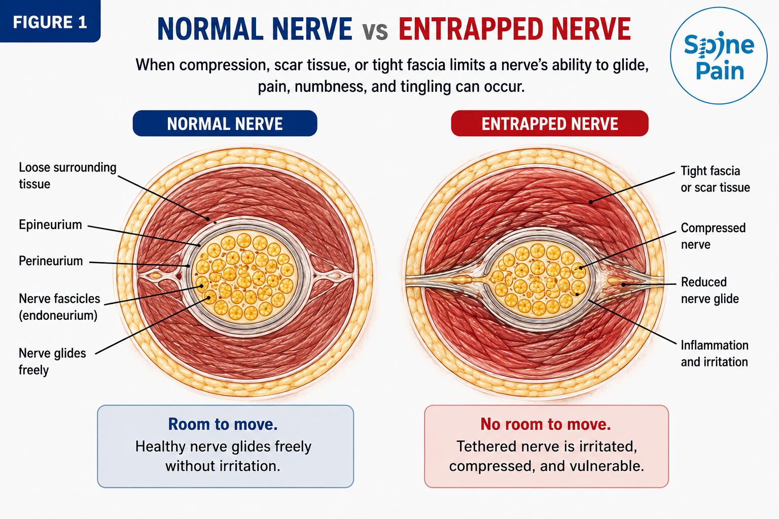
Nerve hydrodissection is an ultrasound-guided technique in which fluid is carefully introduced around a peripheral nerve to separate it from surrounding fascia, scar tissue, muscle, tendon, ligament, or other structures that may restrict normal nerve movement.
Peripheral nerves normally glide through surrounding tissues as the body moves. When a nerve becomes compressed, swollen, tethered by scar tissue, or crowded within a narrow anatomical passage, this normal movement may become restricted. Mechanical irritation can contribute to burning pain, tingling, numbness, electric sensations, hypersensitivity, or symptoms triggered by particular movements.
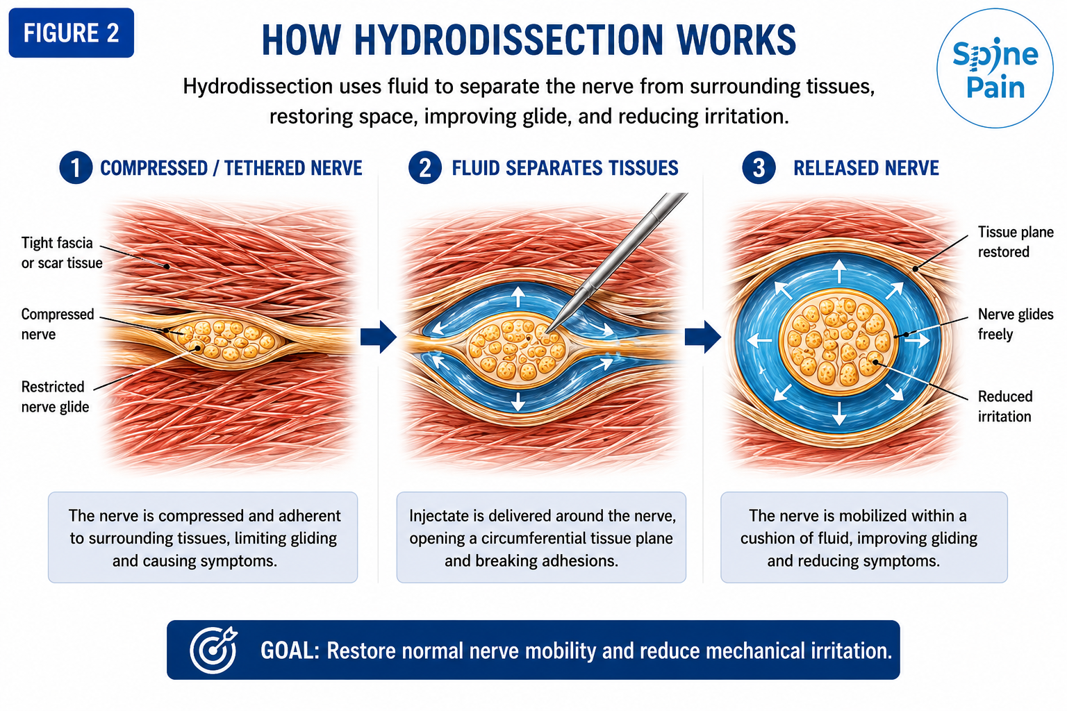
During hydrodissection, the physician identifies the nerve with ultrasound, advances a needle toward the tissue plane surrounding it, and watches fluid spread around the nerve in real time. The fluid rather than the needle is used to open the tissue plane.
The Fluid Is the Dissector
Hydrodissection does not involve cutting scar tissue with the needle. The goal is to place fluid into the correct anatomical plane so that the fluid gently separates the nerve from surrounding tissue.
How Does Nerve Hydrodissection Work?
The exact mechanism by which hydrodissection improves symptoms may differ between patients. In some cases, the most important effect may be mechanical separation of the nerve from surrounding tissue. In others, reducing local irritation or delivering medication around the nerve may also contribute.
Potential effects of hydrodissection include:
- Separating a peripheral nerve from tight fascia or scar tissue.
- Creating additional space around a nerve at a focal entrapment site.
- Reducing mechanical irritation from nearby muscles, tendons, ligaments, or other structures.
- Improving the ability of the nerve to glide during movement.
- Delivering local anesthetic or another selected injectate around an irritated nerve.
- Providing diagnostic information when relief occurs after treatment of a specific nerve.
Clinical Perspective
Hydrodissection is a technique rather than a particular medication. Successful treatment depends first on identifying the correct nerve and the correct site of entrapment. The injectate matters, but accurate diagnosis, ultrasound visualization, and placement within the intended tissue plane are fundamental.
Why Is Ultrasound Guidance Important?
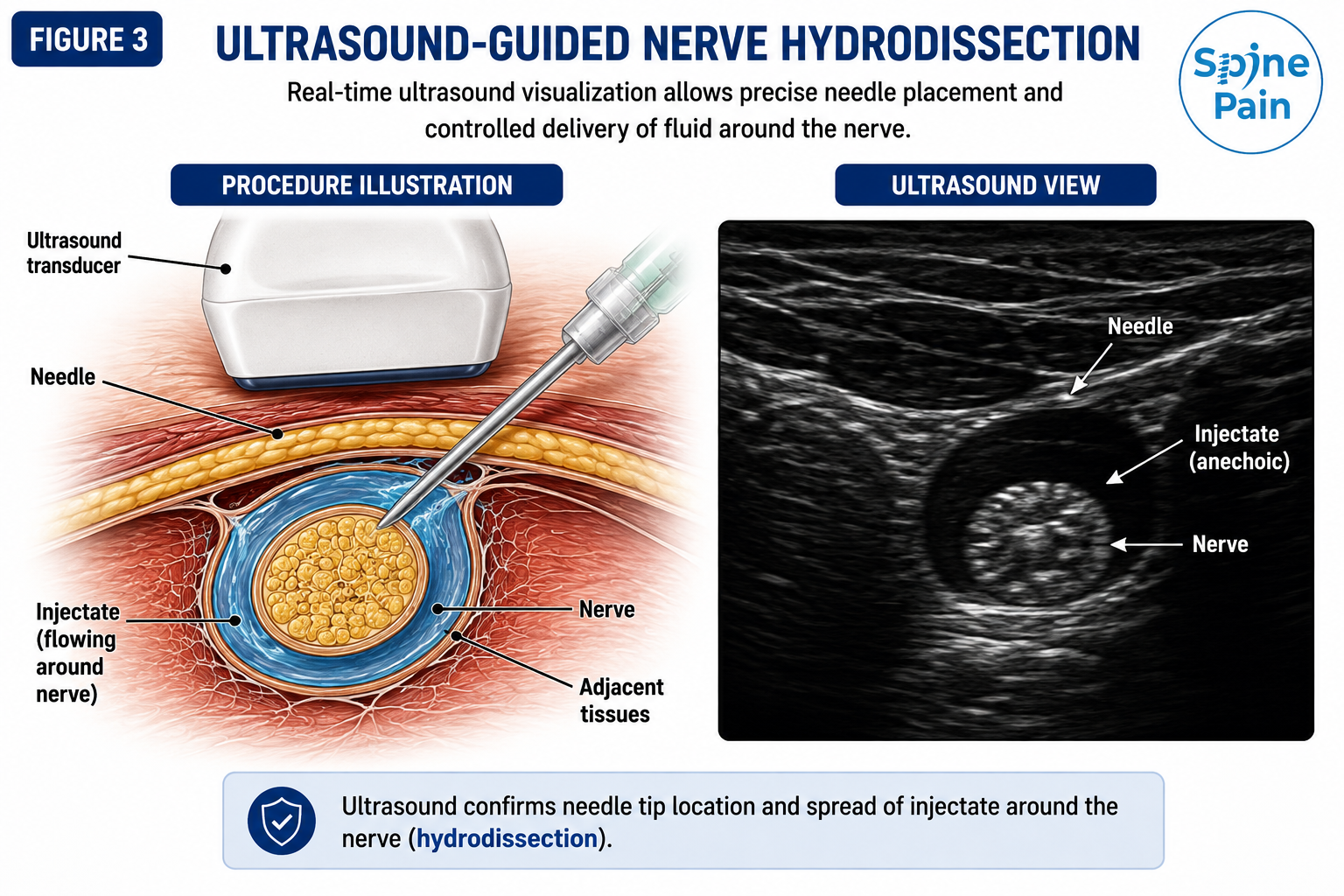
Ultrasound is central to modern nerve hydrodissection because the procedure depends on identifying the nerve, visualizing the surrounding anatomy, following the needle, and observing fluid separation in real time.
Depending on the nerve and anatomical location, ultrasound may help identify:
- The target peripheral nerve.
- Focal nerve enlargement, flattening, or altered appearance when present.
- Restricted movement or abnormal nerve glide in selected locations.
- Scar tissue or altered postoperative anatomy.
- Adjacent arteries and veins.
- Muscles, tendons, fascia, ligaments, and bone.
- The safest pathway for needle advancement.
- The pattern of fluid spread around the nerve.
See the Nerve. See the Needle. See the Fluid.
A useful hydrodissection procedure depends on all three. The physician should be able to identify the nerve, guide the needle safely around it, and watch the injectate open the intended tissue plane rather than relying on surface landmarks alone.
When Is Nerve Hydrodissection Considered?
Nerve hydrodissection may be considered when symptoms and examination findings suggest that a specific peripheral nerve is mechanically irritated, compressed, tethered, or entrapped at a location that can be safely evaluated with ultrasound.
Patients who may be considered for treatment include those with:
- Pain, tingling, burning, numbness, or electric sensations following a recognizable peripheral nerve distribution.
- Focal symptoms corresponding with a known anatomical entrapment site.
- Scar-related nerve pain following surgery or trauma.
- Symptoms triggered by movements that place tension on the involved nerve.
- Ultrasound evidence of focal compression, altered nerve contour, enlargement, or restricted mobility when visible.
- Persistent symptoms despite appropriate activity modification, rehabilitation, bracing, medications, or other conservative treatment.
- A need for diagnostic clarification before considering surgery or another advanced intervention.
Important Distinction
Not every burning, tingling, or numb sensation represents a peripheral nerve entrapment. Similar symptoms may arise from cervical or lumbar radiculopathy, plexus disorders, generalized peripheral neuropathy, metabolic disease, vascular disease, medication effects, or other neurological conditions. Hydrodissection is most appropriate when a specific peripheral nerve and treatment location can be identified.
What Nerves May Be Treated With Hydrodissection?
Nerve hydrodissection may be considered for selected peripheral nerve entrapments when the symptoms, examination, and ultrasound findings support a focal mechanical problem. The strength of evidence is not equal for every nerve, and the procedure should be individualized according to the anatomy and severity of the entrapment.
Median Nerve and Carpal Tunnel Syndrome
Carpal tunnel syndrome is the most extensively studied application of nerve hydrodissection. The median nerve may become compressed beneath the transverse carpal ligament at the wrist. Ultrasound-guided hydrodissection can be used to separate the nerve from adjacent structures and improve the available tissue plane around it.
Ulnar Nerve
The ulnar nerve may become irritated at the elbow, forearm, or wrist. In selected patients with focal entrapment, scar-related tethering, or restricted nerve mobility, hydrodissection may be considered after the exact site of compression is identified.
Radial Nerve
Selected radial nerve or posterior interosseous nerve entrapments may be treated when symptoms correspond with a focal ultrasound-accessible compression site. Scar-related nerve irritation after trauma or surgery may also occasionally be considered.
Lateral Femoral Cutaneous Nerve
The lateral femoral cutaneous nerve may become compressed near the inguinal ligament, producing burning, numbness, or hypersensitivity over the outer thigh known as meralgia paresthetica. Ultrasound-guided perineural treatment may be considered in selected cases.
Cluneal Nerves
Superior, middle, or inferior cluneal nerve irritation may contribute to focal buttock or posterior pelvic pain. When a specific entrapment point or scar-related restriction is identified, hydrodissection may be incorporated into a targeted treatment plan.
Ilioinguinal, Iliohypogastric, and Genitofemoral Nerves
These nerves may become irritated after abdominal, pelvic, inguinal, or hernia surgery. In selected patients with focal scar-related pain or suspected nerve tethering, ultrasound-guided hydrodissection may be considered after careful anatomical localization.
Common Fibular Nerve
The common fibular nerve is vulnerable near the fibular head. Selected patients with focal compression, scar-related irritation, or restricted nerve glide may be candidates for ultrasound-guided perineural treatment.
Tibial Nerve and Tarsal Tunnel
The tibial nerve and its branches may become compressed around the ankle or foot. Ultrasound-guided hydrodissection may be considered selectively when symptoms and imaging support a focal entrapment rather than generalized neuropathy or another source of foot pain.
Clinical Perspective
Carpal tunnel syndrome has the strongest clinical research base for hydrodissection. Treatment of other nerves should be more selective and should depend on a convincing anatomical target, symptom pattern, and ultrasound-accessible site of entrapment.
Nerve Hydrodissection vs a Traditional Nerve Block
Nerve hydrodissection and a conventional nerve block can overlap, but they are not identical procedures. The distinction lies primarily in the treatment goal.
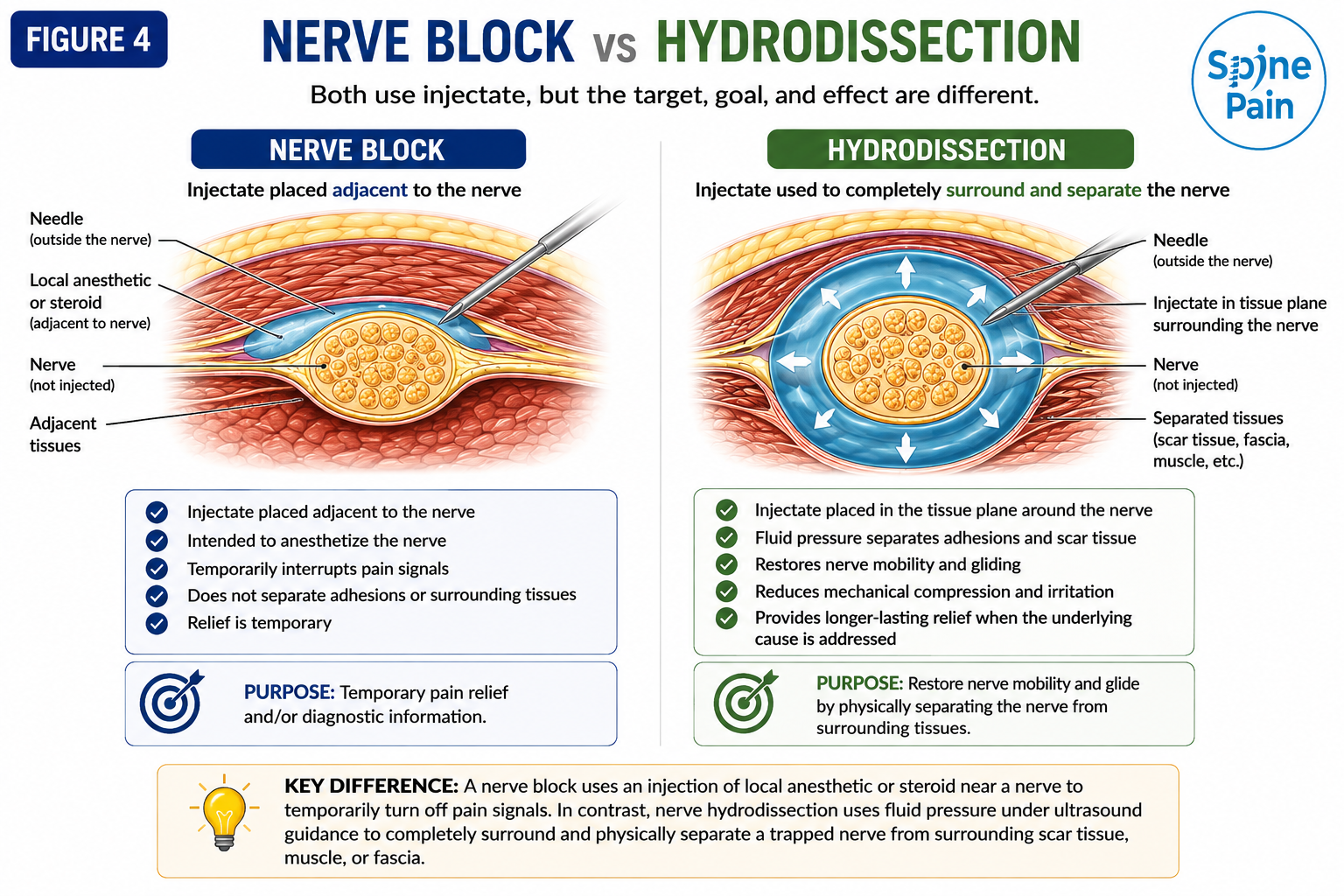
| Procedure | Primary Goal | Typical Role |
|---|---|---|
| Peripheral Nerve Block | Temporarily numb or calm a nerve pathway | Diagnostic clarification, temporary pain relief, or part of a broader treatment pathway |
| Nerve Hydrodissection | Mechanically separate the nerve from surrounding tissue using fluid | Selected entrapment, tethering, scar-related irritation, or restricted nerve glide |
| Hydrodissection With Medication | Open the tissue plane and deliver a selected injectate around the nerve | Cases in which mechanical restriction and local nerve irritation are both clinically relevant |
Key Point
A nerve block asks, “Is this nerve contributing to the pain?” Hydrodissection adds another question: “Is the nerve mechanically restricted by the tissue around it?”
What Fluid or Medication Is Used for Nerve Hydrodissection?
The term hydrodissection describes the technique rather than a specific medication. Different fluids can be used to open the tissue plane around the nerve, and the choice depends on the diagnosis, nerve target, treatment goal, prior response, and available evidence.
Normal Saline
Normal saline can be used primarily as a mechanical hydrodissection fluid. Its role is to create space around the nerve and separate the nerve from surrounding tissue without relying on a specific anti-inflammatory or biologic effect.
Local Anesthetic
Local anesthetic may be included to provide temporary pain relief and diagnostic information. Improvement during the anesthetic window may support that the targeted nerve is contributing to symptoms, although the response should be interpreted together with the clinical examination.
D5W (5% Dextrose in Water)
D5W is commonly discussed in the hydrodissection literature, particularly for carpal tunnel syndrome and selected peripheral nerve entrapments. It provides fluid volume for mechanical separation and has also been studied for possible effects on perineural pain and irritation.
The strength of evidence varies by nerve and condition, so D5W should be considered an option rather than a universal hydrodissection solution.
Corticosteroid
Corticosteroid may be used selectively when inflammation around an entrapped nerve is believed to be clinically important. Repeated steroid exposure near superficial nerves, skin, fat, or nearby tendons should be used thoughtfully because of potential tissue effects.
Platelet-Rich Plasma (PRP)
PRP has been studied for selected peripheral nerve entrapments and may be discussed in carefully chosen cases. It is biologically different from saline, D5W, or corticosteroid and should not be presented as simply another interchangeable hydrodissection fluid.
Evidence varies considerably by nerve and condition, and PRP may not be covered by insurance. It should also never delay surgical evaluation when progressive neurological loss or severe structural compression is present.
Technique First, Injectate Second
The fluid matters, but accurate nerve localization and placement within the correct tissue plane matter more. An expensive or biologically active injectate cannot compensate for treating the wrong nerve or the wrong anatomical plane.
What Happens During Nerve Hydrodissection?
Nerve hydrodissection is generally performed as an outpatient procedure. The exact technique varies according to the nerve involved, the location of entrapment, surrounding anatomy, prior surgery, scar tissue, and the intended treatment goal.
1. Evaluation and Target Selection
The physician reviews the symptom distribution, physical examination, prior surgery or trauma, scar pattern, imaging, ultrasound findings, and electrodiagnostic testing when available. Competing diagnoses should be considered before selecting the nerve target.
2. Ultrasound Mapping
Ultrasound is used to identify the nerve and surrounding arteries, veins, tendons, muscles, fascia, bone, and scar tissue. The safest needle pathway is selected before treatment begins.
3. Needle Placement
The skin is cleansed using sterile technique. A needle is advanced under continuous ultrasound guidance toward the tissue plane surrounding the nerve. The objective is to remain outside the nerve itself.
4. Fluid Separation
Fluid is gradually introduced while the physician watches the tissue plane open around the nerve. The needle can be repositioned as necessary so that hydrodissection occurs around the intended portion of the nerve rather than simply creating a distant fluid pocket.
5. Assessment of Fluid Spread
The pattern of injectate spread is evaluated under ultrasound. In many procedures, the goal is to see fluid separating the nerve from the surrounding tissue along the suspected entrapment plane.
6. Post-Procedure Assessment
After treatment, the patient may be asked to compare the original pain, numbness, tingling, motion sensitivity, or other nerve-related symptoms with the pre-procedure baseline. The early response can provide useful diagnostic information, particularly when local anesthetic is included.
Clinical Perspective
Hydrodissection is not defined by how much fluid is injected. The meaningful endpoint is whether the intended nerve has been safely separated from the surrounding tissue at the clinically relevant entrapment site.
Recovery After Nerve Hydrodissection
Most patients return home the same day after nerve hydrodissection. Recovery depends on the nerve treated, the injectate used, whether the procedure was primarily diagnostic or therapeutic, and the degree of pre-existing nerve irritation.
Temporary numbness, heaviness, warmth, tingling, or mild soreness may occur, particularly when local anesthetic is included. Some patients notice immediate improvement, while others experience a short-lived increase in nerve sensitivity before symptoms settle.
General Recovery Recommendations
- Track the specific symptoms that were present before the procedure, including burning, tingling, numbness, electric pain, scar sensitivity, or motion-related symptoms.
- Avoid aggressive stretching or heavy repetitive use immediately after treatment.
- Do not overuse the area simply because local anesthetic has temporarily reduced pain.
- Resume nerve-gliding exercises, therapy, bracing, or activity modification as directed.
- Follow procedure-specific instructions regarding work, exercise, driving, and medication use.
- Contact the treating physician if severe, progressive, or unexpected neurological symptoms develop.
Track the Nerve Symptoms, Not Just the Pain Score
A useful post-procedure assessment includes changes in numbness, tingling, burning, hypersensitivity, scar pain, grip tolerance, walking tolerance, night symptoms, and symptoms triggered by movement. Functional improvement may be just as important as a change in pain intensity.
What Results Can Be Expected?
The response to nerve hydrodissection varies considerably. Some patients experience substantial improvement in pain and nerve-related symptoms, while others obtain temporary relief, incomplete benefit, or no meaningful change.
The likelihood and durability of improvement depend on several factors, including:
- Accuracy of the diagnosis and nerve localization.
- The severity and duration of nerve compression.
- Whether the nerve is mechanically tethered, inflamed, structurally compressed, or chronically damaged.
- The presence of scar tissue or altered postoperative anatomy.
- Whether weakness or muscle atrophy is already present.
- The injectate and volume selected for the procedure.
- Adherence to bracing, ergonomic changes, rehabilitation, or nerve-gliding recommendations.
- The presence of competing causes of symptoms such as radiculopathy, generalized neuropathy, or another pain generator.
Clinical Perspective
A good response may support that the targeted nerve and surrounding tissue plane are clinically important. It does not necessarily mean that the underlying entrapment has been permanently corrected or that further treatment will never be needed.
What If Nerve Hydrodissection Helps?
When hydrodissection produces meaningful improvement, the response can help guide the next step. The plan depends on the degree of relief, how long the improvement lasts, and whether function also improves.
Possible next steps may include:
- Observation when relief is substantial and durable.
- Physical therapy or nerve-gliding exercises.
- Ergonomic changes, bracing, or activity modification.
- Repeat hydrodissection in carefully selected cases when the first procedure provided meaningful benefit.
- Further electrodiagnostic testing if the degree of nerve injury remains uncertain.
- Surgical evaluation if symptoms recur, progress, or are accompanied by weakness or loss of function.
- Peripheral nerve stimulation in selected chronic neuropathic pain patterns when decompression is no longer the primary issue.
Relief Gives Direction
A favorable response does more than reduce symptoms. It may strengthen the evidence that the treated nerve and entrapment site are important enough to guide rehabilitation, repeat treatment, or the decision to pursue more definitive decompression.
What If Nerve Hydrodissection Does Not Help?
A lack of improvement does not automatically mean that the procedure was technically unsuccessful. It may indicate that the targeted nerve is not the primary pain generator, the entrapment is too severe to respond to fluid separation, the wrong level of the nerve was treated, or the symptoms arise from another neurological or musculoskeletal condition.
When hydrodissection does not help, the next step may include:
- Reassessing the diagnosis and nerve distribution.
- Reviewing the ultrasound findings and the level of entrapment.
- Obtaining EMG or nerve conduction studies when localization or severity remains uncertain.
- Evaluating the cervical or lumbar spine, plexus, joint, vascular system, or systemic neuropathy when clinically appropriate.
- Considering a different targeted nerve block or diagnostic injection.
- Obtaining surgical consultation when structural compression appears significant.
- Considering neuromodulation for selected chronic neuropathic pain after appropriate diagnostic evaluation.
A Negative Procedure Is Still Information
If familiar symptoms do not improve after a carefully targeted hydrodissection, the diagnosis, level of nerve involvement, severity of compression, or overall treatment strategy should be reconsidered rather than simply repeating the same procedure.
When Should Surgical Evaluation Not Be Delayed?
Hydrodissection is not intended to replace surgical decompression when a nerve is severely compressed or neurological function is progressively deteriorating. In these situations, delaying definitive treatment may allow additional nerve damage to occur.
Prompt surgical evaluation should be considered when there is:
- Progressive motor weakness.
- Visible muscle wasting or atrophy.
- Loss of hand, foot, or limb function.
- Persistent or worsening numbness suggesting significant nerve compromise.
- Severe abnormalities on EMG or nerve conduction testing when clinically relevant.
- A mass, ganglion cyst, implant, hardware, or other structural lesion compressing the nerve.
- Fixed anatomical narrowing unlikely to respond adequately to fluid separation alone.
- Progressive symptoms despite appropriately selected conservative and interventional care.
Do Not Hydrodissect Past a Surgical Problem
When a nerve is losing motor function, developing muscle atrophy, or undergoing severe structural compression, the priority is protecting neurological function. Hydrodissection should not become a reason to postpone necessary decompression.
Nerve Hydrodissection vs Surgical Decompression
Hydrodissection and surgery address different levels of nerve compression. Hydrodissection may be useful when soft-tissue restriction, scar tethering, or selected mild-to-moderate entrapment is believed to be contributing to symptoms. Surgery is generally more appropriate when compression is severe, structurally fixed, or threatening nerve function.
| Treatment | Best Fit | Main Limitation |
|---|---|---|
| Nerve Hydrodissection | Selected mild-to-moderate entrapment, scar tethering, or restricted nerve glide | May not correct severe compression, major weakness, or fixed structural narrowing |
| Surgical Decompression | Severe entrapment, progressive weakness, muscle atrophy, or structural compression | More invasive and requires surgical recovery; longstanding nerve damage may not fully reverse |
| Peripheral Nerve Stimulation | Selected chronic peripheral neuropathic pain after appropriate diagnostic evaluation | Modulates pain signaling but does not mechanically decompress the nerve |
Risks and Side Effects of Nerve Hydrodissection
Nerve hydrodissection is generally well tolerated when performed carefully with ultrasound guidance, but it is still a procedure performed near nerves, blood vessels, tendons, muscles, and other sensitive structures.
Potential risks and side effects include:
- Temporary soreness or bruising at the injection site.
- Temporary increase in nerve pain, tingling, or electric sensations.
- Temporary numbness or weakness, particularly when local anesthetic is used.
- Bleeding or hematoma.
- Infection.
- Vascular puncture.
- Nerve irritation or nerve injury.
- Unintended injection into the nerve.
- Medication reaction or allergy.
- Local anesthetic side effects.
- Skin thinning, pigmentation change, or subcutaneous fat changes when corticosteroid is used superficially.
- Temporary elevation in blood sugar when corticosteroid is used, particularly in patients with diabetes.
- Failure to improve or recurrence of symptoms.
Patients taking anticoagulants or antiplatelet medications, those with bleeding disorders, active infection, medication allergies, diabetes, prior surgery, altered anatomy, or significant neurological deficits should discuss these issues with the treating physician before the procedure.
Seek Prompt Evaluation for New Weakness
New or progressive weakness, substantial loss of sensation, rapidly worsening neurological symptoms, fever, significant swelling, or severe unexpected pain after a procedure should be evaluated promptly.
Does Insurance Cover Nerve Hydrodissection?
Insurance coverage varies according to the diagnosis, nerve being treated, payer policy, documentation, procedure performed, and injectate used. Some ultrasound-guided peripheral nerve procedures may be covered when medically necessary, while other treatments may be considered investigational or non-covered by a particular insurance plan.
Coverage for the procedure should also be distinguished from coverage for the injectate. PRP and certain other biologic or regenerative treatments are frequently not covered by insurance and may be offered on a self-pay basis.
When prior authorization is required, coverage does not necessarily guarantee payment. Patients should confirm their individual benefits, deductible, coinsurance, and out-of-pocket responsibilities with their insurance carrier.
Our Approach to Nerve Hydrodissection
At SpinePain Solutions, nerve hydrodissection is considered within a broader evaluation of peripheral nerve pain rather than as an isolated injection. Burning, tingling, numbness, electric pain, scar sensitivity, and movement-related nerve symptoms can arise from several different conditions, and identifying the correct pain generator is essential before treatment.
The evaluation may include the patient’s symptom distribution, neurological examination, prior surgery or trauma, ultrasound findings, diagnostic injections, MRI or other imaging when appropriate, and EMG or nerve conduction testing when the diagnosis or severity remains uncertain.
When hydrodissection is appropriate, ultrasound is used to identify the nerve, surrounding structures, needle trajectory, and fluid spread. The response is then incorporated into the larger treatment plan, which may include rehabilitation, activity modification, repeat treatment in selected cases, additional diagnostic evaluation, peripheral nerve stimulation, or surgical referral.
Clinical Perspective
The most important question is not simply whether a nerve can be hydrodissected. It is whether there is convincing evidence that a specific nerve is being mechanically irritated at a specific anatomical location and whether releasing that tissue plane is likely to change the patient’s symptoms or function.
Key Takeaways
- Nerve hydrodissection uses fluid to separate a peripheral nerve from surrounding tissue.
- The procedure is performed with ultrasound guidance so the nerve, needle, surrounding structures, and fluid spread can be visualized in real time.
- Hydrodissection is different from a traditional nerve block because mechanical tissue separation is part of the treatment goal.
- Carpal tunnel syndrome involving the median nerve has the strongest research base, while evidence for other nerve entrapments varies.
- Saline, local anesthetic, D5W, corticosteroid, and selected biologic injectates may be used depending on the clinical situation.
- The correct diagnosis and anatomical target are more important than simply choosing a particular injectate.
- Meaningful improvement may support that the treated nerve and entrapment site are clinically important.
- A lack of improvement should prompt reconsideration of the diagnosis, target, severity of compression, or treatment strategy.
- Progressive weakness, muscle atrophy, worsening neurological loss, or severe structural compression should prompt surgical evaluation rather than repeated hydrodissection.
Related Nerve Pain Guides
Frequently Asked Questions About Nerve Hydrodissection
What is nerve hydrodissection?
Nerve hydrodissection is an ultrasound-guided procedure in which fluid is introduced around a peripheral nerve to separate it from surrounding fascia, scar tissue, muscle, tendon, ligament, or other structures that may contribute to nerve entrapment or restricted movement.
How is hydrodissection different from a nerve block?
A nerve block primarily places medication near a nerve to temporarily interrupt or reduce pain transmission. Hydrodissection specifically uses fluid to open a tissue plane around the nerve. A hydrodissection procedure may also provide a nerve-blocking effect when local anesthetic is included.
Does hydrodissection break up scar tissue?
Hydrodissection does not cut or mechanically tear scar tissue with the needle. Fluid is placed into the appropriate tissue plane to separate the nerve from surrounding structures. This may reduce mechanical restriction in selected cases of scar-related nerve tethering.
What is D5W hydrodissection?
D5W is 5% dextrose in water. It can provide the fluid volume needed for mechanical hydrodissection and has also been studied as a perineural injectate, particularly in carpal tunnel syndrome and selected peripheral nerve entrapments.
Can PRP be used for nerve hydrodissection?
PRP has been investigated for selected peripheral nerve conditions and may be considered in carefully selected cases. Evidence varies by nerve and diagnosis, and PRP should not delay surgical evaluation when significant neurological compromise is present.
Which condition has the strongest evidence for nerve hydrodissection?
Carpal tunnel syndrome involving the median nerve has the most developed clinical research base. Evidence for hydrodissection of other peripheral nerves is less mature and varies considerably by condition.
How many hydrodissection treatments are needed?
There is no universal treatment series. Some patients may improve after a single procedure, while selected patients may benefit from additional treatment when the initial procedure produced meaningful improvement. Repeated procedures should have a clear clinical purpose rather than being continued automatically.
How long does relief last?
Duration of relief varies according to the nerve, diagnosis, severity and duration of compression, injectate, and individual response. Improvement may be temporary or longer lasting, and some patients may not respond.
Can nerve hydrodissection prevent surgery?
It may provide a nonsurgical treatment option for selected mild-to-moderate entrapments or scar-related nerve restriction, but it cannot reliably replace surgery for severe or fixed structural compression. Progressive weakness, muscle atrophy, or worsening neurological loss should prompt surgical evaluation.
Can hydrodissection be repeated?
Yes, in selected patients. Repeat treatment is most reasonable when the initial procedure produced meaningful improvement and there remains a clear anatomical and clinical reason for additional hydrodissection.
What if hydrodissection does not help?
The diagnosis and treatment target should be reconsidered. Depending on the clinical situation, further evaluation may include EMG or nerve conduction studies, additional imaging, assessment of the spine or plexus, another diagnostic nerve block, or surgical consultation.
Could an Entrapped Nerve Be Contributing to Your Pain?
Burning, tingling, numbness, electric pain, scar sensitivity, and movement-related nerve symptoms can have many causes. The first step is identifying whether a specific peripheral nerve is involved and whether there is a treatable site of entrapment.
This article is intended for educational purposes only and does not replace individualized medical advice. Peripheral nerve pain may result from nerve entrapment, scar tissue, trauma, peripheral neuropathy, radiculopathy, plexopathy, metabolic disease, vascular disease, or other neurological and musculoskeletal conditions. New or progressive weakness, muscle wasting, substantial sensory loss, rapidly worsening neurological symptoms, or other concerning changes should be evaluated promptly. Treatment decisions should be based on an appropriate history, examination, imaging or ultrasound when indicated, electrodiagnostic testing when appropriate, and an individualized discussion of risks, benefits, and alternatives with a qualified healthcare professional.
Medical Evidence and References
Research on nerve hydrodissection is evolving. Carpal tunnel syndrome has the strongest published evidence base, while studies involving other peripheral nerves remain more limited. Outcomes also vary according to technique, injectate, severity of nerve compression, and patient selection.
- Wu YT, et al. Ultrasound-guided hydrodissection for carpal tunnel syndrome: randomized clinical studies evaluating perineural fluid injection and symptom improvement.
- Lam KHS, Hung CY, Chiang YP, et al. Ultrasound-guided nerve hydrodissection for peripheral nerve entrapment: principles, technique, and clinical applications.
- Cass SP. Ultrasound-guided nerve hydrodissection: what is it and when should it be considered?
- Buntragulpoontawee M, et al. Ultrasound-guided perineural dextrose injection and hydrodissection for carpal tunnel syndrome.
- Lin MT, et al. Comparative effectiveness of ultrasound-guided perineural injection therapies for carpal tunnel syndrome.
- American Institute of Ultrasound in Medicine. Practice Parameter for the Performance of a Musculoskeletal Ultrasound Examination.
- American Association of Neuromuscular & Electrodiagnostic Medicine. Clinical guidance regarding electrodiagnostic evaluation of peripheral nerve entrapment.
- American Academy of Orthopaedic Surgeons. Clinical Practice Guideline: Management of Carpal Tunnel Syndrome.



