

Bursitis Treatment: Ultrasound-Guided Bursa Injections for Pain Relief
Bursitis Treatment: Ultrasound-Guided Bursa Injections for Pain Relief
Bursitis is one of the most common causes of pain around the shoulder, hip, elbow, knee, and pelvis. Although many patients assume the pain comes from arthritis or tendon injury, inflammation of a bursa can produce significant discomfort during walking, reaching overhead, kneeling, lying on one side, or performing repetitive activities.
A bursa is a thin, fluid-filled sac that cushions tendons, muscles, ligaments, and skin as they glide over bone. When a bursa becomes inflamed because of overuse, injury, arthritis, or abnormal biomechanics, even routine movement can become painful.
Fortunately, many cases improve with activity modification, rehabilitation, and medications. When symptoms persist, ultrasound-guided bursa injections allow medication to be delivered directly into the inflamed bursa with excellent precision while avoiding nearby tendons, nerves, and blood vessels.
This physician-written guide explains what bursitis is, why it develops, the most common locations, how ultrasound-guided bursa injections work, expected recovery, potential risks, and when surgery may occasionally become necessary.
What Is a Bursa?
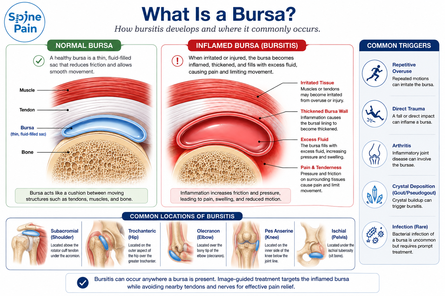
A bursa is a thin, fluid-filled sac that reduces friction between moving tissues such as tendons, muscles, ligaments, skin, and bone. Hundreds of bursae are located throughout the body, particularly around joints that undergo frequent movement. Under normal conditions, these sacs contain only a small amount of lubricating fluid, allowing surrounding structures to glide smoothly during everyday activities.
When a bursa becomes irritated or inflamed, the normally thin lining may thicken and produce excess fluid. This process, known as bursitis, increases friction between adjacent tissues and can produce pain during movement, pressure, or repetitive activity. Although bursitis often develops gradually from repetitive overuse, it may also occur following direct trauma, inflammatory arthritis, crystal deposition disorders such as gout, or, less commonly, infection.
Bursae are located throughout the body, but the areas most frequently requiring treatment include the:
- Shoulder (subacromial-subdeltoid bursa)
- Hip (greater trochanteric bursa)
- Elbow (olecranon bursa)
- Knee (pes anserine and prepatellar bursae)
- Pelvis (ischial bursa)
Although bursitis itself is not usually dangerous, persistent inflammation may interfere with walking, lifting, sleeping, exercise, work, and other daily activities. Fortunately, most patients improve with conservative treatment, and many benefit from precisely performed ultrasound-guided bursa injections when symptoms persist.
Quick Answer
A bursa is a small fluid-filled sac that cushions tendons, muscles, ligaments, and skin as they move over bone. Bursitis occurs when this protective sac becomes inflamed, producing pain during movement or pressure. Ultrasound-guided bursa injections can reduce inflammation while accurately delivering medication into the affected bursa.
Clinical Perspective
Pain attributed to bursitis is not always caused by the bursa alone. Rotator cuff disease, gluteal tendinopathy, arthritis, tendon tears, and nerve disorders frequently coexist with bursal inflammation. Successful treatment depends on identifying the primary pain generator and addressing associated conditions rather than simply injecting every painful bursa.
What Causes Bursitis?
Bursitis develops when repeated friction, excessive pressure, injury, or inflammation irritates the thin lining of a bursa. As the bursa becomes inflamed, it produces excess fluid and thickens, reducing its ability to allow smooth movement between tendons, muscles, ligaments, skin, and bone. The result is pain during movement, tenderness with pressure, localized swelling, and reduced function.
Although bursitis is often associated with repetitive overuse, it may also occur after a single injury or develop as part of a broader musculoskeletal or inflammatory condition. In many patients, the inflamed bursa is only one component of the problem. Tendon degeneration, muscle weakness, arthritis, altered biomechanics, or nerve irritation may all contribute to persistent symptoms.
Common causes of bursitis include:
- Repetitive overuse from occupations, sports, or hobbies involving frequent kneeling, lifting, throwing, reaching, or prolonged pressure.
- Direct trauma such as a fall or impact that causes bleeding or inflammation within the bursa.
- Tendon disorders including chronic tendinopathy or partial tendon tears that increase friction over adjacent bursae.
- Muscle weakness and poor biomechanics that alter joint mechanics and increase stress on surrounding soft tissues.
- Inflammatory arthritis such as rheumatoid arthritis or crystal deposition diseases including gout and pseudogout.
- Osteoarthritis, particularly when altered joint mechanics irritate nearby bursae.
- Infection (septic bursitis), an uncommon but potentially serious condition requiring prompt medical evaluation and treatment.
Not every painful bursa is caused by the same underlying problem. Identifying why the bursa became inflamed is essential because successful treatment often requires addressing the contributing tendon, joint, muscle, or biomechanical abnormality rather than simply reducing inflammation.
Clinical Perspective
Many patients are told they have bursitis when the inflamed bursa is actually reacting to an underlying tendon disorder. Trochanteric bursitis frequently accompanies gluteal tendinopathy, while subacromial bursitis often occurs alongside rotator cuff disease. Treating both the inflamed bursa and the underlying tendon pathology generally produces better long-term outcomes than treating the bursa alone.

What Are the Symptoms of Bursitis?
Although symptoms vary depending on which bursa is affected, bursitis typically causes pain that worsens with movement or direct pressure over the inflamed area. Some patients develop symptoms gradually from repetitive overuse, while others experience sudden pain after a fall, prolonged kneeling, or increased physical activity.
The discomfort may be mild during the early stages but can progressively interfere with work, exercise, sleep, and everyday activities if inflammation persists.
Common symptoms include:
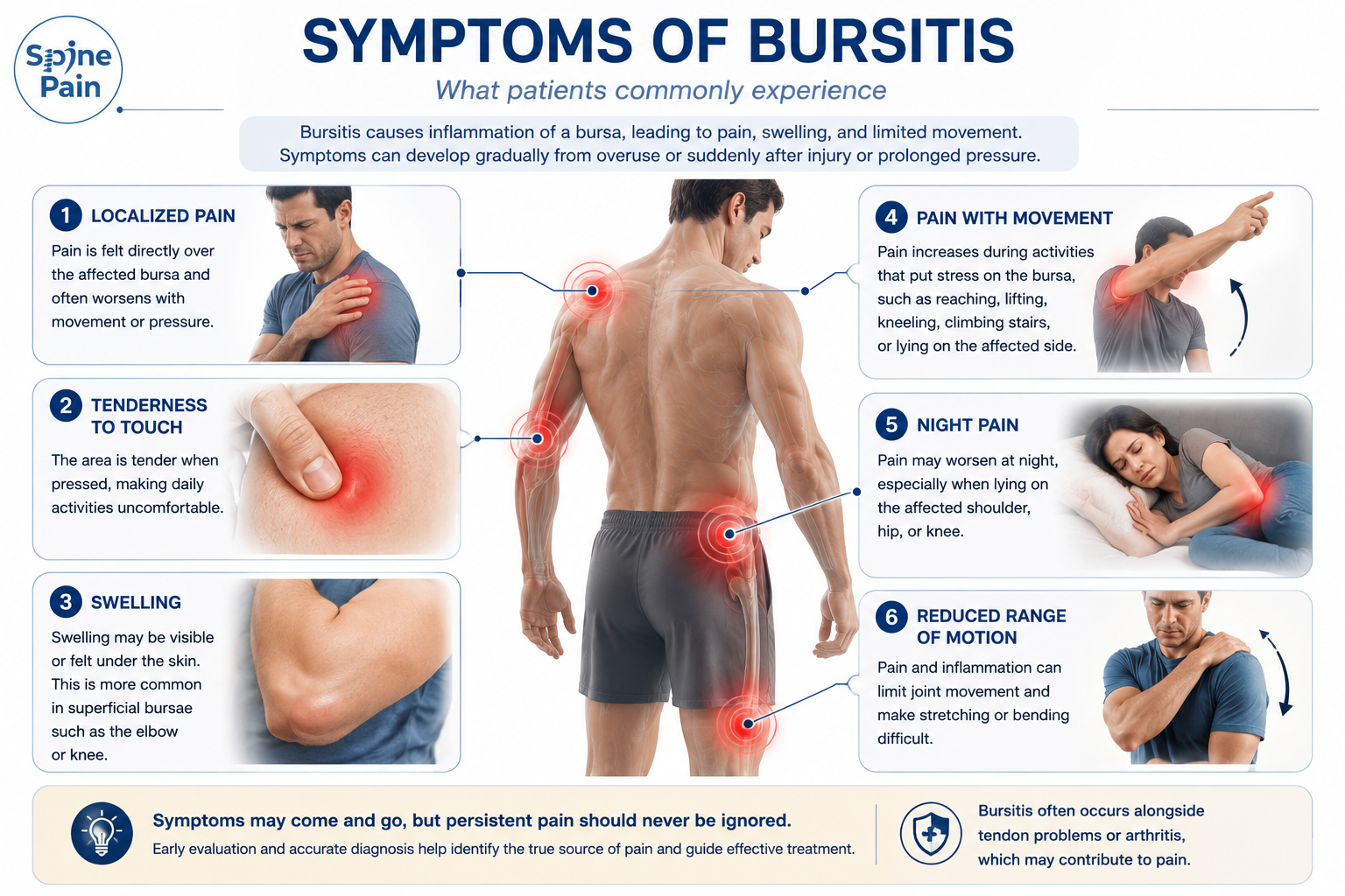
- Localized pain directly over the affected bursa.
- Tenderness when pressing on the inflamed area.
- Pain with movement, particularly during activities that repeatedly compress or stretch the bursa.
- Swelling that may be visible or palpable, especially in superficial bursae such as the elbow or knee.
- Warmth and redness, which are more common with acute inflammation or infection.
- Reduced range of motion caused by pain rather than mechanical joint stiffness.
- Night pain, particularly when lying on the affected shoulder or hip.
Because bursitis frequently occurs alongside tendon disorders, arthritis, or muscle imbalance, patients may experience symptoms arising from more than one structure. Careful examination helps determine whether the inflamed bursa is the primary source of pain or part of a larger musculoskeletal problem.
Clinical Perspective
Pain directly over a bony prominence often points toward bursitis, but location alone is not enough to establish the diagnosis. Rotator cuff disease, gluteal tendinopathy, arthritis, lumbar radiculopathy, and other conditions can produce very similar symptoms. Successful treatment begins by identifying the true pain generator rather than assuming every painful shoulder, hip, elbow, or knee is caused by bursitis.
How Is Bursitis Diagnosed?
Diagnosing bursitis begins with understanding the patient’s symptoms rather than relying on imaging alone. Although X-rays, ultrasound, and MRI provide valuable information, the diagnosis is established by combining the patient’s history, physical examination, and imaging findings. Because many musculoskeletal conditions produce similar symptoms, accurately identifying the true pain generator is essential before recommending treatment.
Pain around a joint does not always originate from an inflamed bursa. Tendon disorders, arthritis, ligament injuries, nerve compression, stress fractures, referred pain from the spine, and muscle injuries may closely mimic bursitis. In many patients, bursitis develops alongside another condition rather than occurring in isolation.
A comprehensive evaluation typically includes:
- Detailed medical history including symptom onset, aggravating activities, prior injuries, occupation, sports participation, and previous treatments.
- Physical examination to identify localized tenderness, swelling, range of motion, muscle strength, gait abnormalities, and provocative maneuvers that reproduce symptoms.
- X-rays to evaluate arthritis, bone spurs, fractures, calcification, or other structural abnormalities contributing to pain.
- Musculoskeletal ultrasound to visualize the inflamed bursa, evaluate surrounding tendons, detect fluid collections, and perform dynamic assessment during movement.
- MRI when deeper structures such as tendons, ligaments, cartilage, muscles, or occult bone injuries require further evaluation.
- Ultrasound-guided diagnostic injections in selected patients to confirm that the inflamed bursa is the primary source of pain before proceeding with additional treatment.
Because bursitis frequently coexists with tendon degeneration or arthritis, identifying every structure contributing to pain allows treatment to be individualized rather than simply focusing on the inflamed bursa.
Clinical Perspective
Modern musculoskeletal ultrasound has transformed the evaluation of bursitis. Unlike static imaging studies, ultrasound allows physicians to examine the bursa, tendons, and surrounding soft tissues in real time while the joint is moving. This dynamic assessment improves diagnostic accuracy and helps guide precisely targeted treatments when an injection is indicated.
Quick Answer
Bursitis is diagnosed through a combination of medical history, physical examination, and imaging when necessary. Ultrasound is particularly valuable because it can identify inflamed bursae, evaluate nearby tendons, and guide accurate injections. Since many conditions mimic bursitis, establishing the correct diagnosis is the first step toward effective treatment.
Treatment Options for Bursitis
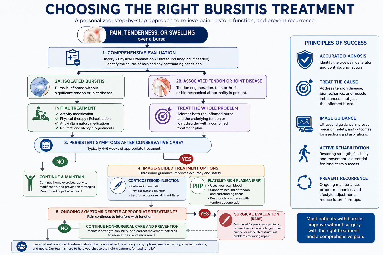
Treatment for bursitis depends on the underlying cause of inflammation, the duration of symptoms, the affected bursa, and whether other conditions such as tendon disease or arthritis are also contributing to pain. While many patients improve with conservative treatment, persistent or recurrent bursitis may benefit from image-guided procedures that target the inflamed bursa with greater precision.
The goal of treatment is not only to reduce inflammation, but also to correct the factors that caused the bursa to become irritated in the first place. Addressing muscle weakness, tendon disorders, biomechanical abnormalities, or repetitive overuse often provides more durable relief than treating inflammation alone.
Treatment typically progresses from the least invasive options to more advanced interventions based on the patient’s symptoms and response to previous care.
Activity Modification and Physical Therapy
Most patients begin with conservative treatment. Reducing repetitive pressure on the affected bursa allows inflammation to settle while rehabilitation addresses the mechanical factors that contributed to its development.
Physical therapy may include:
- Stretching tight muscles and surrounding soft tissues.
- Strengthening muscles that stabilize the affected joint.
- Improving posture and movement patterns.
- Correcting gait or biomechanical abnormalities.
- Gradually returning to work, sports, and recreational activities.
Simple modifications such as avoiding prolonged kneeling, using protective padding, improving workstation ergonomics, or temporarily reducing repetitive activities often allow symptoms to improve without additional intervention.
Medications
Anti-inflammatory medications may reduce pain during acute flare-ups by decreasing inflammation within the bursa. Depending on the clinical situation, treatment may include nonsteroidal anti-inflammatory drugs (NSAIDs), acetaminophen for pain control, or topical anti-inflammatory medications. Medication alone rarely addresses the underlying mechanical cause of recurrent bursitis and is generally combined with rehabilitation or other treatments.
Ultrasound-Guided Bursa Injections
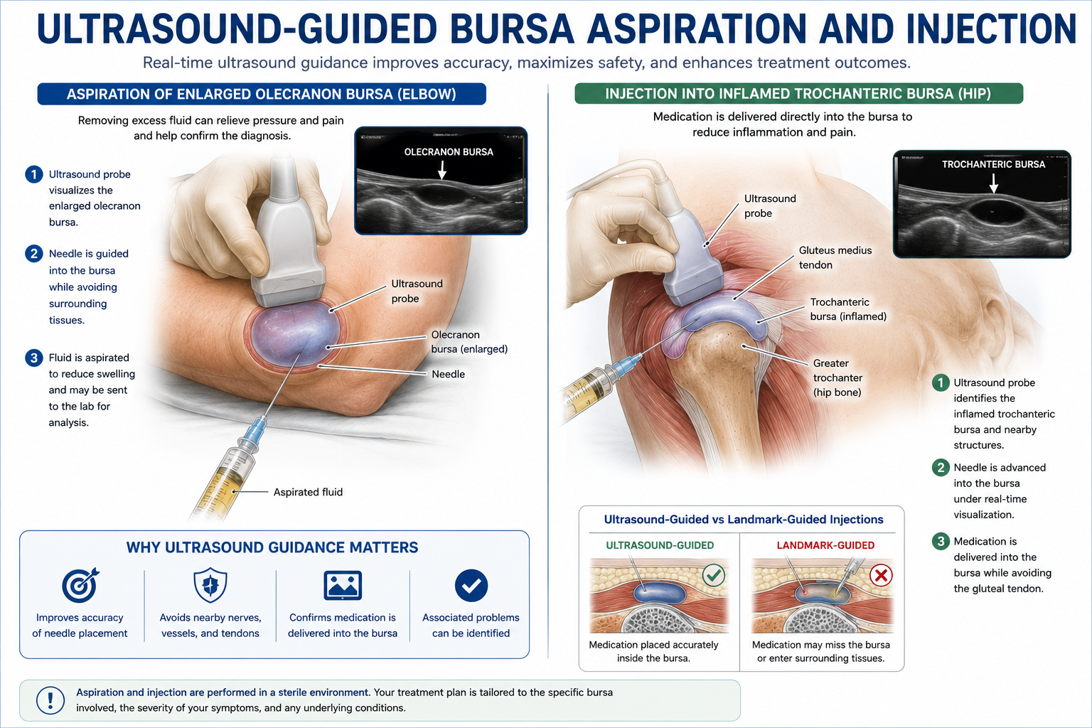
When conservative treatment fails to provide adequate relief, an image-guided bursa injection may reduce inflammation and improve function. At SpinePain Solutions, bursa injections are performed under ultrasound guidance, allowing the physician to visualize the inflamed bursa, surrounding tendons, nearby nerves, and blood vessels in real time.
Compared with landmark-guided injections, ultrasound guidance improves placement accuracy while helping avoid unintended injection into adjacent tendons or soft tissues.
Depending on the diagnosis, the injection may include:
- Local anesthetic for immediate pain relief.
- Corticosteroid medication to reduce inflammation.
- Occasionally aspiration of excess fluid when clinically appropriate.
The choice of medication is individualized based on the affected bursa, underlying diagnosis, and previous response to treatment.
Clinical Perspective
Image guidance is especially important when treating bursae that lie adjacent to major tendons. Ultrasound allows medication to be delivered accurately into the inflamed bursa while avoiding unintended injection into nearby tendon tissue.
Platelet-Rich Plasma (PRP) for Selected Patients
Not every patient with bursitis requires corticosteroid treatment. In selected individuals, particularly those with chronic bursitis associated with underlying tendon degeneration, platelet-rich plasma (PRP) may be considered as part of a comprehensive treatment strategy.
PRP is prepared from the patient’s own blood and contains a concentrated population of platelets that release naturally occurring growth factors involved in tissue repair. Rather than simply suppressing inflammation, PRP is intended to improve the biological environment surrounding chronically injured tendon tissue.
Examples include:
- Trochanteric bursitis with gluteal tendinopathy.
- Subacromial bursitis with rotator cuff tendinosis.
- Pes anserine bursitis associated with chronic hamstring tendon disease.
- Ischial bursitis occurring alongside proximal hamstring tendinopathy.
Because the scientific evidence continues to evolve, PRP is recommended selectively after careful clinical evaluation rather than as a routine treatment for every inflamed bursa.
When Is Bursa Aspiration Recommended?
Not every case of bursitis requires aspiration. While many inflamed bursae contain only a small amount of fluid, others become significantly swollen, creating a visible or palpable fluid collection that may be painful or interfere with daily activities. In these situations, removing excess fluid with a needle, a procedure known as bursa aspiration, may provide relief while also helping establish the correct diagnosis.
Aspiration is performed under sterile conditions and is often guided by ultrasound, allowing the physician to visualize the bursa, avoid nearby tendons and nerves, and remove fluid with greater precision.
Bursa aspiration may be recommended when:
- A large fluid collection causes pain, pressure, or limits movement.
- Infection is suspected, allowing fluid to be analyzed for bacteria, white blood cells, or crystals.
- Recurrent swelling persists despite conservative treatment.
- Crystal arthritis, such as gout or pseudogout, is suspected.
- The diagnosis remains uncertain after clinical evaluation.
Fluid obtained during aspiration may be sent to a laboratory for analysis when infection, gout, or other inflammatory conditions are being considered.
Although aspiration can reduce swelling, it does not correct the underlying reason the bursa became inflamed. For many patients, additional treatment such as rehabilitation, correction of biomechanical abnormalities, ultrasound-guided injections, or treatment of associated tendon disorders is necessary to reduce the risk of recurrence.
Clinical Perspective
Superficial bursae, such as the olecranon (elbow) and prepatellar (kneecap) bursae, are more commonly aspirated because fluid collections are easier to access and evaluate. In contrast, deeper bursae such as the trochanteric or subacromial bursae are rarely aspirated unless there is a specific clinical indication.
Recovery After Bursitis Treatment
Recovery depends on the underlying cause of bursitis, the duration of symptoms, the affected bursa, and the type of treatment performed. Acute bursitis often improves within several weeks with activity modification and conservative care, whereas chronic bursitis associated with tendon degeneration or biomechanical abnormalities may require a more gradual recovery.
Most patients notice improvement as inflammation subsides and surrounding muscles regain strength. Returning to normal activities too quickly, however, may delay healing or contribute to recurrent symptoms.
General recovery expectations include:
- Activity modification is often recommended for several days while inflammation begins to settle.
- Physical therapy helps restore flexibility, strength, and normal movement patterns while reducing stress on the affected bursa.
- After corticosteroid injection, many patients experience meaningful pain relief within several days, although full improvement may continue over one to two weeks.
- After PRP treatment, improvement is typically more gradual. Mild soreness is common during the first several days, with benefits often developing over several weeks to months as tendon healing progresses.
- Following aspiration, swelling frequently decreases immediately, although recurrence may occur if the underlying mechanical problem is not corrected.
- Most patients gradually return to normal daily activities, while higher-impact sports and repetitive occupational activities should be resumed according to physician recommendations.
Successful recovery depends not only on reducing inflammation but also on correcting the factors that contributed to bursitis. Continuing strengthening exercises, improving flexibility, and modifying repetitive activities help reduce the likelihood of future flare-ups.
Clinical Perspective
The inflamed bursa is often only part of the problem. Long-term success depends on restoring normal biomechanics through rehabilitation and addressing associated tendon disease, muscle weakness, or joint dysfunction. Treating the underlying cause reduces the risk of recurrent bursitis far more effectively than repeated injections alone.
When Is Surgery Necessary for Bursitis?
Fortunately, most patients with bursitis improve without surgery. The majority respond to a combination of activity modification, rehabilitation, medications, image-guided injections, or treatment of the underlying tendon or biomechanical disorder. Surgical intervention is generally reserved for the relatively small number of patients whose symptoms persist despite comprehensive conservative care or when another structural problem requires operative treatment.
In many cases, persistent pain previously attributed to bursitis is ultimately found to arise from an associated tendon tear, advanced arthritis, chronic tendon degeneration, or another musculoskeletal condition. Identifying these contributing factors helps ensure that surgery is recommended only when it offers the greatest likelihood of improving pain and function.
Surgical evaluation may be appropriate when:
- Symptoms persist despite appropriate non-surgical treatment including rehabilitation and image-guided injections.
- Recurrent bursitis continues to interfere with daily activities despite correcting contributing mechanical factors.
- Large chronic bursae remain painful or repeatedly refill after aspiration.
- Septic bursitis fails to respond adequately to antibiotics and drainage.
- Associated structural pathology, such as a significant rotator cuff tear or chronic gluteal tendon tear, requires surgical repair.
- Mechanical abnormalities create ongoing friction that cannot be adequately corrected with conservative treatment.
Depending on the underlying condition, surgery may involve removal of the chronically inflamed bursa (bursectomy), repair of injured tendons, correction of mechanical impingement, or treatment of associated joint pathology. Fortunately, isolated bursectomy alone is relatively uncommon because many patients improve once the underlying cause of inflammation has been addressed.
Clinical Perspective
Surgery should address the underlying cause of persistent bursitis rather than simply removing the inflamed bursa. For example, repairing a torn rotator cuff or treating chronic gluteal tendinopathy may eliminate the abnormal friction responsible for recurrent bursitis. Treating the entire pain generator often produces more durable results than focusing on the bursa alone.
Quick Answer
Most patients with bursitis never require surgery. Surgical treatment is typically considered only when symptoms persist despite appropriate conservative care, recurrent swelling continues to interfere with function, infection fails to respond to treatment, or associated tendon or joint damage requires repair.
Can Bursitis Be Prevented?
Although not every case of bursitis can be prevented, many patients can substantially reduce their risk by minimizing repetitive stress on vulnerable bursae and maintaining healthy joint mechanics. Because bursitis often develops gradually from repetitive friction, prolonged pressure, muscle imbalance, or tendon dysfunction, early intervention frequently prevents symptoms from progressing.
Preventive strategies include:
- Maintaining flexibility and strength around the affected joint.
- Gradually increasing activity levels rather than making sudden changes.
- Using proper lifting, throwing, and exercise techniques.
- Avoiding prolonged kneeling or leaning on hard surfaces whenever possible.
- Using protective padding during occupational or athletic activities.
- Maintaining a healthy body weight to reduce stress on weight-bearing joints.
- Treating tendon disorders and biomechanical abnormalities before they become chronic.
For patients who have experienced bursitis previously, continuing a home exercise program and correcting the underlying mechanical cause may substantially reduce the likelihood of recurrence.
Clinical Perspective
Preventing recurrent bursitis often requires more than avoiding overuse. Addressing muscle weakness, tendon disease, joint stiffness, and abnormal movement patterns reduces the excessive friction responsible for repeated inflammation.
Key Takeaways
- Bursae are small fluid-filled sacs that reduce friction between tendons, muscles, ligaments, skin, and bone.
- Bursitis develops when a bursa becomes irritated by repetitive motion, prolonged pressure, trauma, inflammation, or underlying tendon disease.
- Common locations include the shoulder, hip, elbow, knee, and pelvis.
- Many conditions mimic bursitis, making an accurate diagnosis essential before treatment.
- Ultrasound improves both diagnosis and the accuracy of bursa injections.
- Most patients improve with activity modification, rehabilitation, and image-guided treatment without surgery.
- PRP may benefit selected patients when chronic bursitis is associated with tendon degeneration.
- Surgery is rarely necessary and is generally reserved for persistent symptoms or significant associated structural pathology.
Related Articles
- Trochanteric Bursitis
- Subacromial Bursitis
- Olecranon Bursitis
- Pes Anserine Bursitis
- Ischial Bursitis
- Joint Preservation
- Platelet-Rich Plasma (PRP) for Arthritis
- TenJet Percutaneous Needle Tenotomy
- Ultrasound-Guided Joint Injections
About This Guide
This educational guide from Dr. Amit Sharma is intended to help patients better understand bursitis, its causes, diagnosis, and available treatment options. Because every patient’s condition is unique, treatment recommendations should always be individualized following evaluation by a qualified healthcare professional.
Frequently Asked Questions
What is bursitis?
Bursitis is inflammation of a bursa, a small fluid-filled sac that reduces friction between tendons, muscles, ligaments, skin, and bone. When inflamed, the bursa may become painful, swollen, and tender, particularly during movement or direct pressure.
What causes bursitis?
Bursitis most commonly develops from repetitive overuse, prolonged pressure, direct injury, tendon disorders, arthritis, crystal deposition diseases such as gout, or, less commonly, infection.
How do I know if I have bursitis or arthritis?
Both conditions may produce joint pain, but bursitis usually causes localized tenderness over a specific bursa and pain during movement or direct pressure. Arthritis affects the joint itself and often produces stiffness, reduced range of motion, and pain deep within the joint. Clinical examination and imaging help distinguish between these conditions.
Can bursitis go away on its own?
Yes. Mild cases often improve with rest, activity modification, ice, and rehabilitation. Persistent or recurrent symptoms should be evaluated because underlying tendon disease or other conditions may also require treatment.
How long does bursitis last?
Acute bursitis may improve within several weeks, while chronic bursitis associated with tendon degeneration or repetitive overuse may require several months of rehabilitation and other treatments.
When should I see a physician?
You should seek medical evaluation if pain persists despite conservative care, limits normal activities, is associated with significant swelling, redness, fever, weakness, or follows a major injury.
Is ultrasound better than X-ray for bursitis?
Each imaging study provides different information. X-rays evaluate bones and arthritis, while musculoskeletal ultrasound visualizes bursae, tendons, fluid collections, and surrounding soft tissues in real time. Ultrasound also guides accurate injections.
Are corticosteroid injections safe?
When appropriately selected and performed under image guidance, corticosteroid injections are generally safe and may provide significant relief. Repeated injections into certain tendons should be avoided because corticosteroids may weaken tendon tissue.
When is PRP considered?
PRP may be considered for selected patients with chronic bursitis associated with tendon degeneration, particularly when conservative treatment has not provided sufficient improvement.
Can bursitis come back?
Yes. Symptoms may recur if repetitive friction, tendon disease, muscle weakness, or biomechanical abnormalities are not corrected. Treating the underlying cause reduces the risk of recurrence.
Is aspiration painful?
Most patients tolerate aspiration well using local anesthetic. Ultrasound guidance helps improve comfort while accurately positioning the needle within the bursa.
Will I need surgery?
Most patients never require surgery. Operative treatment is generally reserved for persistent symptoms, recurrent septic bursitis, large chronic bursae, or associated structural problems requiring repair.
Can exercise make bursitis worse?
Exercises that repeatedly compress the inflamed bursa may temporarily worsen symptoms. A structured rehabilitation program focuses on improving flexibility, strength, and biomechanics while avoiding excessive irritation.
How can I prevent bursitis from returning?
Maintaining flexibility, strengthening supporting muscles, improving movement patterns, avoiding repetitive pressure, and treating tendon disorders early all reduce the likelihood of recurrent bursitis.
References
- American Academy of Orthopaedic Surgeons (AAOS). Bursitis.
- American Academy of Family Physicians. Evaluation of Musculoskeletal Pain.
- American College of Rheumatology. Musculoskeletal Disorders.
- American Medical Society for Sports Medicine (AMSSM). Sports Medicine Guidelines.
- American Institute of Ultrasound in Medicine (AIUM). Practice Parameters for Musculoskeletal Ultrasound.
- European Society of Musculoskeletal Radiology (ESSR). Ultrasound Guidelines.
- Gray’s Anatomy: The Anatomical Basis of Clinical Practice.
- Netter FH. Atlas of Human Anatomy.
- Brukner & Khan’s Clinical Sports Medicine.
- Journal of Bone and Joint Surgery.
- American Journal of Sports Medicine.
- PM&R KnowledgeNow.
- National Institute for Health and Care Excellence (NICE). Musculoskeletal Conditions.
- UpToDate. Bursitis in Adults.
- Manchikanti L, et al. Evidence-Based Guidelines for Interventional Pain Procedures.



