
Joint Radiofrequency Ablation (RFA): A Complete Guide to Knee, Shoulder & Hip Pain
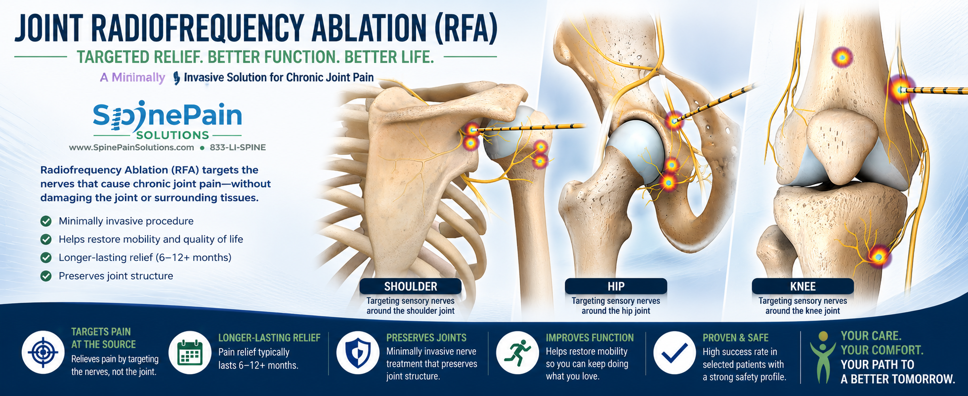
Living with chronic joint pain can be frustrating, especially when medications, physical therapy, and joint injections no longer provide lasting relief. Many patients are not ready for joint replacement surgery, while others may not be good surgical candidates because of age, medical conditions, or personal preference. Joint radiofrequency ablation (RFA) offers a minimally invasive treatment option that may reduce chronic pain by targeting the sensory nerves responsible for transmitting pain signals from an arthritic joint.
Unlike joint replacement surgery, radiofrequency ablation does not remove cartilage, replace the joint, or alter the mechanical stability of the joint itself. Instead, it interrupts pain transmission from carefully selected sensory nerve branches while preserving the muscles and major nerves responsible for movement. By reducing pain, many patients are better able to walk, participate in physical therapy, remain active, and improve their overall quality of life.
Today, joint RFA is most commonly performed for chronic knee, shoulder, and hip pain related to osteoarthritis. It may also help carefully selected patients who continue to experience persistent pain after knee, shoulder, or hip replacement surgery, provided infection, implant loosening, fracture, instability, and other correctable causes have first been excluded.
This comprehensive guide from Dr. Amit Sharma explains how joint radiofrequency ablation works, which joints can be treated, who may be an appropriate candidate, what results patients can realistically expect, and how RFA compares with joint injections, regenerative medicine, and joint replacement surgery.
What Is Joint Radiofrequency Ablation?
Quick Answer
Joint radiofrequency ablation (RFA) is a minimally invasive procedure that treats chronic joint pain by interrupting pain signals carried through small sensory nerve branches surrounding the joint. It does not replace the joint, repair damaged cartilage, or weaken the muscles responsible for movement. Instead, RFA is designed to reduce pain and improve function in carefully selected patients with chronic knee, shoulder, or hip pain, including some individuals who continue to experience pain after joint replacement surgery once mechanical complications and infection have been excluded.
Most patients first undergo a comprehensive evaluation to determine whether the painful joint is the true source of symptoms. Diagnostic nerve blocks may be performed before radiofrequency ablation to confirm that the targeted sensory nerves are contributing to the patient’s pain. When appropriately selected, many patients experience meaningful improvements in pain, walking tolerance, sleep, daily activities, and overall quality of life.
Clinical Perspective
One of the most common misconceptions about radiofrequency ablation is that it “burns the joint” or permanently destroys important nerves. In reality, joint RFA targets tiny sensory nerve branches responsible for transmitting pain signals while avoiding the major nerves that control muscle strength and movement. The procedure is intended to reduce pain, not eliminate normal joint function, and it does not prevent patients from pursuing future treatments such as joint replacement if those options become appropriate later.
Why Do Arthritic Joints Hurt?
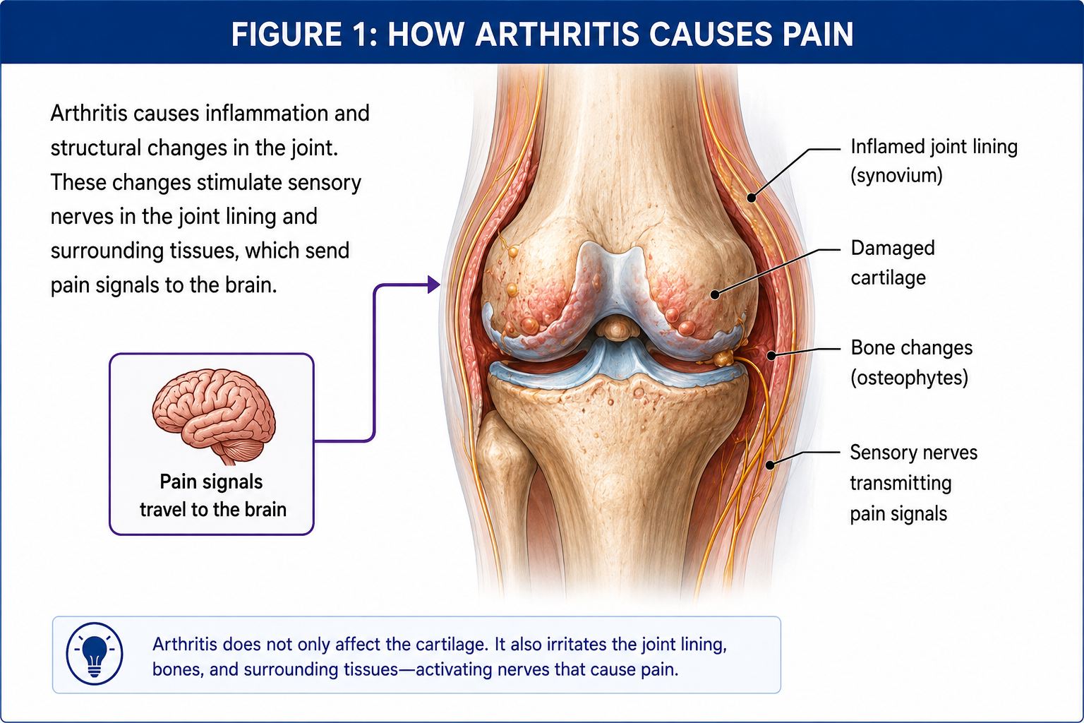
Many people believe arthritis hurts simply because cartilage wears away and bone rubs directly against bone. While cartilage loss is an important feature of osteoarthritis, cartilage itself contains very few pain-sensitive nerve endings. Instead, pain usually arises from several surrounding structures that become irritated, inflamed, or mechanically stressed as arthritis progresses.
An arthritic joint functions as a complete organ rather than a single piece of cartilage. The joint capsule, synovial lining, ligaments, subchondral bone, surrounding muscles, tendons, and small sensory nerves all contribute to normal movement. When arthritis develops, one or more of these structures may become an important source of pain, often long before complete cartilage loss occurs.
Radiofrequency ablation is based on an important principle of pain medicine: although arthritis may involve many different tissues, much of the pain they generate ultimately travels through a relatively small number of sensory articular nerve branches before reaching the spinal cord and brain. By treating these nerves, radiofrequency ablation attempts to reduce pain without altering the structural stability or movement of the joint.
Common Sources of Arthritic Joint Pain
Several structures may contribute to arthritis-related pain simultaneously:
- Synovium: Inflammation of the joint lining may produce pain, swelling, stiffness, and excess joint fluid.
- Joint capsule: Stretching and chronic inflammation of the capsule can produce pain during movement or at the extremes of motion.
- Subchondral bone: The bone beneath the cartilage contains abundant pain-sensitive nerve endings and often becomes an important source of discomfort as arthritis progresses.
- Ligaments: Chronic instability or abnormal joint mechanics can place excessive stress on supporting ligaments.
- Muscles and tendons: Weakness, altered movement patterns, and compensation frequently lead to secondary muscle fatigue and tendon pain.
- Peripheral sensory nerves: Small articular nerve branches surrounding the joint transmit pain signals toward the spinal cord, where they are ultimately interpreted by the brain as joint pain.
Key Point: Radiofrequency ablation does not repair cartilage or reverse arthritis. Instead, it reduces pain by treating the sensory nerves that carry pain signals from the arthritic joint.
How Does Joint Radiofrequency Ablation Work?
Radiofrequency ablation uses precisely controlled thermal energy to treat carefully selected sensory nerve branches that transmit pain from an arthritic joint. Using fluoroscopic or ultrasound guidance, specialized needles are positioned adjacent to these small articular nerves without entering the joint itself. Radiofrequency energy is then applied through the needle tip, creating a controlled treatment zone that interrupts the nerve’s ability to transmit pain signals.
Unlike surgical nerve cutting, radiofrequency ablation does not remove the nerve or permanently eliminate all sensation. The treated nerve gradually undergoes healing and regeneration over time, which is why pain relief is often temporary rather than permanent. For many patients, however, this period of reduced pain may last several months and significantly improve walking, sleep, physical therapy participation, and overall function.
Because the procedure targets small sensory articular branches rather than the major motor nerves controlling muscles, normal joint movement and strength are preserved. Patients remain able to walk, exercise, and participate in rehabilitation while benefiting from reduced pain transmission.
Clinical Pearl
Think of radiofrequency ablation as lowering the volume of pain coming from the joint rather than repairing the joint itself. The arthritis remains, but many patients experience less pain, making it easier to stay active, strengthen surrounding muscles, and improve quality of life.
Which Joints Can Be Treated with Radiofrequency Ablation?
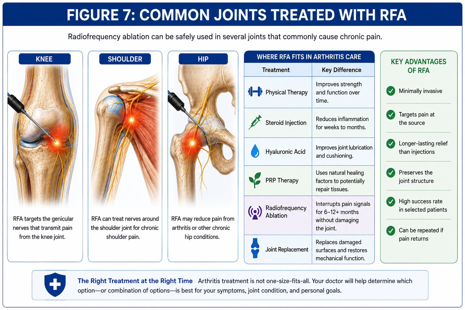
Joint radiofrequency ablation is most commonly performed for chronic pain involving the knee, shoulder, and hip. Although each joint has its own anatomy and unique sensory nerve supply, the treatment principle remains the same: carefully selected sensory articular nerve branches are targeted to reduce pain while preserving muscle strength, joint stability, and normal movement.
The specific nerves treated, the imaging guidance used, and the expected outcomes vary according to the joint involved. Careful evaluation is necessary to determine whether the pain is truly originating from the joint itself or from surrounding muscles, tendons, bursae, peripheral nerves, or the spine.
Knee Radiofrequency Ablation
Knee radiofrequency ablation is currently the most extensively studied application of joint RFA. The procedure primarily targets the genicular sensory nerves, which transmit pain from the knee capsule, surrounding ligaments, periosteum, and other pain-sensitive structures. Because these nerves are primarily sensory, treatment is intended to reduce pain while preserving normal movement and muscle strength.
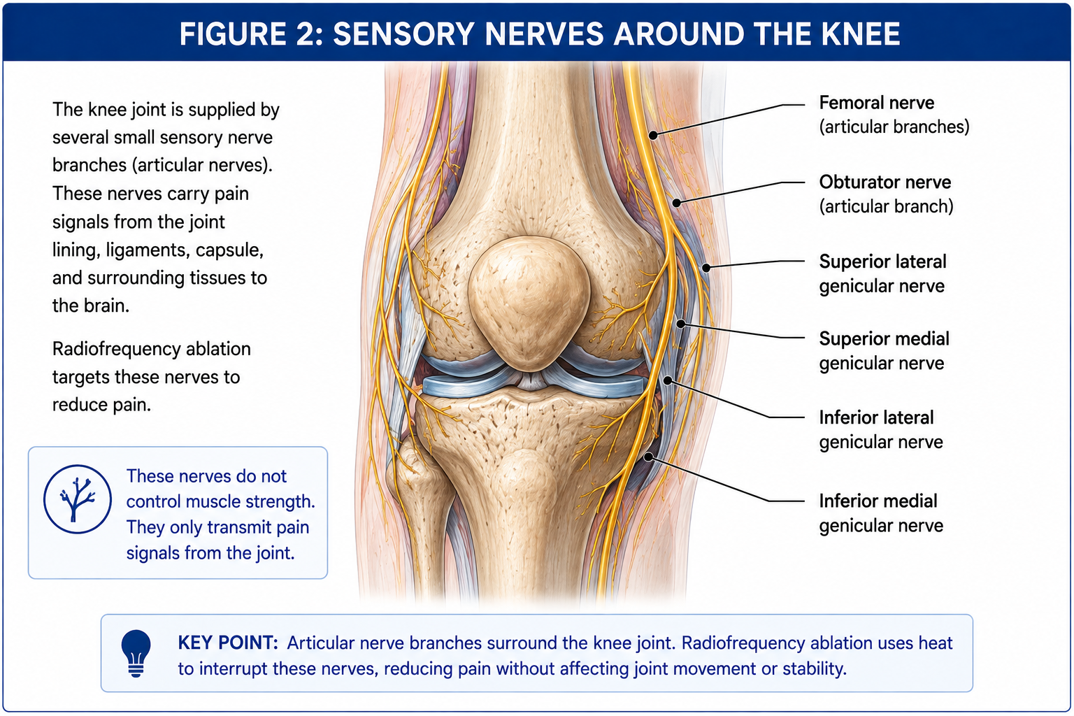
Patients with symptomatic knee osteoarthritis who continue to experience pain despite exercise, physical therapy, medications, or joint injections may be candidates for genicular nerve radiofrequency ablation. It may also benefit carefully selected patients with persistent pain after total knee replacement once infection, implant loosening, instability, fracture, and other mechanical complications have been excluded.
Read more in our comprehensive guide to Knee Radiofrequency Ablation.
Shoulder Radiofrequency Ablation
Shoulder radiofrequency ablation treats chronic shoulder pain by targeting sensory articular branches that commonly arise from the suprascapular, axillary, and lateral pectoral nerves. These nerves transmit pain from the glenohumeral joint while leaving the major motor pathways responsible for shoulder movement intact.
The procedure may be considered for carefully selected patients with glenohumeral osteoarthritis, rotator cuff arthropathy, chronic degenerative shoulder pain, or persistent pain after shoulder replacement when evaluation confirms that no surgically correctable complication is present.
A dedicated guide to Shoulder Radiofrequency Ablation will explore these techniques in greater detail.
Hip Radiofrequency Ablation
Hip radiofrequency ablation typically targets sensory articular branches arising from the femoral and obturator nerves, which provide pain sensation to the anterior hip capsule. These small sensory branches can be treated while preserving the major motor nerves that control walking and leg strength.
Hip RFA may be considered for patients with painful hip osteoarthritis who continue to have significant symptoms despite conservative treatment, as well as selected patients who experience persistent pain after total hip replacement once infection, implant loosening, fracture, instability, and other mechanical causes have been excluded.
Learn more on our upcoming Hip Radiofrequency Ablation service page.
What Joint Radiofrequency Ablation Cannot Treat
Although joint radiofrequency ablation can reduce pain arising from an arthritic joint, it does not correct the underlying structural problem causing the arthritis. The procedure is designed to interrupt pain transmission through small sensory nerve branches, not repair damaged cartilage, restore joint stability, or correct mechanical abnormalities. For this reason, careful evaluation is essential before considering treatment.
Patients with severe joint pain should first be evaluated to determine whether another condition is responsible for their symptoms. In some situations, treating the pain-transmitting nerves alone could delay more appropriate treatment without addressing the underlying problem.
Active Joint Infection
A painful, swollen, warm joint caused by infection requires urgent medical treatment. Radiofrequency ablation has no role in treating septic arthritis or prosthetic joint infection and should never be performed until infection has been fully evaluated and appropriately managed.
Loose or Failing Joint Replacement
Persistent pain after joint replacement may result from implant loosening, component failure, malalignment, instability, polyethylene wear, or other mechanical problems. These conditions require orthopedic evaluation rather than treatment of the sensory nerves alone.
Fractures and Structural Instability
Acute fractures, stress fractures, severe ligament instability, and significant mechanical deformity require treatment directed at the underlying structural abnormality. While pain relief is important, eliminating pain without correcting an unstable joint may increase the risk of further injury.
Major Tendon or Muscle Injury
Complete tendon ruptures, large rotator cuff tears, extensor mechanism disruption around the knee, or other major soft tissue injuries usually require a different treatment strategy. These conditions may coexist with arthritis but are not corrected by radiofrequency ablation.
Pain Originating Outside the Joint
Not every painful knee, shoulder, or hip originates from the joint itself. Pain may arise from the lumbar spine, cervical spine, sacroiliac joint, peripheral nerves, muscles, tendons, bursae, or other surrounding structures. Diagnostic evaluation is necessary to identify the true pain generator before proceeding with joint RFA.
Important: Joint radiofrequency ablation treats pain, not the underlying arthritis. Persistent or worsening pain should always be evaluated to ensure that infection, fracture, implant failure, instability, or another treatable condition is not being overlooked.
Who May Be a Candidate for Joint Radiofrequency Ablation?
Joint radiofrequency ablation is not intended for every patient with arthritis. The procedure is generally considered after a comprehensive evaluation confirms that pain is arising from the joint and that appropriate conservative treatments have not provided adequate or lasting relief. Successful outcomes depend not only on the severity of arthritis but also on identifying the correct pain generator and selecting the appropriate patient.
Patients who may benefit include those with chronic knee, shoulder, or hip pain that continues to interfere with walking, work, sleep, or daily activities despite exercise, physical therapy, medications, or image-guided joint injections.
- Moderate to severe osteoarthritis
- Persistent pain despite conservative treatment
- Pain that limits walking or daily activities
- Patients wishing to postpone joint replacement
- Patients who are poor surgical candidates because of medical conditions
- Persistent pain after joint replacement once mechanical causes have been excluded
- Positive response to diagnostic articular nerve blocks when performed
Clinical Pearl: The best predictor of success is not simply the severity of arthritis seen on an X-ray. Patients tend to do best when the painful joint has been carefully evaluated, alternative pain generators have been excluded, and diagnostic nerve blocks confirm that the targeted sensory nerves are responsible for the pain.
Why Are Diagnostic Nerve Blocks Performed First?
Before proceeding with radiofrequency ablation, many physicians perform diagnostic articular nerve blocks. During this procedure, a small amount of local anesthetic is placed around the sensory nerve branches that would later be treated with radiofrequency ablation.
If these nerves are responsible for the patient’s pain, temporary pain relief is expected while the anesthetic remains active. Although a successful diagnostic block cannot guarantee long-term success with radiofrequency ablation, it increases confidence that the targeted nerves are contributing to the patient’s symptoms.
Some insurance carriers require one or more diagnostic blocks before approving radiofrequency ablation, while others do not. The protocol varies depending on the joint being treated, the clinical situation, and insurance requirements.
How Is Joint Radiofrequency Ablation Performed?
Joint radiofrequency ablation is performed as an outpatient procedure using image guidance to accurately position specialized needles adjacent to the targeted sensory nerves. Fluoroscopy is commonly used for knee procedures, while ultrasound or fluoroscopy may be selected for the shoulder and hip depending on physician preference and anatomy.
- The skin is cleaned using sterile technique.
- Local anesthetic is administered to numb the skin.
- Specialized radiofrequency needles are advanced using imaging guidance.
- Motor and sensory stimulation may be performed to confirm safe positioning.
- Radiofrequency energy is delivered for a controlled period of time.
- The needles are removed and small bandages are applied.
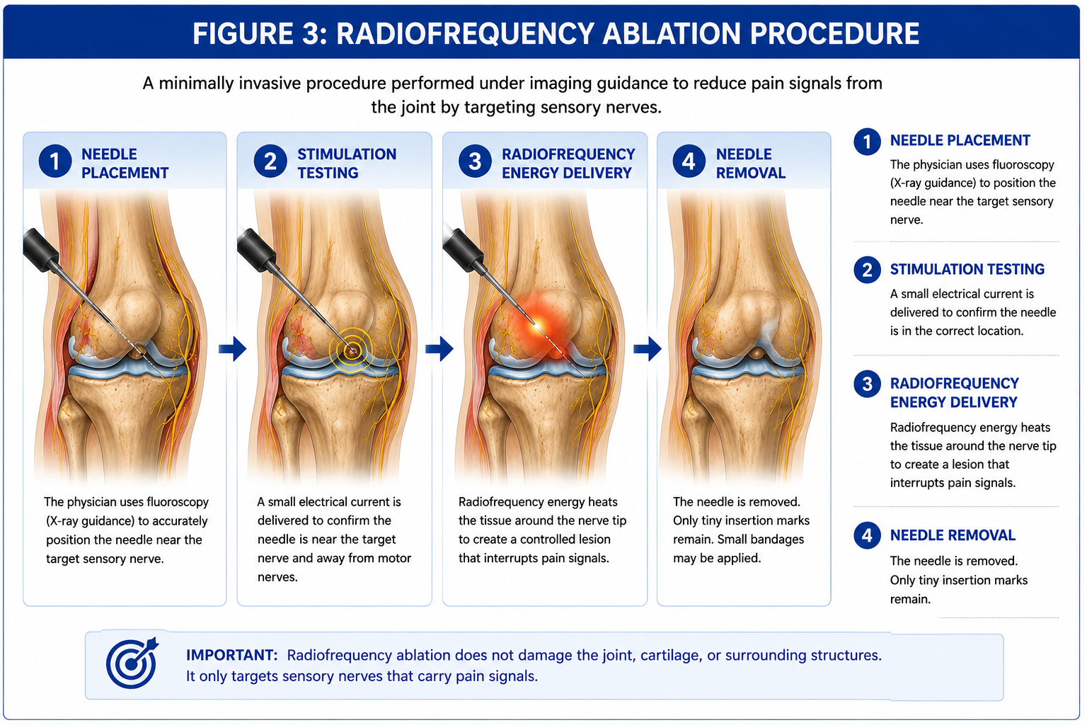
Most procedures are completed within 30 to 60 minutes, and patients generally return home the same day.
Types of Radiofrequency Ablation
| Technology | Electrode Design | Treatment Zone | Typical Clinical Use |
|---|---|---|---|
| Conventional RFA | Single active-tip electrode | Focused thermal lesion adjacent to the needle tip | Well-defined target nerves; multiple lesions may be created to improve coverage. |
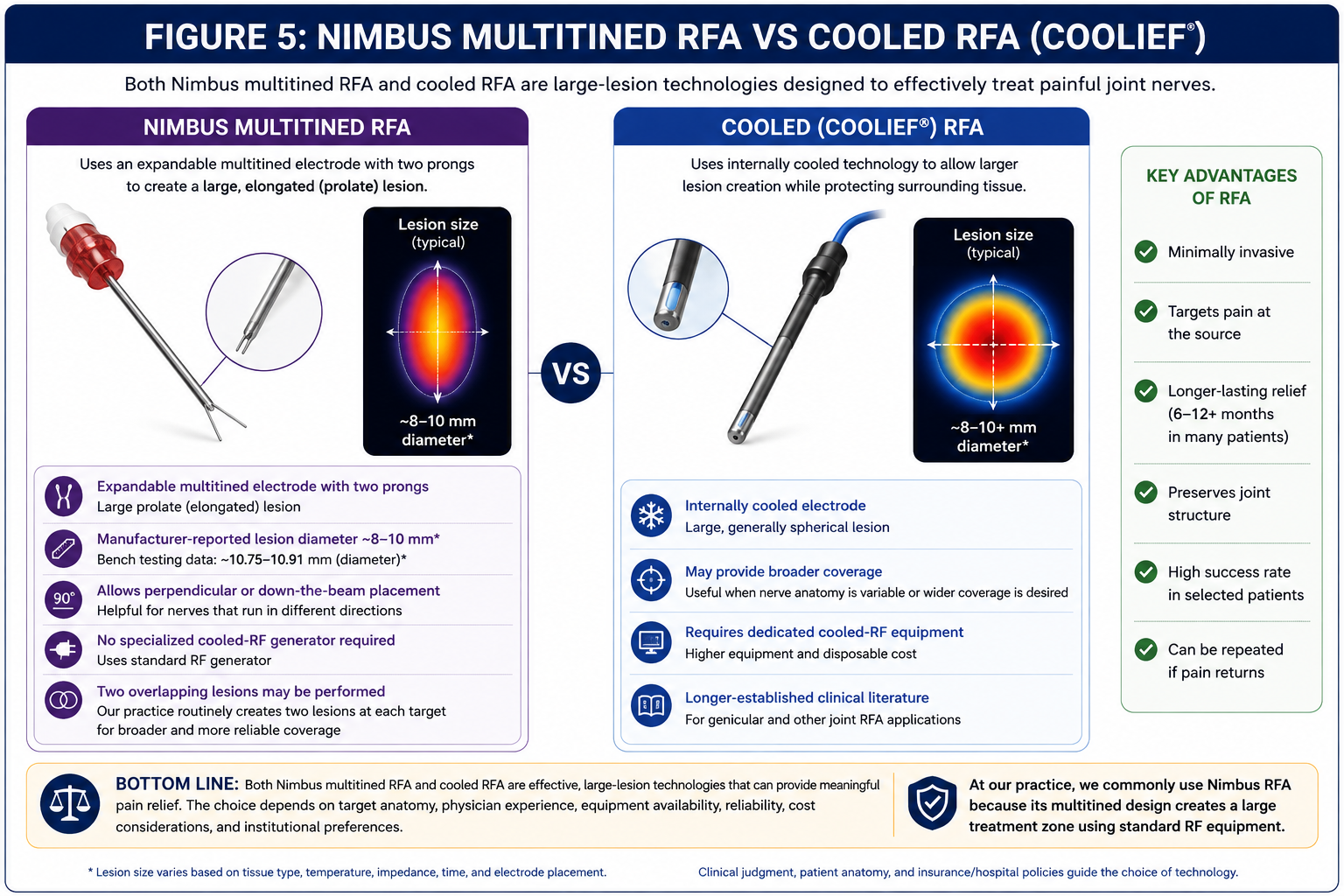
| Nimbus® Multitined RFA | Expandable two-tined electrode | Broad elongated treatment zone created by the deployed tines | Commonly used for articular nerve ablation. At our practice, two overlapping lesions are routinely created at each target to maximize coverage. |
| Cooled RFA | Internally cooled electrode | Broad treatment zone extending beyond the electrode tip | Useful when larger coverage is desired; requires dedicated cooled-RF equipment. |
| Bipolar RFA | Two electrodes | Treatment zone forms between the electrodes | Used selectively when anatomy favors bipolar lesioning. |
| Pulsed RFA | Intermittent radiofrequency energy | Neuromodulation rather than continuous thermal ablation | Selected pain conditions where thermal lesioning is not the objective. |
Several radiofrequency technologies are available for treating chronic joint pain. Conventional, multitined (Nimbus®), cooled, bipolar, and pulsed radiofrequency each create treatment effects through different electrode designs and energy delivery strategies. No single technology has been proven universally superior for every patient. The optimal choice depends on the target anatomy, physician experience, procedural technique, equipment availability, and individual clinical circumstances.
Clinical Pearl
Patients often ask whether one radiofrequency system is “better” than another. The reality is that successful joint radiofrequency ablation depends far more on accurate diagnosis, appropriate patient selection, precise needle placement, thoughtful lesioning strategy, and physician experience than on the brand of equipment used. At SpinePain Solutions, we routinely perform two overlapping lesions with the Nimbus® multitined system to maximize treatment coverage while preserving surrounding structures. We also offer cooled radiofrequency ablation (Coolief®) when it is the preferred option based on a patient’s anatomy, clinical circumstances, or personal preference. Rather than favoring one technology over another, our philosophy is to match the right technique to the right patient to achieve the safest and most durable pain relief possible.
Recovery After Joint Radiofrequency Ablation
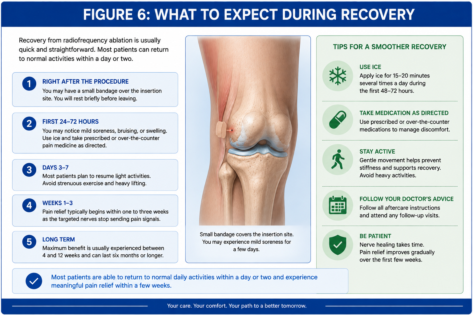
Joint radiofrequency ablation is performed on an outpatient basis, allowing most patients to return home shortly after the procedure. Because only small needles are used, recovery is generally much faster than after joint replacement surgery. Mild soreness at the treatment sites is common during the first several days and usually resolves with conservative care.
Pain relief is rarely immediate. Although the local anesthetic used during the procedure may temporarily reduce discomfort, this effect typically wears off within several hours. The treated sensory nerves require time to stop transmitting pain signals, and meaningful improvement often develops gradually over the following two to six weeks.
General Recovery Timeline
- First 24 hours: Mild soreness at the needle insertion sites is common.
- First week: Some patients experience temporary discomfort as the treated nerves respond to the procedure.
- Two to six weeks: Pain relief typically becomes more noticeable.
- Following months: Many patients experience improved walking, sleep, and participation in rehabilitation or exercise.
Clinical Pearl: Radiofrequency ablation should not be viewed as a stand-alone treatment. Patients often achieve the best long-term results by combining pain reduction with strengthening exercises, weight management, physical therapy, and other joint-preserving strategies.
Potential Benefits of Joint Radiofrequency Ablation
When performed in appropriately selected patients, joint radiofrequency ablation may provide meaningful improvements in pain and physical function. The goal is not simply to reduce pain scores but to help patients return to activities that arthritis has gradually taken away.
- Reduced chronic joint pain
- Improved walking tolerance
- Greater participation in physical therapy
- Improved sleep quality
- Reduced dependence on pain medications
- Improved ability to perform daily activities
- Delay or avoidance of joint replacement surgery for selected patients
- Improved quality of life
The degree of improvement varies among individuals and depends on several factors, including the severity of arthritis, the accuracy of diagnosis, overall health, rehabilitation efforts, and whether other painful conditions are also present.
How Long Does Radiofrequency Ablation Last?
Pain relief following joint radiofrequency ablation varies from one patient to another. Many patients experience meaningful improvement for approximately six to twelve months, while some continue to benefit for considerably longer. Others may experience a shorter duration of relief depending on the severity of arthritis, nerve regeneration, activity level, and progression of the underlying joint disease.
Radiofrequency ablation does not permanently destroy the treated sensory nerves. Over time, these nerves gradually regenerate and may once again transmit pain signals. If substantial pain returns and the original procedure was successful, repeat radiofrequency ablation may be considered in appropriately selected patients.
Important: Even if pain relief decreases over time, radiofrequency ablation does not reduce future treatment options. Patients may still undergo joint injections, regenerative medicine procedures, or joint replacement surgery if clinically appropriate.
Risks and Possible Complications
Joint radiofrequency ablation is generally considered a safe procedure when performed by experienced physicians using image guidance. As with any medical procedure, however, potential risks and complications should be discussed before treatment.
- Temporary soreness at the treatment site
- Bruising or minor bleeding
- Temporary numbness or altered skin sensation
- Post-procedural neuritis or nerve irritation
- Incomplete pain relief
- Recurrence of pain as nerves regenerate
- Rare infection
- Rare allergic reaction to medications or contrast material
- Extremely rare injury to nearby nerves or blood vessels
Serious complications are uncommon because the procedure targets small sensory nerve branches located outside the joint while using fluoroscopic or ultrasound guidance to improve procedural accuracy.
Persistent Pain After Joint Replacement: Can Radiofrequency Ablation Help?
Joint replacement surgery has transformed the lives of millions of patients suffering from advanced arthritis. Modern knee, shoulder, and hip replacements are among the most successful procedures in orthopedic surgery, with most patients experiencing substantial pain relief and improved function. Despite these excellent results, a meaningful number of patients continue to experience chronic pain months or even years after an otherwise technically successful joint replacement.
Persistent pain after joint replacement can be frustrating for both patients and physicians. Some patients are told that their X-rays look normal and that the implants appear well positioned, yet they continue to struggle with pain during walking, climbing stairs, reaching overhead, or routine daily activities. In carefully selected individuals, radiofrequency ablation may provide meaningful pain relief, but only after a thorough evaluation has ruled out problems that require orthopedic treatment rather than pain management.
Important: Persistent pain after joint replacement should never be assumed to be “normal.” Before considering radiofrequency ablation, infection, implant loosening, fracture, instability, and other surgically correctable conditions must first be excluded.
How Common Is Persistent Pain?
Although most patients do very well following joint replacement surgery, studies suggest that approximately 10% to 20% continue to report chronic pain despite an apparently successful operation. The severity of symptoms varies considerably. Some patients notice discomfort only during strenuous activity, while others experience persistent pain that interferes with walking, sleep, work, or quality of life.
The First Step Is Finding the Cause
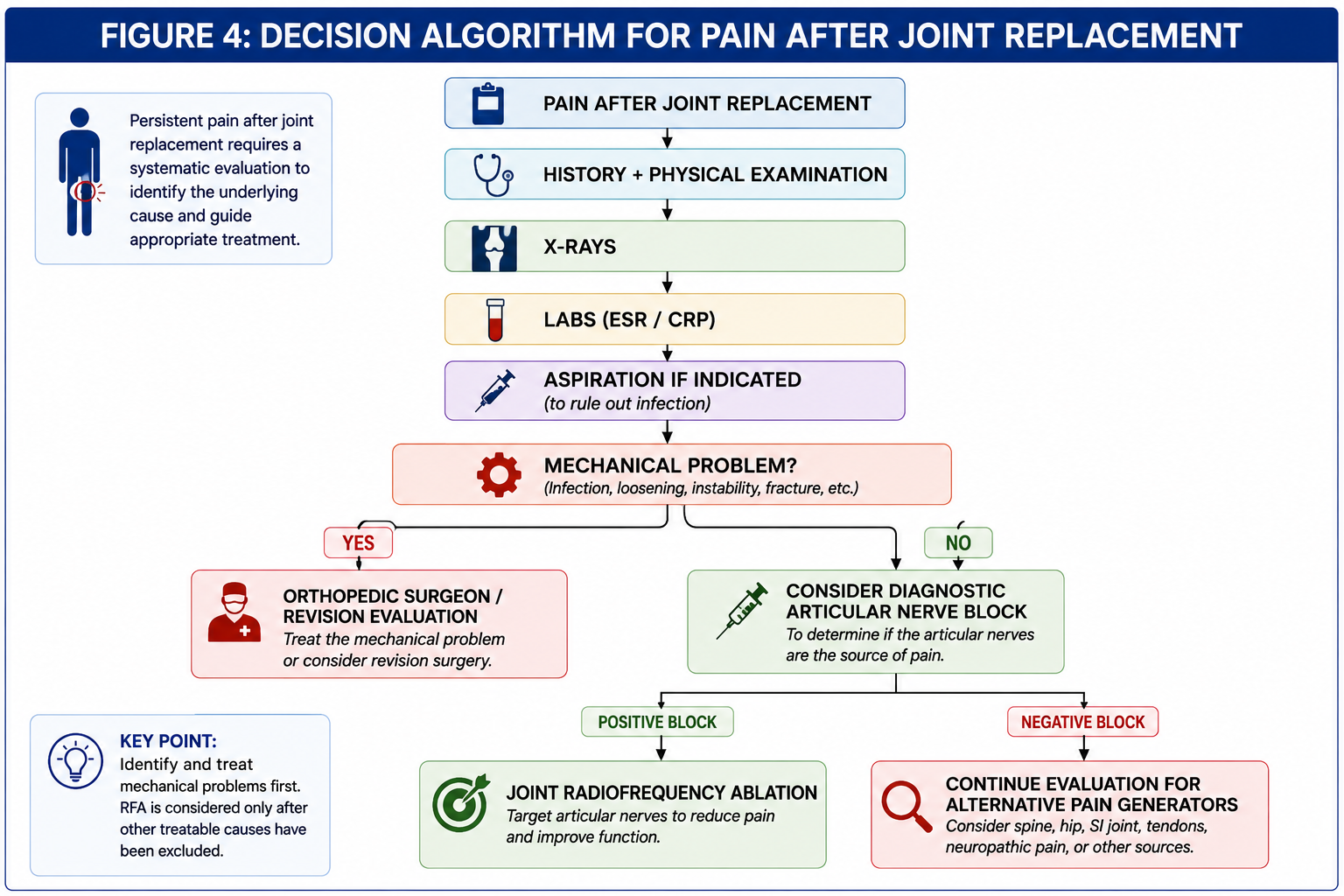
Radiofrequency ablation should never be used simply because pain persists after surgery. The first priority is determining why the joint remains painful. A careful history, physical examination, imaging studies, and, when appropriate, laboratory testing are often necessary before deciding whether pain is arising from the replaced joint or from another source.
- Periprosthetic joint infection
- Implant loosening or mechanical failure
- Component malalignment or malposition
- Instability of the replaced joint
- Periprosthetic fracture
- Patellofemoral disorders following knee replacement
- Soft tissue impingement or tendon disorders
- Complex regional pain syndrome (CRPS)
- Pain referred from the cervical or lumbar spine
- Peripheral nerve disorders
Only after these conditions have been appropriately evaluated can persistent pain be considered for treatment with radiofrequency ablation.
Persistent Pain After Total Knee Replacement
Total knee replacement is highly successful for relieving pain caused by advanced arthritis, yet some patients continue to experience discomfort despite well-positioned implants. Once infection, loosening, instability, fracture, and other orthopedic complications have been excluded, persistent pain may continue to originate from sensory nerve pathways surrounding the knee.
In carefully selected patients, genicular nerve radiofrequency ablation may reduce chronic pain and improve function without affecting the stability or integrity of the knee replacement itself.
Persistent Pain After Shoulder Replacement
Most shoulder replacements provide substantial pain relief, but persistent pain occasionally occurs because of implant-related problems, rotator cuff pathology, infection, instability, or stiffness. Following a comprehensive orthopedic evaluation, radiofrequency ablation of selected articular branches may be considered for carefully selected patients whose pain continues despite appropriate treatment.
Persistent Pain After Hip Replacement
Persistent pain after total hip replacement requires careful evaluation because symptoms may originate from the hip implant itself, surrounding soft tissues, the lumbar spine, sacroiliac joint, or peripheral nerves. Once mechanical causes have been excluded, radiofrequency ablation of the articular branches supplying the anterior hip capsule may provide pain relief in selected patients.
When Should Radiofrequency Ablation Be Considered?
Joint radiofrequency ablation is generally considered only after a comprehensive evaluation demonstrates that the painful joint replacement is mechanically stable and free of infection. Diagnostic nerve blocks may help confirm that the targeted sensory nerves are contributing to the patient’s symptoms before proceeding with treatment.
For patients who are not candidates for revision surgery, prefer to avoid another major operation, or continue to experience chronic pain despite appropriate orthopedic care, radiofrequency ablation may offer a minimally invasive option that improves pain and function while preserving future treatment choices.
Clinical Perspective: Radiofrequency ablation should not be viewed as an alternative to revision surgery when a correctable mechanical problem exists. Rather, it serves as an additional treatment option for carefully selected patients whose implants are functioning appropriately but who continue to experience chronic pain arising from the sensory nerves surrounding the joint.
How Does Radiofrequency Ablation Compare with Other Arthritis Treatments?
Radiofrequency ablation is one of several non-surgical treatments available for chronic joint pain. Each option works differently, and no single treatment is appropriate for every patient. Factors such as the severity of arthritis, the source of pain, previous treatments, overall health, activity goals, and patient preferences all influence the most appropriate treatment plan.
Rather than competing with one another, physical therapy, medications, joint injections, regenerative medicine, radiofrequency ablation, and joint replacement often complement each other during different stages of arthritis.
| Treatment | Primary Goal | Typical Duration | Best For |
|---|---|---|---|
| Physical Therapy | Improve strength and mechanics | Long-term with continued exercise | Nearly all stages of arthritis |
| Corticosteroid Injection | Reduce inflammation | Weeks to several months | Inflammatory flare-ups |
| Hyaluronic Acid | Improve joint lubrication | Several months | Selected knee osteoarthritis |
| Platelet-Rich Plasma (PRP) | Support biologic healing | Variable | Selected patients with mild to moderate arthritis |
| Radiofrequency Ablation | Reduce pain transmission | Often 6-12 months or longer | Persistent chronic pain despite conservative care |
| Joint Replacement | Replace damaged joint surfaces | Many years | Advanced arthritis with severe disability |
Because each treatment addresses arthritis differently, many patients receive more than one of these therapies during the course of their condition. A personalized treatment plan is often more effective than relying on a single intervention alone.
When Is Joint Replacement a Better Option?
Although radiofrequency ablation can provide meaningful pain relief for many patients, it is not intended to replace joint replacement surgery in individuals with advanced structural joint damage who are appropriate surgical candidates. Instead, the two treatments address different aspects of arthritis and may be appropriate at different stages of the disease.
Joint replacement is often considered when severe arthritis causes persistent pain, significant loss of function, and reduced quality of life despite comprehensive non-surgical treatment. Unlike radiofrequency ablation, joint replacement removes damaged joint surfaces and restores mechanical function by replacing the arthritic joint with prosthetic components.
- Severe bone-on-bone arthritis with marked functional limitation
- Progressive joint deformity or instability
- Failure of comprehensive non-surgical treatment
- Pain that significantly limits work, sleep, or daily activities
- Patients who are medically appropriate surgical candidates
Clinical Perspective: Choosing between joint injections, regenerative medicine, radiofrequency ablation, and joint replacement is rarely an either-or decision. These treatments often complement one another, and the best approach depends on the patient’s symptoms, goals, imaging findings, overall health, and response to previous therapies.
Frequently Asked Questions
Does radiofrequency ablation cure arthritis?
No. Radiofrequency ablation does not repair damaged cartilage or reverse arthritis. It reduces pain by interrupting sensory nerve signals originating from the arthritic joint.
How long does joint radiofrequency ablation last?
Many patients experience meaningful pain relief for approximately six to twelve months, although some benefit for considerably longer and others for a shorter period. Results vary depending on the severity of arthritis, the treated joint, and individual healing.
Can radiofrequency ablation be repeated?
Yes. If pain returns after an initially successful procedure, repeat radiofrequency ablation may be considered after reassessment by your physician.
Is the procedure painful?
Most patients tolerate the procedure well using local anesthetic, with or without mild sedation depending on the clinical situation. Temporary soreness after the procedure is common and generally resolves within several days.
Will radiofrequency ablation make my joint weak?
No. Joint radiofrequency ablation targets small sensory nerve branches responsible for pain transmission rather than the motor nerves that control muscle strength.
Can I still have joint replacement surgery later?
Yes. Radiofrequency ablation does not prevent future joint replacement surgery if it later becomes medically appropriate.
Which joints can be treated?
The strongest evidence currently exists for knee osteoarthritis. Radiofrequency ablation is also increasingly used for carefully selected patients with chronic shoulder and hip pain, including some patients with persistent pain after joint replacement.
Can radiofrequency ablation help after knee replacement?
It may. Before treatment, infection, implant loosening, instability, fracture, and other mechanical problems must first be excluded. For carefully selected patients, genicular nerve radiofrequency ablation may reduce persistent postoperative pain.
Is radiofrequency ablation better than steroid injections?
They serve different purposes. Corticosteroid injections primarily reduce inflammation, whereas radiofrequency ablation interrupts pain transmission through sensory nerves. Depending on the patient, one or both treatments may be appropriate.
Does insurance cover joint radiofrequency ablation?
Coverage varies depending on the insurance carrier, the joint being treated, and medical necessity. Some insurers require diagnostic nerve blocks before approving treatment.
Is cooled radiofrequency ablation better than conventional radiofrequency ablation?
Both techniques have demonstrated benefit in selected patients. The most appropriate approach depends on the treated joint, physician experience, available equipment, and current clinical evidence.
Can radiofrequency ablation delay joint replacement?
For some patients, yes. By reducing pain and improving function, radiofrequency ablation may postpone the need for joint replacement. However, it does not stop arthritis from progressing.
Key Takeaways
- Joint radiofrequency ablation treats chronic pain by interrupting sensory nerve signals rather than repairing the joint.
- The procedure is most commonly performed for knee osteoarthritis and is increasingly used for selected shoulder and hip conditions.
- Patients should undergo a comprehensive evaluation to confirm that the joint is the primary pain source.
- Diagnostic articular nerve blocks may help predict treatment success.
- Pain relief commonly lasts six to twelve months or longer, although individual results vary.
- Joint radiofrequency ablation does not prevent future injections, regenerative medicine treatments, or joint replacement surgery.
- Persistent pain after joint replacement should always be evaluated for infection, loosening, fracture, or instability before considering radiofrequency ablation.
Related Services
- Arthritis Treatment
- Knee Radiofrequency Ablation
- Shoulder Radiofrequency Ablation (Coming Soon)
- Hip Radiofrequency Ablation (Coming Soon)
- Corticosteroid Joint Injections
- Hyaluronic Acid Joint Injections
- PRP for Arthritis
- Joint Preservation (Coming Soon)
References
- American Academy of Orthopaedic Surgeons (AAOS). Clinical Practice Guideline: Management of Osteoarthritis of the Knee.
- American Society of Pain and Neuroscience (ASPN). Best Practice Guidelines for Radiofrequency Ablation of Articular Branches.
- National Institute for Health and Care Excellence (NICE). Radiofrequency Denervation for Osteoarthritic Knee Pain.
- Chen AF, et al. Persistent Pain After Total Knee Arthroplasty: Evaluation and Management.
- Kapural L, et al. Genicular Nerve Radiofrequency Ablation for Chronic Knee Pain.
- Recent systematic reviews and meta-analyses on knee, shoulder, and hip radiofrequency ablation.
Medical Disclaimer
This educational guide is intended for informational purposes only and should not be considered medical advice. Every patient is unique, and treatment recommendations depend on individual symptoms, physical examination findings, imaging studies, medical history, and personal goals. Consultation with a qualified physician is essential before making decisions regarding diagnosis or treatment.



