

Pes Anserine Bursa Injection: Ultrasound-Guided Knee Treatment
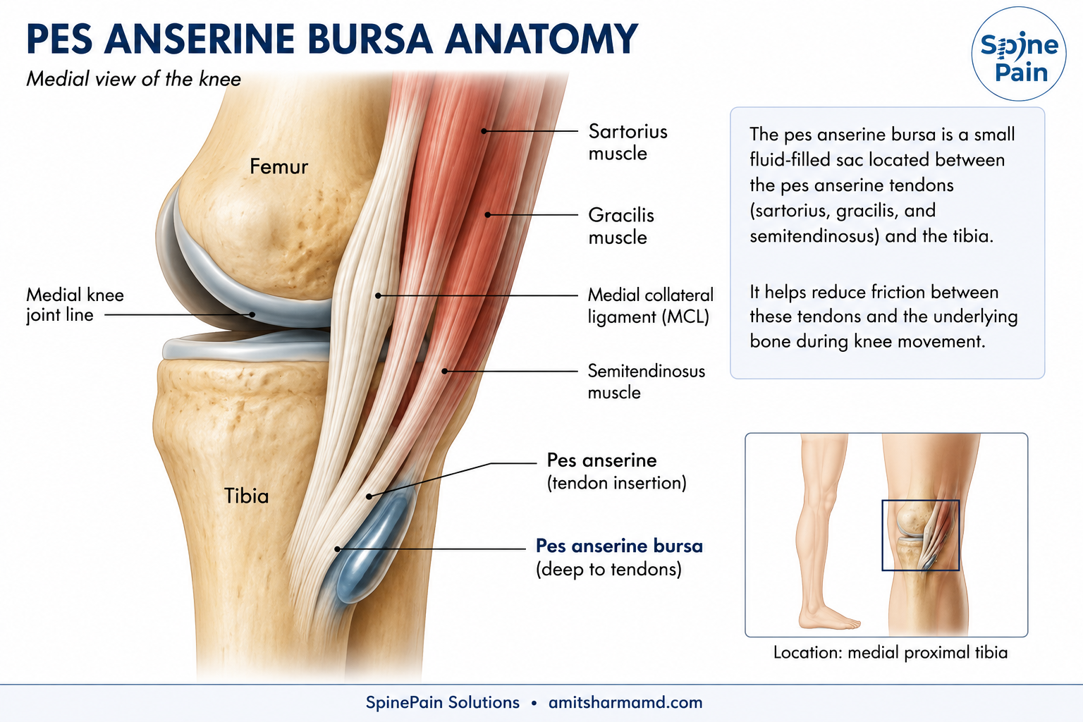
A Pes Anserine Bursa Injection is an ultrasound-guided procedure used to treat selected causes of pain along the inner aspect of the knee. The pes anserine bursa lies along the upper medial tibia beneath the tendons of the sartorius, gracilis, and semitendinosus muscles. When this region becomes inflamed or painful, precisely targeted treatment may help reduce symptoms and improve function.
At SpinePain Solutions, treatment is individualized according to the structure responsible for pain. Some patients have predominantly inflammatory bursitis and may benefit from corticosteroid injection, while others have associated tendon degeneration or tendinobursitis that may require a different treatment strategy. Ultrasound allows the bursa, adjacent tendons, medial collateral ligament, tibia, and nearby soft tissues to be evaluated before treatment is delivered.
Depending on the clinical findings, treatment may include local anesthetic, corticosteroid injection, platelet-rich plasma (PRP) for selected tendon-related pathology, rehabilitation, or treatment of another pain generator when the pes anserine bursa is not the primary source of symptoms.
Quick Answer
A pes anserine bursa injection is an ultrasound-guided treatment for selected cases of medial knee pain arising from the pes anserine bursa or closely related soft tissues. Corticosteroid may be used when inflammation is the primary problem, while PRP may be considered selectively when chronic tendon-related pathology is more prominent.
Clinical Perspective
A referral for a “pes anserine bursa injection” does not automatically mean the bursa is the only treatment target. Medial knee osteoarthritis, tendon disease, medial collateral ligament pathology, and other conditions may produce pain in the same region. Ultrasound helps determine which structure should actually be treated.
Related Guides
When Is a Pes Anserine Bursa Injection Considered?
A pes anserine bursa injection may be considered when pain along the inner aspect of the knee remains functionally limiting despite appropriate activity modification, physical therapy, medications, weight-management strategies, or other conservative treatment.
The pes anserine region contains several closely positioned structures. The bursa lies beneath the tendons of the sartorius, gracilis, and semitendinosus muscles near the upper medial tibia. Pain in this region may arise from the bursa itself, the adjacent tendons, nearby soft tissues, or another medial knee pain generator. For this reason, treatment should be directed toward the structure believed to be responsible for symptoms rather than based only on the location of pain.
Patients may be considered for ultrasound-guided intervention when they experience:
- Persistent pain along the upper inner tibia despite appropriate conservative treatment.
- Pain with walking, climbing stairs, rising from a chair, running, exercise, or prolonged activity.
- Localized tenderness corresponding with the pes anserine bursa or adjacent tendon insertions.
- Symptoms that interfere with sleep, work, sports, or normal daily activities.
- Temporary or incomplete improvement following previous treatment.
- A need for a precisely targeted diagnostic or therapeutic injection.
- Suspected associated tendon disease requiring ultrasound evaluation.
Quick Answer
A pes anserine bursa injection is generally considered when persistent medial knee pain has been localized to the pes anserine region and conservative treatment has not provided adequate relief. Ultrasound helps determine whether the primary target is the bursa, adjacent tendons, or another nearby structure before treatment is performed.
Clinical Perspective
A patient referred for a “pes anserine bursa injection” may ultimately have more prominent tendon disease, medial knee osteoarthritis, or another source of medial knee pain. The referral name should not determine the procedure. Treatment should follow the clinical examination and ultrasound findings.
For information about the condition itself, including symptoms, causes, and diagnosis, visit our
Pes Anserine Bursitis
resource.
Why Is Ultrasound Guidance Used?
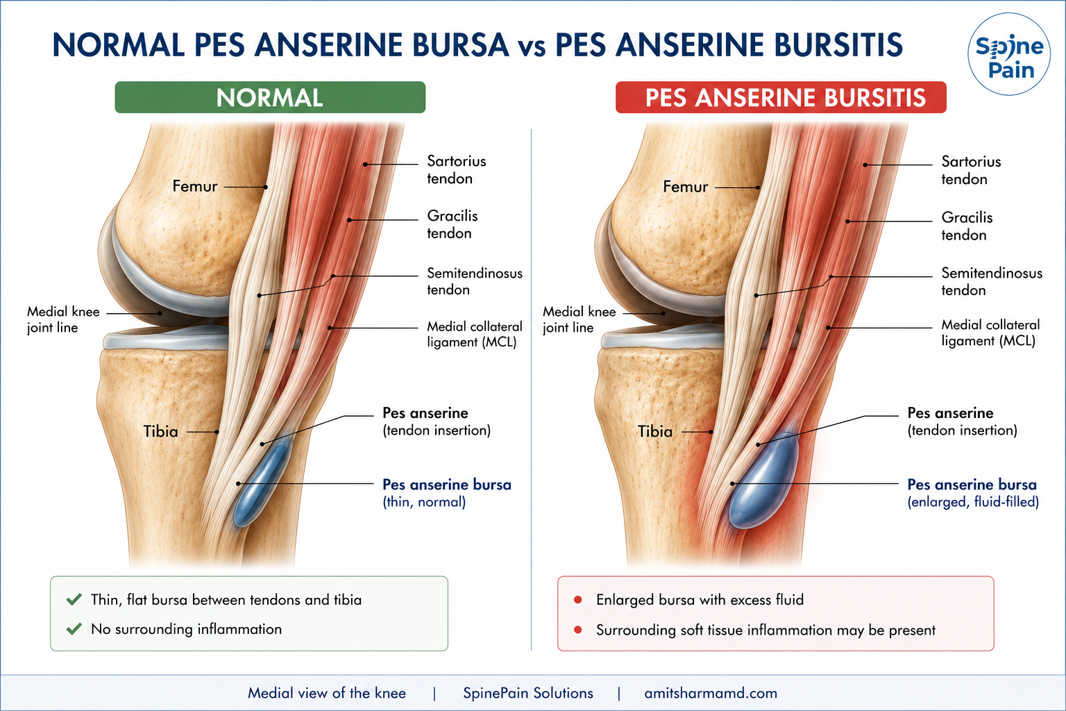
Ultrasound provides real-time visualization of the pes anserine bursa, the sartorius, gracilis, and semitendinosus tendons, the medial collateral ligament, the tibial cortex, and the needle pathway. It allows the physician to distinguish these closely positioned structures before medication is delivered.
The pes anserine bursa is often thin and may not contain a large visible fluid collection. Its location can also vary slightly between patients. Ultrasound therefore provides an advantage over relying solely on surface landmarks because the treatment target can be identified according to the patient’s individual anatomy.
Structures Evaluated With Ultrasound
- Pes anserine bursa and any visible fluid or thickening.
- Sartorius tendon.
- Gracilis tendon.
- Semitendinosus tendon.
- Medial collateral ligament.
- Medial tibial cortex.
- Nearby soft tissues and neurovascular structures when clinically relevant.
Clinical Perspective
Ultrasound is useful not simply because it shows the needle. It allows the physician to determine whether the painful structure is actually the bursa, one of the pes anserine tendons, the medial collateral ligament, or another nearby tissue before deciding where treatment should be placed.
Diagnostic and Therapeutic Pes Anserine Bursa Injection
A pes anserine bursa injection may serve both diagnostic and therapeutic purposes. When the source of medial knee pain is uncertain, a small amount of local anesthetic can be placed precisely into the suspected painful region. Meaningful temporary improvement may help support the pes anserine region as a contributor to the patient’s symptoms.
For therapeutic treatment, medication is selected according to the underlying pathology. Corticosteroid may be used when inflammation of the bursa is the predominant finding. When chronic tendon degeneration is more prominent, treatment may instead be directed toward the involved tendon rather than simply injecting the bursa.
Key Point
The pes anserine bursa lies outside the knee joint. A pes anserine bursa injection treats the medial extra-articular soft tissues and should not be confused with an injection performed inside the knee joint for arthritis or other intra-articular conditions.
Corticosteroid Injection for Pes Anserine Bursitis
Corticosteroid injection is one of the principal interventional treatments for persistent inflammatory pain arising from the pes anserine bursa. Under ultrasound guidance, medication can be placed into the bursal plane while minimizing unintended injection into the adjacent tendons or other medial knee structures.
The goal is to reduce local inflammation and pain so that walking, stair climbing, exercise, and rehabilitation become more comfortable. Improvement may also allow patients to participate more effectively in strengthening and flexibility exercises intended to address contributing mechanical factors.
Corticosteroid treatment does not repair degenerative tendon tissue or treat arthritis within the knee joint. When either of these is a substantial contributor to symptoms, additional or alternative treatment may be necessary.
Clinical Perspective
The objective is not to place corticosteroid somewhere near the tender area. Ultrasound allows the medication to be directed into the intended bursal plane while avoiding unnecessary corticosteroid exposure to the adjacent sartorius, gracilis, and semitendinosus tendons.
PRP and Other Advanced Treatment Options
Not every patient with persistent pain in the pes anserine region has an isolated inflammatory bursitis. In some patients, ultrasound examination and clinical findings suggest that chronic tendon disease is an important component of the pain. In these situations, a tendon-directed strategy may be considered rather than repeatedly treating the bursa with corticosteroid.
Platelet-Rich Plasma (PRP)
Platelet-rich plasma may be considered selectively when chronic tendinopathy or combined tendon-bursal pathology is believed to be contributing to persistent symptoms. PRP is prepared from the patient’s own blood and concentrated platelets are delivered under ultrasound guidance to the selected treatment target.
PRP is not routinely necessary for straightforward inflammatory pes anserine bursitis. Its potential role becomes more relevant when chronic tendon pathology is present, symptoms have persisted despite appropriate rehabilitation, and a regenerative rather than primarily anti-inflammatory strategy is desired.
Dry Needling
Dry needling is rarely required for isolated pes anserine bursitis. It may occasionally be incorporated when associated hamstring, adductor, or other myofascial pain is contributing to the patient’s symptoms. Dry needling addresses muscular or myofascial pain and does not directly treat an inflamed bursa.
Percutaneous Tendon Treatment
Percutaneous tendon procedures are not routine treatments for pes anserine bursitis. In unusual cases involving clearly demonstrated chronic degenerative tendon disease that has failed rehabilitation and other appropriate treatment, a more advanced tendon-directed procedure may be considered based on the specific tendon and imaging findings.
Clinical Perspective
Bursa-directed and tendon-directed treatments should not be used interchangeably. Corticosteroid is generally considered when inflammation within the bursa is the principal target, while PRP or other advanced procedures may be considered selectively when chronic tendon pathology is the more important problem.
What Else Can Cause Pain in the Pes Anserine Region?
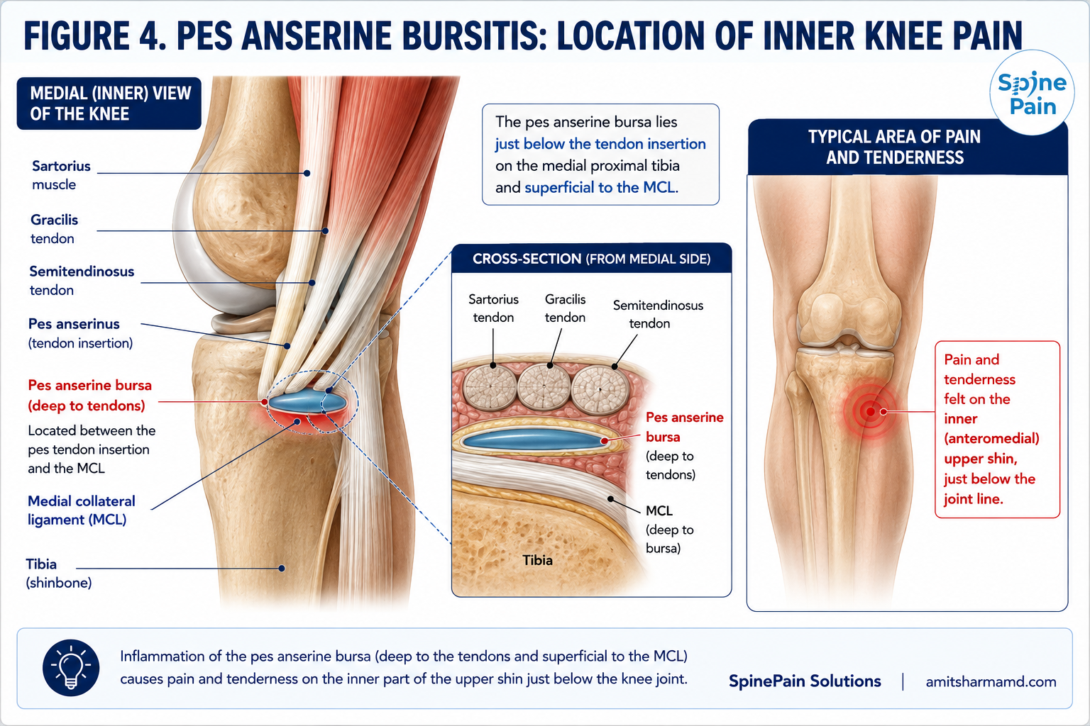
Pain along the inner knee does not always originate from the pes anserine bursa. Several conditions can produce symptoms in the same general region, and more than one pain generator may be present simultaneously.
Other potential sources include:
- Medial compartment knee osteoarthritis.
- Medial meniscus pathology.
- Medial collateral ligament injury or degeneration.
- Sartorius, gracilis, semitendinosus, or other hamstring tendon pathology.
- Semimembranosus tendon pathology.
- Saphenous nerve or infrapatellar branch irritation.
- Other superficial soft-tissue or muscular pain generators.
- Referred pain from the hip or lumbar spine.
Important Distinction
Medial knee osteoarthritis and pes anserine pain commonly coexist. Treating the pes anserine bursa does not treat arthritis inside the knee joint, and treating the knee joint does not necessarily eliminate pain arising from the pes anserine region. When both are clinically significant, they may require separate treatment strategies.
Comparing Pes Anserine Treatment Options
Treatment should be matched to the underlying pain generator. An inflamed bursa, chronic tendon disease, associated muscular pain, and knee-joint arthritis represent different problems and may require different approaches.
| Treatment | Primary Target | Primary Goal | Typical Role |
|---|---|---|---|
| Physical therapy and rehabilitation | Strength, flexibility, gait, and biomechanics | Reduce mechanical stress and restore function | Foundation of treatment for most patients |
| Local anesthetic injection | Suspected pes anserine pain generator | Diagnostic clarification and temporary relief | Selected diagnostically uncertain cases |
| Corticosteroid injection | Inflamed pes anserine bursa | Reduce inflammation and pain | Common interventional option for persistent bursitis |
| PRP | Selected chronic tendon or tendon-bursal pathology | Biologic treatment of selected degenerative tissue | Considered selectively when tendon disease is prominent |
| Dry needling | Associated muscular or myofascial pain | Address contributing muscular pain | Occasional adjunct; not a treatment for the bursa itself |
| Advanced tendon treatment | Confirmed chronic tendon pathology | Treat refractory degenerative tendon tissue | Uncommon and highly selected |
What Happens During a Pes Anserine Bursa Injection?
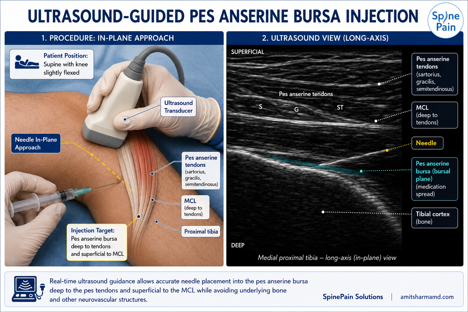
A pes anserine bursa injection is generally performed as a brief outpatient procedure. Ultrasound is used throughout the procedure to identify the relevant anatomy and guide the needle to the intended treatment target.
Positioning and Ultrasound Examination
The patient is positioned comfortably with the medial aspect of the knee accessible. Before needle placement, ultrasound is used to examine the pes anserine region and identify the tibia, medial collateral ligament, sartorius, gracilis and semitendinosus tendons, and the bursal plane.
Sterile Needle Placement
The skin is cleansed using sterile technique. The needle is then advanced under continuous ultrasound visualization toward the selected treatment target. Real-time imaging allows the physician to follow the needle while avoiding unintended placement into adjacent tendons or other structures.
Medication Delivery
For a conventional bursa injection, local anesthetic and corticosteroid may be delivered into the bursal plane. The medication can often be seen spreading through the intended tissue plane under ultrasound.
When PRP or another tendon-directed treatment is selected, the target and technique are modified according to the specific tendon pathology rather than simply reproducing a standard bursa injection.
The needle is removed and a small bandage is applied. Sedation is generally unnecessary, and patients typically return home shortly afterward.
Clinical Perspective
The procedural endpoint is not simply seeing the needle near the medial tibia. Ultrasound allows the physician to confirm the intended tissue plane and visualize the distribution of medication around the treatment target.
Recovery After a Pes Anserine Bursa Injection
Recovery is usually straightforward after a corticosteroid injection. Most patients can walk immediately and resume light daily activities, although strenuous exercise and activities that substantially reproduce medial knee pain may be temporarily limited.
- Mild soreness at the injection site may occur for one or several days.
- Temporary numbness may occur if local anesthetic was used.
- Relative rest is generally recommended for the remainder of the procedure day.
- Walking and light daily activity can usually continue as tolerated.
- More strenuous exercise is resumed gradually according to symptoms and physician guidance.
- Physical therapy or a home exercise program may be continued or restarted as directed.
Recovery After PRP or Tendon-Directed Treatment
Recovery after PRP differs from recovery after a corticosteroid injection. Temporary soreness is common, improvement is generally gradual, and activity progression is based on the tendon or tissue treated. Anti-inflammatory medications may also be restricted around the procedure when clinically appropriate.
Key Point
Pain reduction from an injection should be used as an opportunity to address strength, flexibility, gait, weight-related loading, and other factors that may have contributed to persistent stress on the medial knee.
What Results Can Be Expected?
The goal of treatment is to reduce medial knee pain, improve walking and stair tolerance, facilitate rehabilitation, and help patients return to normal activities. The degree and duration of improvement vary according to the underlying diagnosis and contributing mechanical factors.
When active bursal inflammation is the primary pain generator, corticosteroid may provide relatively rapid improvement. If symptoms arise predominantly from chronic tendon disease, recovery may be slower and a tendon-focused rehabilitation or biologic treatment strategy may be more appropriate.
Treatment results may be influenced by:
- Accuracy of the diagnosis and treatment target.
- Whether bursitis, tendinopathy, or both are present.
- Coexisting medial compartment knee osteoarthritis.
- Hamstring tightness and lower-extremity weakness.
- Gait and lower-extremity biomechanics.
- Body weight and repetitive loading of the knee.
- Occupational, athletic, and recreational demands.
- Participation in rehabilitation and correction of contributing factors.
Clinical Perspective
Incomplete relief does not automatically mean that an accurately performed injection failed. Persistent pain may indicate that another structure, such as the knee joint, meniscus, medial collateral ligament, tendon, or saphenous nerve, is also contributing to the patient’s symptoms.
What Are the Risks of a Pes Anserine Bursa Injection?
Ultrasound-guided pes anserine bursa injections are generally well tolerated. As with any injection, however, potential risks and side effects should be considered before treatment.
- Temporary soreness or bruising at the injection site.
- Temporary increase in pain following the procedure.
- Bleeding or hematoma.
- Infection.
- Allergic reaction to medication, antiseptic, or adhesive.
- Temporary numbness related to local anesthetic.
- Skin thinning, pigment change, or local soft-tissue atrophy following corticosteroid exposure.
- Temporary elevation of blood glucose following corticosteroid injection.
- Unintended irritation or injury to a nearby tendon, ligament, nerve, or blood vessel.
- Incomplete, temporary, or absent pain relief.
- Need for additional diagnostic evaluation or treatment.
When Should You Contact Your Physician?
Contact your physician promptly for fever, progressive redness or warmth, drainage, rapidly increasing swelling, severe or worsening pain, new weakness, persistent numbness, significant calf swelling, difficulty bearing weight, or other unexpected symptoms following the procedure.
Who May Not Be a Candidate for a Pes Anserine Bursa Injection?
A pes anserine bursa injection is not appropriate for every patient with medial knee pain. Before treatment, the diagnosis, medical history, medications, infection risk, imaging findings, and other potential pain generators should be reviewed.
Treatment may need to be postponed, modified, or avoided in patients with:
- Active infection at or near the treatment site.
- Systemic infection or significant acute illness.
- Uncontrolled bleeding risk or anticoagulation that cannot be safely managed for the planned procedure.
- Known allergy to a required medication without an appropriate alternative.
- Suspected fracture, tumor, or another condition requiring a different diagnostic approach.
- Symptoms primarily arising from advanced knee-joint disease or another pain generator rather than the pes anserine region.
- Progressive weakness, significant instability, or mechanical symptoms requiring additional evaluation.
- Medical conditions that substantially increase procedural or healing risk.
When Is Surgery Considered?
Surgery is rarely required for isolated pes anserine bursitis. Most patients can be managed with rehabilitation, modification of contributing activities, medications when appropriate, and selective image-guided intervention.
Orthopedic evaluation may become appropriate when persistent medial knee pain is caused by a structural abnormality that cannot be adequately treated with non-surgical care. Examples may include:
- A significant associated tendon injury.
- A symptomatic bony prominence or other structural abnormality irritating the pes anserine region.
- A space-occupying lesion or unusual persistent bursal enlargement.
- Significant ligamentous or meniscal pathology requiring surgical treatment.
- Another surgically correctable cause of persistent medial knee pain.
Clinical Perspective
Persistent symptoms after a pes anserine bursa injection do not automatically indicate a need for surgery. More commonly, the diagnosis should be reconsidered to determine whether knee osteoarthritis, meniscal pathology, tendon disease, ligament pathology, nerve irritation, or another condition is contributing to the remaining pain.
Our Approach at SpinePain Solutions
Our approach begins by determining which structure is actually responsible for the patient’s medial knee pain. The pes anserine bursa, adjacent tendons, medial collateral ligament, knee joint, and nearby neural structures can produce overlapping symptoms, and treatment should be matched to the identified pain generator.
At SpinePain Solutions, treatment may include:
- Ultrasound evaluation of the pes anserine bursa and adjacent tendons.
- Assessment of the medial collateral ligament, tibia, and surrounding soft tissues.
- Diagnostic injection when clarification of the pain generator is helpful.
- Ultrasound-guided corticosteroid injection for selected inflammatory bursitis.
- PRP for carefully selected chronic tendon-related pathology.
- Dry needling when a separate muscular or myofascial pain component is identified.
- Integration of physical therapy, stretching, strengthening, and biomechanical correction.
- Separate evaluation and treatment of knee osteoarthritis when the joint is an independent pain generator.
- Referral for orthopedic evaluation when significant structural pathology is identified.
The objective is not simply to inject the area where the patient points to pain. It is to identify the responsible structure, place treatment accurately, and combine the procedure with rehabilitation or other therapies when necessary to improve function and reduce recurrence.
Key Takeaways
- The pes anserine bursa lies along the upper medial tibia beneath the sartorius, gracilis, and semitendinosus tendons.
- Ultrasound allows the bursa, adjacent tendons, medial collateral ligament, tibia, and surrounding soft tissues to be evaluated before treatment.
- A pes anserine bursa injection is different from an injection performed inside the knee joint.
- Corticosteroid may be used for selected cases in which bursal inflammation is the principal treatment target.
- PRP may be considered selectively when chronic tendon-related pathology is an important component of symptoms.
- Dry needling addresses associated muscular pain and does not directly treat the bursa.
- Medial knee osteoarthritis and pes anserine pain can coexist and may require separate treatments.
- Rehabilitation, strengthening, flexibility, and correction of contributing biomechanics remain important even when an injection provides substantial relief.
- Surgery is rarely required for isolated pes anserine bursitis.
Related Services and Resources
Bursitis Treatment
Learn about conservative care and interventional treatment options for painful or inflamed bursae.
Pes Anserine Bursitis
Learn about the condition itself, including symptoms, causes, diagnosis, and common sources of medial knee pain.
Knee Joint Injections
Learn about image-guided injections performed inside the knee joint for arthritis and other intra-articular conditions.
PRP for Knee Arthritis
Learn how platelet-rich plasma may be used for selected patients with symptomatic knee osteoarthritis.
Hyaluronic Acid Joint Injections
Learn about image-guided gel injections for selected patients with symptomatic osteoarthritis.
Frequently Asked Questions
Where is the pes anserine bursa located?
The pes anserine bursa is located along the upper inner portion of the tibia, beneath the sartorius, gracilis, and semitendinosus tendons. Pain from this region is typically felt below the medial knee joint line rather than directly inside the knee joint.
Is a pes anserine bursa injection the same as a knee injection?
No. A pes anserine bursa injection is performed outside the knee joint along the medial tibia. A knee-joint injection places medication inside the joint and is used for different conditions, including knee osteoarthritis.
Why use ultrasound guidance?
Ultrasound allows the physician to identify the bursal plane, pes anserine tendons, medial collateral ligament, tibia, and surrounding soft tissues while following the needle in real time. It also allows medication spread to be observed during the procedure.
What medication is used for a pes anserine bursa injection?
A conventional therapeutic injection may contain local anesthetic and corticosteroid. The medication selected depends on the diagnosis, medical history, previous treatment response, and procedural goals.
How quickly does a corticosteroid injection work?
Local anesthetic may provide temporary improvement shortly after the procedure. Corticosteroid benefit typically develops more gradually over the following several days, although response varies between patients.
How long does the relief last?
The duration of relief varies considerably. Some patients experience prolonged improvement, while others have temporary or incomplete benefit when tendon disease, knee osteoarthritis, altered biomechanics, or repetitive loading continues to contribute to symptoms.
Can the injection be repeated?
Repeat corticosteroid injection is not automatic. The diagnosis, duration of benefit, condition of the adjacent tendons, cumulative corticosteroid exposure, and other treatment options should be reconsidered before repeating the procedure.
Is PRP used for pes anserine bursitis?
PRP is not routinely required for uncomplicated inflammatory bursitis. It may be considered selectively when chronic tendon-related pathology or combined tendinobursitis is an important component of persistent symptoms.
Is PRP better than corticosteroid?
They serve different purposes, and current evidence does not establish one treatment as universally superior. Corticosteroid is primarily used to reduce inflammation, while PRP may be considered when chronic tendon-related pathology is a more important treatment target.
Can a pes anserine bursa injection treat knee arthritis?
No. The pes anserine bursa is outside the knee joint. When knee osteoarthritis and pes anserine pain coexist, the two conditions may require separate treatments.
Does the procedure require sedation?
Sedation is generally unnecessary. The procedure is typically performed using ultrasound guidance and local measures for comfort, and patients usually return home shortly afterward.
When can I exercise after the injection?
Light activity can usually resume quickly after a conventional bursa injection, while strenuous exercise is reintroduced gradually. PRP or other tendon-directed procedures may require a different rehabilitation schedule.
What if the injection does not help?
Persistent pain should prompt reassessment of the diagnosis. Knee osteoarthritis, meniscal disease, medial collateral ligament pathology, tendon disease, saphenous nerve irritation, or referred pain from the hip or lumbar spine may produce symptoms in the same region.
References
- American Academy of Family Physicians. Evaluation and treatment of common causes of knee pain and bursitis.
- American Institute of Ultrasound in Medicine. Practice Parameter for the Performance of a Musculoskeletal Ultrasound Examination.
- Lee JH, Lee JU, Yoo SW. Accuracy and efficacy of ultrasound-guided pes anserinus bursa injection.
- Rennie WJ, Saifuddin A. Pes anserine bursitis: incidence in symptomatic knees and clinical presentation.
- Yoon HS, et al. Correlation between ultrasonographic findings and response to local corticosteroid injection in pes anserinus tendinitis-bursitis syndrome.
- Brukner & Khan’s Clinical Sports Medicine. Medial knee pain and pes anserine disorders.
- Gray’s Anatomy: The Anatomical Basis of Clinical Practice. Anatomy of the medial knee and pes anserinus.
About This Guide
This article from Dr. Amit Sharma is provided for educational purposes only and should not replace personalized medical advice, diagnosis, or treatment from a qualified healthcare professional. The appropriate treatment depends on the cause of medial knee pain, clinical examination, ultrasound findings, medical history, medications, previous treatment response, and individual treatment goals.



