
Shoulder Radiofrequency Ablation (RFA): Non-Surgical Shoulder Pain Relief
Chronic shoulder pain can interfere with reaching, lifting, dressing, driving, sleeping, working, and many of the ordinary movements people perform without thinking. Physical therapy, medications, activity modification, and image-guided injections help many patients, but some continue to experience substantial pain despite an appropriate course of conservative care.
Shoulder radiofrequency ablation (Shoulder RFA) is a minimally invasive procedure designed to reduce pain by treating selected sensory nerve branches that carry signals from the shoulder joint. The procedure does not remove the joint, repair damaged cartilage, reconnect a torn tendon, or replace the shoulder. Instead, it alters pain transmission while attempting to preserve the motor nerves responsible for shoulder strength and movement.
Shoulder RFA may be considered for carefully selected patients with glenohumeral osteoarthritis, rotator cuff tear arthropathy, chronic degenerative shoulder pain, or persistent pain after shoulder replacement when infection, implant failure, instability, fracture, and other correctable causes have been excluded.
This physician-written guide explains the sensory anatomy of the shoulder, how shoulder RFA works, who may be a candidate, what the procedure can and cannot treat, recovery expectations, potential risks, and how RFA compares with injections and shoulder replacement.
Quick Answer
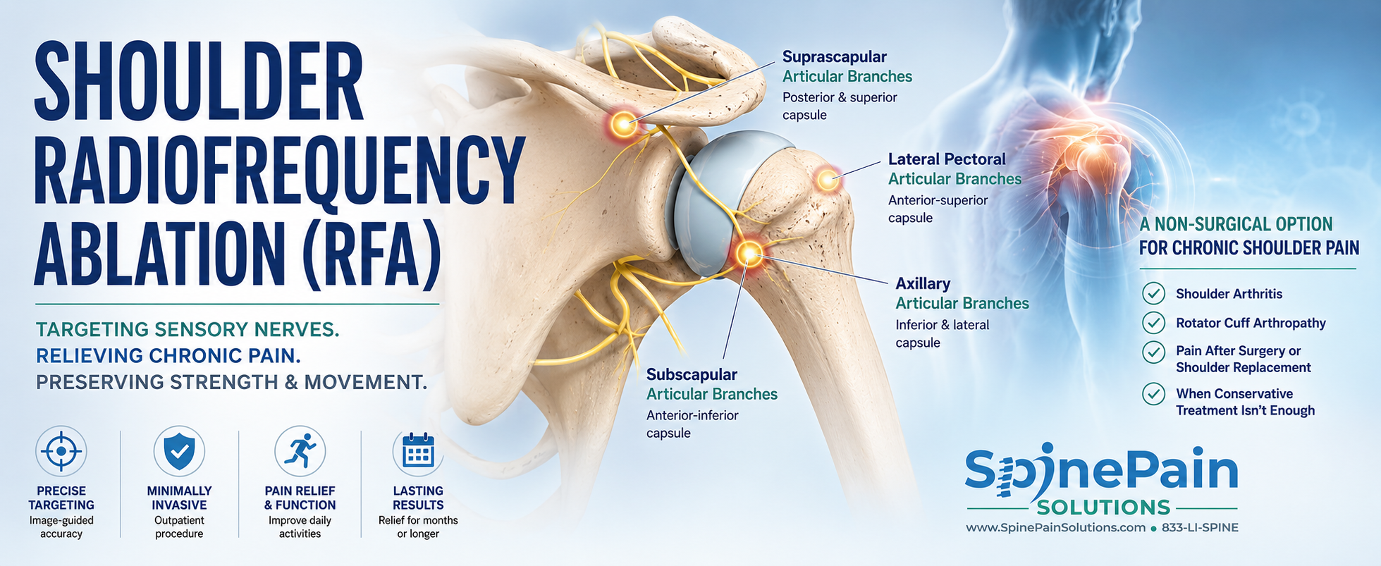
Shoulder radiofrequency ablation is an image-guided outpatient procedure that treats selected sensory articular branches carrying pain from the shoulder joint. Common targets may include branches associated with the suprascapular, axillary, lateral pectoral, and selected subscapular nerves. The goal is to reduce chronic shoulder pain while preserving the major motor pathways responsible for strength and movement.
What Is Shoulder Radiofrequency Ablation?
Shoulder radiofrequency ablation uses controlled radiofrequency energy to alter the ability of selected sensory nerve branches to transmit pain. Specialized electrodes are positioned near carefully chosen articular nerve targets using fluoroscopic or ultrasound guidance. Radiofrequency energy then creates a controlled treatment zone around the electrode.
The procedure is performed outside the joint. It does not burn the cartilage, remove bone, enter a shoulder implant, or destroy the major nerves that activate the deltoid, rotator cuff, or other shoulder muscles.
Treated sensory branches may recover over time, which is why pain relief is not necessarily permanent. When a patient experiences substantial benefit and symptoms later return, repeat treatment may be considered after reassessment.
Clinical Perspective
Shoulder RFA is not simply “burning the suprascapular nerve.” Modern selective techniques attempt to identify and treat terminal sensory articular branches while avoiding the larger mixed nerves that also carry motor fibers. Because shoulder sensory anatomy varies between individuals, treatment planning may require several targets rather than relying on a single nerve.
Understanding the Shoulder Joint
The shoulder is not one simple hinge. It is a coordinated system involving the glenohumeral joint, acromioclavicular joint, scapulothoracic articulation, rotator cuff, deltoid, biceps tendon, capsule, labrum, bursae, and surrounding nerves.
The glenohumeral joint is formed by the humeral head and the shallow glenoid socket. Articular cartilage permits smooth movement, while the labrum deepens the socket. The capsule and ligaments provide passive stability, and the rotator cuff muscles center the humeral head during motion.
This remarkable mobility comes with a tradeoff. Shoulder pain may arise from many overlapping structures, and visible arthritis on an X-ray does not automatically prove that every symptom is coming from the glenohumeral joint.
Where Does Chronic Shoulder Pain Come From?
Pain associated with shoulder arthritis or degeneration can arise from several pain-sensitive tissues. Cartilage deterioration is important, but cartilage itself is not the only structure capable of producing symptoms.
- Joint capsule and synovium: Inflammation, contracture, and stretching of the capsule may produce pain and stiffness.
- Subchondral bone: The bone beneath damaged cartilage can remodel and become painful under load.
- Labrum: Degeneration or tearing may contribute to deep joint pain, catching, or mechanical symptoms.
- Rotator cuff: Tendinopathy, partial tearing, massive tearing, or cuff-tear arthropathy may coexist with glenohumeral degeneration.
- Subacromial bursa: Bursal irritation may produce pain with overhead movement or lying on the affected side.
- Long head of the biceps tendon: Biceps disease can generate anterior shoulder pain.
- Acromioclavicular joint: Arthritis at the top of the shoulder can be painful but may require a different treatment target.
- Sensory articular nerve branches: These small branches carry pain information from the joint toward the spinal cord and brain.
Key Point: A technically accurate shoulder RFA may provide incomplete relief when the dominant pain source is a torn tendon, inflamed bursa, unstable joint, cervical nerve root, or another structure not supplied by the treated articular branches.
Shoulder Pain Is Not Always Glenohumeral Arthritis
Accurate diagnosis is particularly important in the shoulder because pain location and movement restriction can overlap across several conditions.
Rotator Cuff Disease
Rotator cuff tendinopathy or tearing may cause pain with reaching, lifting, or lying on the affected side. A massive tear can also cause true weakness or loss of active elevation. RFA may reduce pain in selected patients, but it does not reconnect a torn tendon or restore lost tendon mechanics.
Cervical Radiculopathy
A pinched nerve in the neck may produce shoulder or arm pain, numbness, tingling, or weakness. Symptoms extending below the elbow, neck-provoked pain, altered reflexes, or neurologic deficits may suggest a cervical source.
Adhesive Capsulitis
Frozen shoulder causes pain and substantial restriction of both active and passive movement. Pain treatment alone may not restore motion unless it is combined with an appropriate mobility and rehabilitation strategy.
Acromioclavicular Joint Pain
Pain centered over the top of the shoulder, especially during cross-body movement, may arise from the acromioclavicular joint rather than the glenohumeral joint.
Peripheral Nerve Disorders
Suprascapular neuropathy, axillary neuropathy, brachial plexus disease, or other peripheral nerve disorders may cause pain and weakness. These conditions require neurologic evaluation and should not automatically be treated as ordinary joint pain.
Sensory Articular Nerves of the Shoulder
The sensory supply of the shoulder is variable and overlapping. The glenohumeral capsule receives articular branches from several nerves rather than one isolated pathway.
Suprascapular Articular Branches
The suprascapular nerve contributes sensory branches to portions of the posterior and superior shoulder capsule. Because the parent nerve also supplies the supraspinatus and infraspinatus muscles, treatment should focus on selected articular branches or anatomically appropriate targets rather than indiscriminate destruction of the main nerve.
Axillary Articular Branches
The axillary nerve contributes sensory branches to portions of the inferior and lateral capsule. Its main trunk also supplies the deltoid and teres minor muscles, making selective targeting important.
Lateral Pectoral Articular Branches
The lateral pectoral nerve may contribute articular branches to the anterior-superior shoulder region. These branches have become part of several contemporary multi-target shoulder RFA protocols.
Subscapular Articular Branches
Recent anatomical protocols also recognize variable contributions from upper and lower subscapular nerve branches. These targets may be considered selectively when anatomy and the treatment plan support their inclusion.
How Does Shoulder Radiofrequency Ablation Work?
Under image guidance, specialized radiofrequency electrodes are positioned near selected shoulder articular nerve targets. Sensory and motor stimulation may be used to confirm proximity to the intended pain pathway while reducing the likelihood of affecting important motor fibers.
Controlled radiofrequency energy creates a treatment zone that alters pain-signal transmission. The shoulder joint, cartilage, rotator cuff, implant components, and major motor nerves are not intentionally treated.
Because articular branches are small and anatomically variable, the physician may create more than one lesion or treat several target regions. Successful treatment depends on anatomy, electrode orientation, lesion geometry, and procedural judgment rather than simply placing one needle near the shoulder.
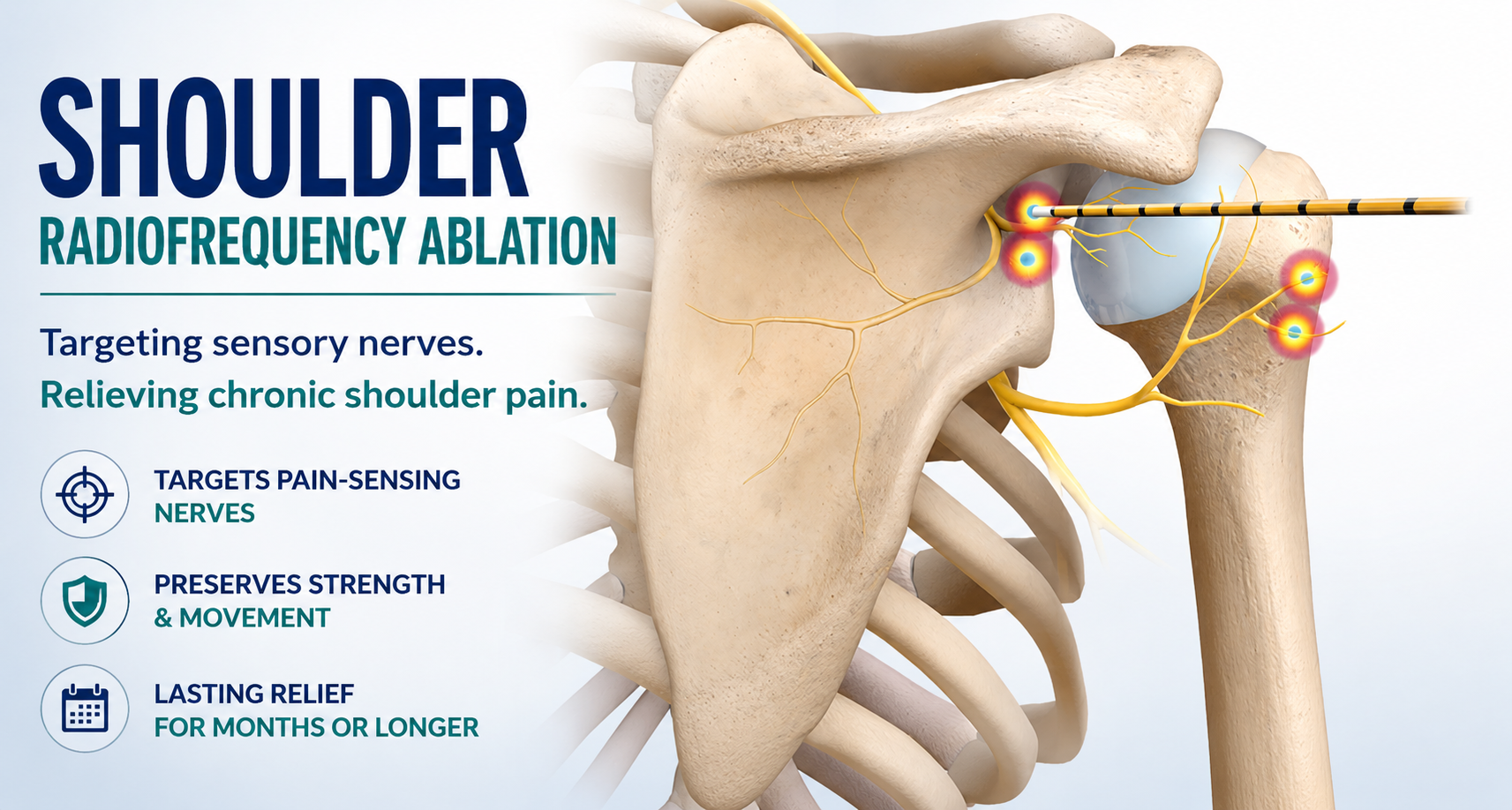
Shoulder radiofrequency ablation interrupts pain transmission by treating selected sensory articular branches of the suprascapular, axillary, and lateral pectoral nerves. The procedure is designed to preserve the major motor pathways responsible for shoulder strength, stability, and range of motion.
Who May Be a Candidate for Shoulder Radiofrequency Ablation?
Shoulder RFA may be considered when chronic pain continues to limit sleep, reaching, lifting, dressing, rehabilitation, or daily activities despite appropriate non-surgical care.
- Symptomatic glenohumeral osteoarthritis that has not responded adequately to physical therapy, medication, or injections
- Rotator cuff tear arthropathy in patients who are not undergoing corrective surgery
- Chronic degenerative shoulder pain in patients who are poor surgical candidates
- Patients who prefer to postpone shoulder replacement after informed orthopedic consultation
- Patients who cannot safely undergo major surgery because of medical conditions
- Selected patients with persistent pain after shoulder replacement when infection and mechanical complications have been excluded
- Patients who obtain substantial temporary relief from appropriately performed diagnostic articular branch blocks
The appearance of an X-ray or MRI does not determine candidacy by itself. The pain pattern, functional limitations, examination, prior treatment response, diagnostic blocks, and presence of other pain generators all matter.
What Shoulder Radiofrequency Ablation Cannot Fix
Shoulder RFA treats pain transmission. It does not restore damaged anatomy or correct a mechanical problem that requires another form of treatment.
Massive Rotator Cuff Tear
RFA may reduce pain associated with cuff-tear arthropathy in selected patients, but it does not reconnect a torn tendon, reverse muscle atrophy, or restore strength lost because the rotator cuff is mechanically nonfunctional.
Instability or Recurrent Dislocation
RFA does not tighten the capsule, repair the labrum, or stabilize a shoulder that repeatedly dislocates.
Active Infection
A septic native joint or infected shoulder prosthesis requires urgent diagnostic and medical or surgical treatment. RFA has no role in treating infection.
Fracture or Implant Failure
Acute fractures, periprosthetic fractures, loose components, implant dislocation, component breakage, or other mechanical problems require orthopedic evaluation.
Motor-Nerve Injury
RFA cannot restore strength lost because of axillary neuropathy, suprascapular neuropathy, brachial plexus injury, cervical radiculopathy, or another motor-nerve disorder.
Important: New trauma, fever, a hot or rapidly swelling joint, sudden loss of movement, visible deformity, progressive weakness, or severe pain after shoulder replacement requires prompt evaluation rather than routine scheduling for RFA.
Why Are Diagnostic Shoulder Articular Branch Blocks Performed?
A diagnostic block places a small amount of local anesthetic near the sensory articular branches that may later be treated with RFA. The purpose is to determine whether temporarily interrupting those pain pathways produces meaningful relief.
After the block, the patient may be asked to perform movements that ordinarily provoke pain, such as reaching overhead, reaching behind the back, dressing, lifting a light object, or lying in a normally painful position.
A favorable response increases confidence that the targeted articular branches contribute to the pain. It does not guarantee the degree or duration of relief after RFA. A poor response may prompt reassessment of the diagnosis, block targets, shoulder anatomy, or alternative pain generators.
Clinical Perspective
A shoulder contains several overlapping pain generators. Diagnostic blocks should therefore be interpreted as part of the full clinical picture rather than as an isolated pass-or-fail test. Temporary relief supports the targeted sensory pathway, but persistent weakness, instability, or mechanical symptoms still require separate evaluation.
What Happens During Shoulder Radiofrequency Ablation?
Shoulder RFA is performed as an outpatient procedure. The exact positioning, imaging method, number of electrodes, and treatment targets depend on the patient’s anatomy and the technique selected.
- The patient is positioned to provide safe access to the selected shoulder targets.
- The skin is cleaned using sterile technique.
- Local anesthetic is administered to numb the skin and deeper tissues.
- Fluoroscopy, ultrasound, or a combined imaging approach is used to identify the relevant anatomical landmarks.
- Specialized radiofrequency electrodes are positioned near the selected sensory articular branches.
- Sensory and motor stimulation may be performed to confirm appropriate positioning and avoid unintended motor-nerve treatment.
- Additional local anesthetic may be administered before lesioning.
- Controlled radiofrequency energy is delivered according to the electrode system and lesioning strategy.
- The electrodes are removed, small bandages are applied, and the patient is monitored before discharge.
Sedation may be used according to the planned procedure, patient comfort, medical history, and facility protocol. Patients receiving sedation must follow fasting and transportation instructions.
Radiofrequency Technologies Used for Shoulder RFA
Conventional thermal RFA, lesion-enhanced or multitined electrodes, cooled RFA, bipolar configurations, and pulsed radiofrequency have all been described for shoulder pain. These technologies do not create identical treatment effects.
| Technology | General Approach | Clinical Considerations |
|---|---|---|
| Conventional Thermal RFA | Creates a controlled thermal lesion adjacent to the active electrode tip | Accurate electrode orientation and proximity to the target are important; multiple lesions may improve coverage |
| Multitined or Lesion-Enhanced RFA | Uses an electrode design intended to create a broader treatment zone | May help address variable terminal-branch anatomy; technique remains important |
| Cooled RFA | Uses internal electrode cooling to create a relatively large treatment zone | Used in several contemporary shoulder articular-branch protocols; requires dedicated equipment |
| Bipolar RFA | Creates a treatment zone between two electrodes | May be selected when anatomy favors an intervening lesion |
| Pulsed Radiofrequency | Delivers intermittent energy at lower target temperatures | Produces a different neuromodulatory effect and should not be treated as equivalent to continuous thermal ablation |
No single platform has been established as universally superior for every shoulder condition. The choice should reflect the target anatomy, intended lesion geometry, physician technique, available equipment, patient factors, and current evidence.
Recovery After Shoulder Radiofrequency Ablation
Most patients return home the same day. Mild soreness, bruising, or sensitivity around the electrode sites may occur during the first several days.
The local anesthetic used during the procedure may provide temporary early relief. This can wear off before the radiofrequency treatment reaches its full effect. Some patients experience a short-lived increase in discomfort or nerve irritation during the early recovery period.
Meaningful improvement may develop gradually over several weeks. Patients should follow their specific instructions regarding driving, bathing, lifting, exercise, and return to work.
| Time | What Patients May Experience |
|---|---|
| Procedure day | Temporary numbness, local soreness, or short-lived relief from anesthetic |
| First several days | Bruising, aching, or sensitivity around the treatment sites |
| First one to two weeks | Activity is advanced gradually; temporary nerve irritation may occur |
| Two to six weeks | Pain reduction and improved tolerance for movement may become more apparent |
| Following months | Reduced pain may support rehabilitation, sleep, and improved use of the arm |
What Results Can Be Expected?
The goal of shoulder RFA is meaningful improvement in pain and function, not permanent elimination of every symptom. Potential benefits may include:
- Reduced shoulder pain at rest and during movement
- Improved sleep, particularly when shoulder pain disrupts lying positions
- Greater tolerance for physical therapy or home exercise
- Improved ability to dress, reach, lift light objects, and perform daily activities
- Reduced reliance on selected pain medications
- A less invasive option for patients unable or unwilling to undergo shoulder replacement
- Possible postponement of surgery in selected patients
- Pain relief without preventing later surgery
Published studies report encouraging improvements in pain and function, but results vary and the shoulder evidence remains less mature than the evidence for knee genicular RFA. Some patients obtain several months of relief, while others experience shorter benefit, longer improvement, or no meaningful response.
The treated sensory branches may regenerate. When substantial initial relief eventually wears off, repeat RFA may be considered after confirming that the pain source and shoulder anatomy have not changed.
Treatment Tip: Pain reduction is often most valuable when it creates an opportunity to improve range of motion, scapular control, rotator-cuff conditioning when appropriate, and functional use of the arm. RFA should support rehabilitation rather than replace it.
What Are the Risks of Shoulder Radiofrequency Ablation?
Shoulder RFA is generally performed using image guidance and selective nerve targeting. Potential risks nevertheless exist.
- Temporary soreness or increased pain
- Bruising or bleeding
- Temporary numbness or altered skin sensation
- Post-procedural neuritis or nerve irritation
- Failure to obtain meaningful relief
- Recurrence of pain as sensory nerves recover
- Infection
- Allergic or medication reaction
- Unintended injury to a nearby sensory or motor nerve
- Weakness if an important motor pathway is inadvertently affected
- Rare injury to a nearby blood vessel, lung, or other structure depending on the target and approach
Careful imaging, stimulation testing, anatomical planning, and selective treatment are used to reduce these risks.
When Should You Contact Your Physician?
Patients should promptly report fever, increasing redness or drainage, severe or rapidly worsening pain, shortness of breath, new major numbness, progressive weakness, or another unexpected symptom after the procedure.
Shoulder RFA for Persistent Pain After Shoulder Replacement
Most patients undergo shoulder replacement to reduce pain and improve function, but some continue to experience significant discomfort. Persistent pain after anatomic total shoulder replacement, reverse shoulder replacement, or hemiarthroplasty should be evaluated carefully before nerve treatment is considered.
Potential causes include:
- Periprosthetic joint infection
- Implant loosening or component failure
- Instability or dislocation
- Periprosthetic fracture
- Rotator cuff or subscapularis failure after anatomic replacement
- Acromial or scapular-spine stress fracture after reverse replacement
- Component malposition or mechanical impingement
- Deltoid dysfunction
- Nerve injury
- Adhesive capsulitis or postoperative stiffness
- Cervical spine or peripheral nerve pain
- Complex regional pain syndrome or altered pain processing
RFA should not be used to conceal a surgically correctable prosthetic complication. The operating surgeon or another qualified shoulder specialist may need to review imaging, laboratory studies, and the clinical course.
When the prosthesis is stable, infection has been excluded, no correctable mechanical problem is identified, and the pain pattern remains compatible with the treated sensory pathway, diagnostic articular branch blocks and RFA may be considered. Evidence for post-arthroplasty shoulder RFA remains limited, and expectations should be individualized.
Clinical Perspective
Radiofrequency ablation is not an alternative to revision surgery when infection, loosening, instability, fracture, tendon failure, or another correctable mechanical problem exists. Its role is limited to carefully selected patients whose replacement appears structurally acceptable but who continue to have chronic pain carried through treatable sensory pathways.
Shoulder RFA vs Injections, PRP, and Shoulder Replacement
| Treatment | Primary Goal | Potential Advantages | Important Limitations |
|---|---|---|---|
| Corticosteroid Injection | Reduce inflammation and joint or bursal pain | May act relatively quickly; little downtime | Relief may be temporary; does not repair structural damage |
| PRP | Influence the painful tissue environment using an autologous blood preparation | Avoids corticosteroid; may be considered for selected joint or tendon conditions | Evidence varies by diagnosis; does not restore a massive tear or advanced deformity |
| Shoulder RFA | Reduce pain transmission through selected sensory articular branches | Minimally invasive; preserves future treatment options | Does not repair tendons, cartilage, instability, or mechanical failure |
| Shoulder Replacement | Replace severely damaged joint surfaces and address advanced structural disease | Can provide substantial pain and functional improvement in appropriate candidates | Major surgery with rehabilitation, perioperative risks, and prosthetic considerations |
These treatments solve different problems. RFA reduces sensory pain transmission, while replacement addresses advanced mechanical joint failure. Repeated temporary procedures should not indefinitely postpone surgery when reconstruction offers a more appropriate opportunity to restore function.
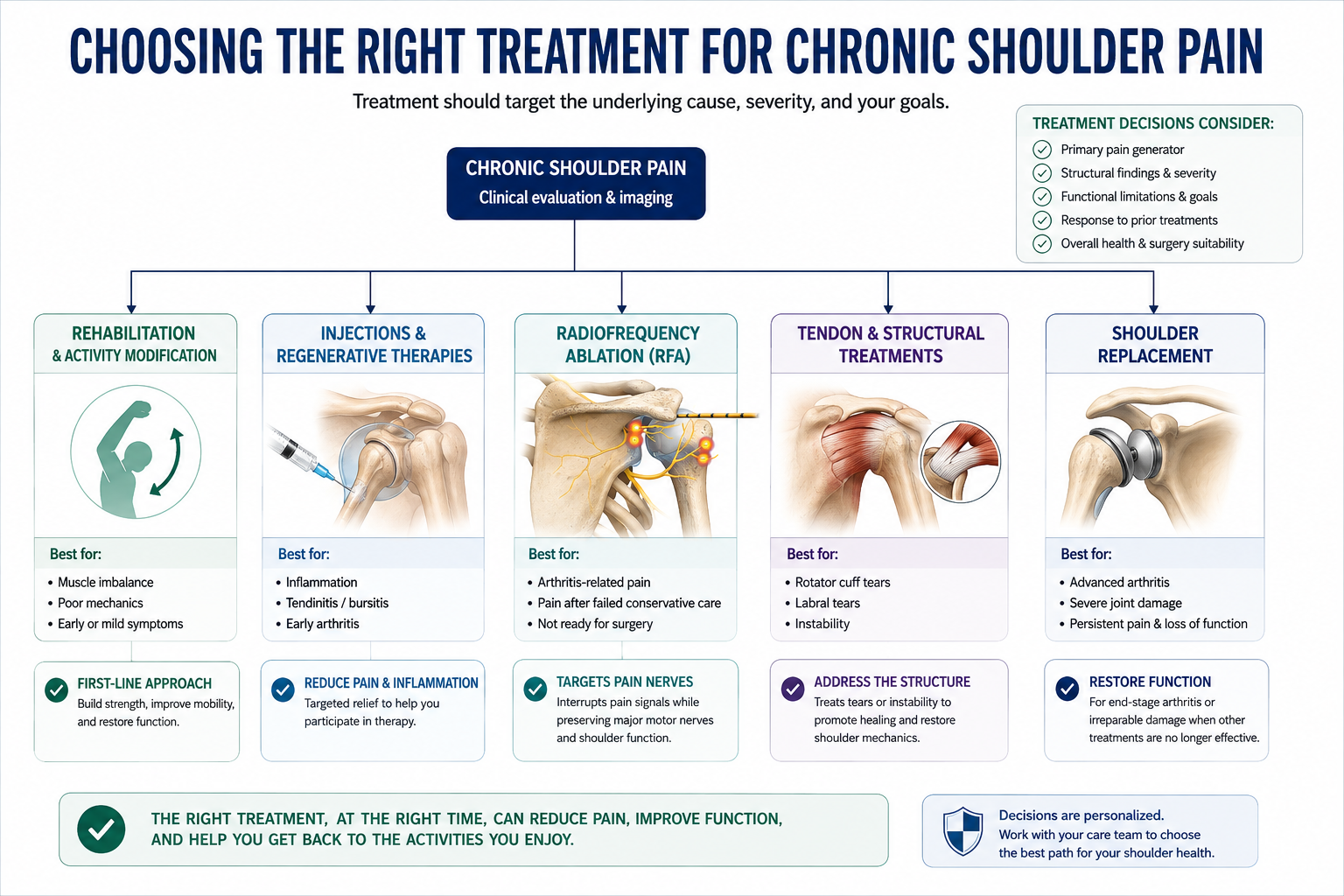
The most effective treatment for chronic shoulder pain depends on identifying the primary pain generator. Depending on the diagnosis, patients may benefit from rehabilitation, image-guided injections, regenerative therapies, radiofrequency ablation, or shoulder replacement surgery. Careful clinical evaluation helps match the treatment to the underlying condition rather than simply treating symptoms.
An Individualized Approach to Shoulder RFA
A successful shoulder RFA program begins with diagnosis rather than technology. The decision to proceed should consider:
- The structure or structures most likely causing pain
- The presence of arthritis, rotator cuff disease, instability, stiffness, or neurologic symptoms
- Active and passive range of motion
- True weakness versus pain-limited effort
- Imaging findings and whether they match the symptoms
- Prior response to therapy, medication, and injections
- Response to diagnostic articular branch blocks
- Age, health, activity goals, and surgical candidacy
- Whether pain relief is likely to improve meaningful function
Clinical Perspective
The most advanced radiofrequency equipment cannot compensate for treating the wrong pain generator. Successful shoulder RFA depends on accurate diagnosis, appropriate patient selection, precise targeting, thoughtful lesioning strategy, and a rehabilitation plan that converts pain relief into improved function.
Key Takeaways
- Shoulder RFA treats selected sensory articular branches carrying pain from the shoulder joint.
- Potential targets include branches associated with the suprascapular, axillary, lateral pectoral, and selected subscapular nerves.
- The procedure is designed to preserve the major motor pathways responsible for shoulder strength and movement.
- Shoulder RFA does not repair a rotator cuff tear, stabilize a dislocating shoulder, treat infection, or correct a failed prosthesis.
- Diagnostic articular branch blocks may help determine whether the targeted sensory pathways contribute meaningfully to pain.
- Pain reduction may improve sleep, daily function, and participation in rehabilitation.
- Evidence is encouraging but still evolving, and results vary among patients.
- Shoulder RFA does not prevent future injections, tendon treatment, or shoulder replacement.
Frequently Asked Questions About Shoulder Radiofrequency Ablation
Does shoulder RFA cure arthritis?
No. Shoulder RFA reduces pain transmission but does not rebuild cartilage, reverse arthritis, or restore the joint to normal.
Which nerves are treated during shoulder RFA?
Targets may include sensory articular branches associated with the suprascapular, axillary, lateral pectoral, and selected subscapular nerves. The exact targets depend on anatomy and technique.
Is the entire suprascapular nerve destroyed?
Selective articular-branch RFA is intended to treat pain-carrying sensory branches while avoiding important motor fibers. Technique matters because the main suprascapular nerve also supplies shoulder muscles.
Will shoulder RFA make my arm weak?
Weakness is not the intended effect. Image guidance and stimulation may be used to avoid major motor pathways. New or progressive weakness after treatment should be evaluated promptly.
Can RFA repair a torn rotator cuff?
No. RFA may reduce pain in selected patients but does not reconnect a torn tendon, restore tendon tension, or reverse muscle atrophy.
Can shoulder RFA help rotator cuff tear arthropathy?
It may help selected patients with painful cuff-tear arthropathy who are not undergoing reconstruction or replacement. It treats pain transmission rather than the mechanical consequences of the tear.
How long does shoulder RFA last?
Duration varies. Some patients benefit for several months or longer, while others obtain shorter relief or no meaningful improvement. The treated sensory branches may recover over time.
Can shoulder RFA be repeated?
Yes. Repeat treatment may be considered when pain returns after substantial initial benefit and reassessment confirms that the same pain pathway remains responsible.
How quickly will I notice improvement?
The anesthetic may provide temporary early relief, but the radiofrequency effect commonly develops gradually over several weeks.
Is shoulder RFA better than a steroid injection?
They work differently. Steroid injections primarily reduce inflammation, while RFA alters sensory pain transmission. The best option depends on the diagnosis, prior response, structural condition, and treatment goals.
Can shoulder RFA delay replacement surgery?
Pain reduction may allow some patients to postpone surgery. RFA does not stop structural degeneration, however, and should not indefinitely delay replacement when surgery offers a more appropriate path to restoring function.
Can I still have shoulder replacement after RFA?
Yes. Shoulder RFA generally does not prevent future shoulder replacement when surgery becomes appropriate.
Can RFA help after shoulder replacement?
It may help carefully selected patients after infection, loosening, instability, fracture, tendon failure, component problems, and other correctable causes have been evaluated. Evidence in this setting remains limited.
Does insurance cover shoulder RFA?
Coverage varies by payer, policy, diagnosis, and technique. Some insurers may consider shoulder articular-branch RFA investigational.
How do I know whether I am a candidate?
Potential candidates have chronic shoulder-joint pain despite appropriate conservative treatment, no urgent surgically correctable problem, and a clinical response suggesting that targeted articular sensory pathways contribute substantially to the pain.
Related Services
Joint Radiofrequency Ablation
A broader guide to radiofrequency treatment for chronic knee, shoulder, and hip pain.
Arthritis Treatment
An overview of exercise, medications, joint injections, PRP, RFA, joint preservation, and surgery.
Hip Radiofrequency Ablation
How femoral and obturator articular branches may be treated for selected chronic hip pain.
Knee Radiofrequency Ablation
A detailed guide to genicular nerve ablation for chronic knee pain.
Corticosteroid Joint Injections
How image-guided cortisone injections may reduce inflammation and shoulder-joint pain.
PRP for Arthritis
A guide to platelet-rich plasma treatment for selected painful arthritic joints.
References
- Burnham TR, et al. Shoulder terminal sensory articular nerve radiofrequency ablation: an updated treatment protocol accounting for variable terminal sensory anatomy. 2024.
- Jain E, et al. Radiofrequency ablation for shoulder pain: an updated review. Annals of Palliative Medicine. 2024.
- Tran A, et al. Selective denervation of the shoulder joint for chronic glenohumeral osteoarthritis pain using image-guided radiofrequency ablation.
- Santi C, et al. Two-centre retrospective analysis of selective sensory shoulder denervation using cooled radiofrequency. 2024.
- Pushparaj H, et al. A systematic review and meta-analysis of radiofrequency procedures for chronic shoulder pain. 2021.
- Sag AA, et al. CT-guided cooled radiofrequency denervation for chronic shoulder pain. 2022.
- Eckmann MS, et al. Current understanding and gaps in nerve ablation for chronic shoulder pain. 2021.
- Hagedorn JM, et al. The painful shoulder arthroplasty: appropriate work-up and interventional pain management. 2022.
- Gabriel S, et al. A narrative review of the non-infected painful total shoulder arthroplasty. 2023.
- American Academy of Orthopaedic Surgeons. Management of Glenohumeral Joint Osteoarthritis: Evidence-Based Clinical Practice Guideline. 2020.
- American Society of Regional Anesthesia and Pain Medicine. Shoulder articular nerve blockade and radiofrequency ablation: technical considerations.
- American Society of Pain and Neuroscience. Best-practice guidance for radiofrequency neurotomy.
About This Guide
This article is provided for educational purposes only and should not replace personalized medical advice, diagnosis, or treatment from a qualified healthcare professional. Every patient is unique, and treatment recommendations should be based on symptoms, examination findings, imaging, medical history, response to prior care, and individual treatment goals.



