
Subacromial Bursa Injection: Image-Guided Shoulder Treatment
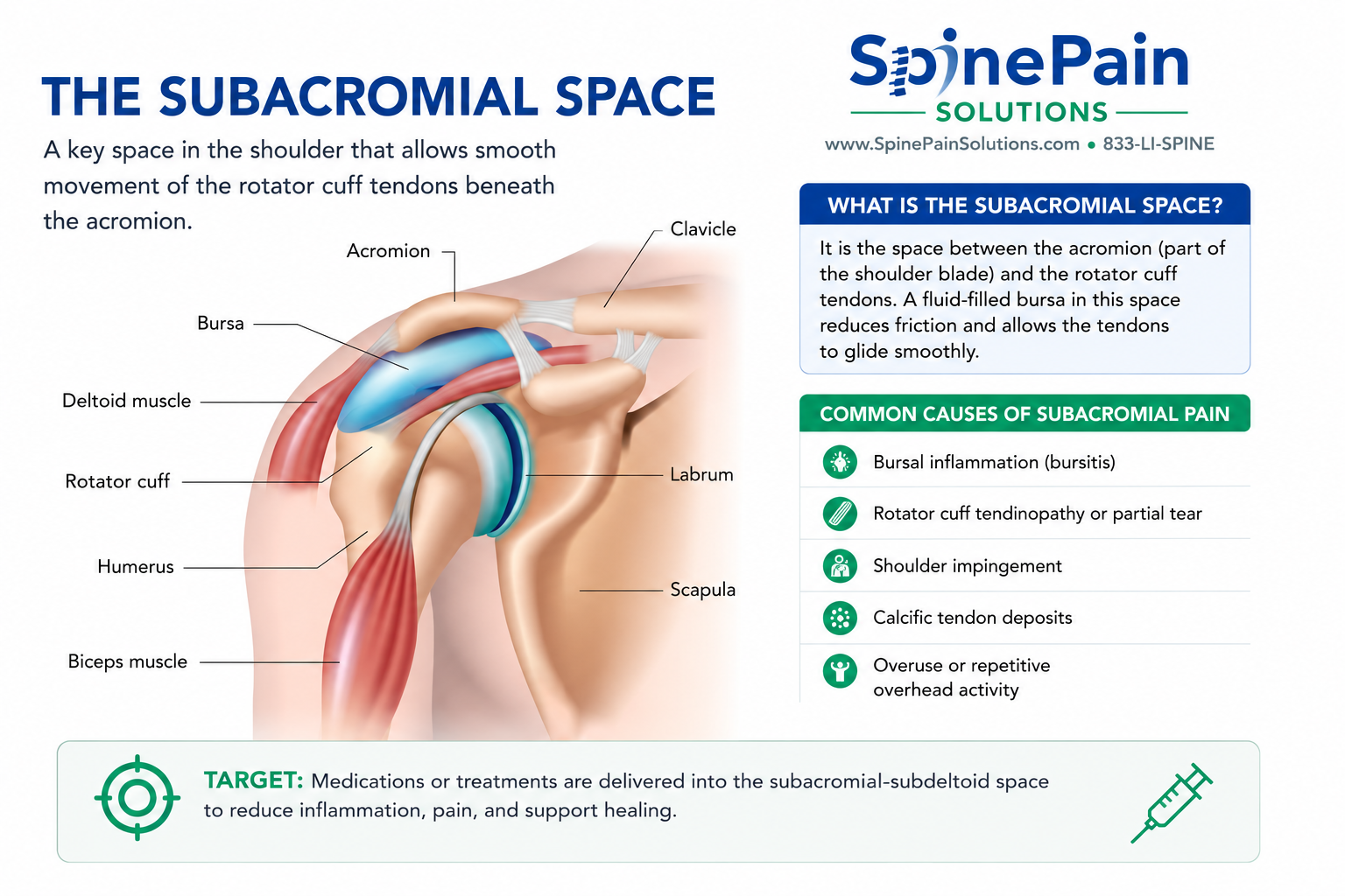
A subacromial bursa injection is an image-guided procedure used to deliver treatment into the space between the rotator cuff tendons and the overlying acromion and deltoid. It may be considered when shoulder pain remains functionally limiting despite appropriate rehabilitation, activity modification, medications, or other conservative treatment.
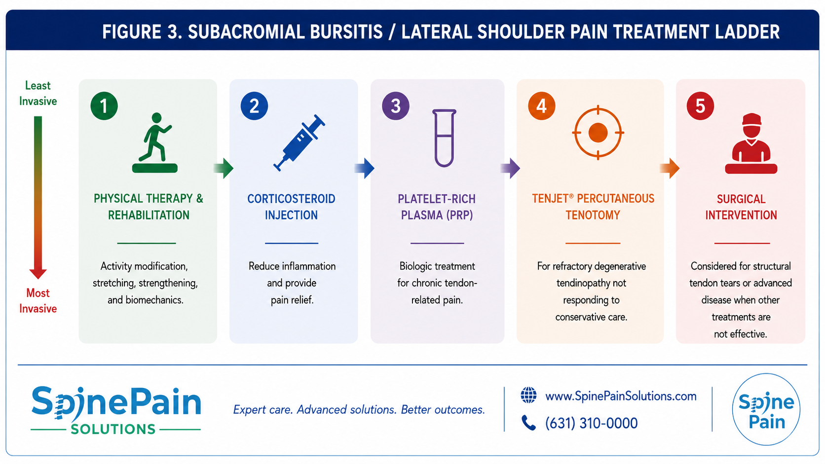
At SpinePain Solutions, subacromial treatment is individualized rather than limited to one medication or one imaging method. Depending on the patient’s anatomy, imaging findings, previous treatment response, and the tissue believed to be responsible for pain, management may include corticosteroid injection, platelet-rich plasma (PRP), TenJet® percutaneous tenotomy for qualifying rotator cuff tendinopathy, dry needling for selected muscular pain, rehabilitation, or referral for surgical evaluation.
Ultrasound is commonly selected when the subacromial bursa and rotator cuff tendons can be clearly visualized. In patients with a larger body habitus or less favorable ultrasound visualization, fluoroscopic guidance may provide a more dependable route using bony landmarks and contrast confirmation. The objective is not to use one technique for every patient, but to select the imaging method and intervention best suited to the treatment target.
Quick Answer
A subacromial bursa injection is an image-guided shoulder procedure used to reduce inflammation and pain within the subacromial space. Depending on the patient’s anatomy and treatment target, ultrasound or fluoroscopy may be used. Treatment may involve corticosteroid medication, PRP for selected tendon-related cases, or more advanced procedures when chronic rotator cuff pathology is present.
Clinical Perspective
The phrase “subacromial bursa injection” describes an access route, not necessarily the entire treatment strategy. Some patients have predominantly inflammatory bursitis and may benefit from corticosteroid medication. Others have chronic rotator cuff tendinopathy, partial tendon tearing, calcific disease, or associated muscular pain that requires a different intervention. Treatment should be matched to the tissue responsible for symptoms rather than determined by the referral label alone.
When Is a Subacromial Bursa Injection Considered?
A subacromial bursa injection may be considered when shoulder pain continues to interfere with reaching, lifting, dressing, sleeping, exercise, work, or other daily activities despite appropriate conservative treatment. The procedure allows medication or another treatment to be delivered directly into the subacromial-subdeltoid space using image guidance rather than relying on surface landmarks alone.
Although patients are often referred specifically for a “subacromial bursa injection,” the most appropriate intervention depends on the tissue responsible for pain. Treatment may be directed toward active bursal inflammation, a degenerative rotator cuff tendon, a partial tendon tear, calcific tendon disease, or associated muscular pain. The name of the referral should not automatically determine the treatment performed.
Patients may be considered for an image-guided intervention when they have:
- Persistent shoulder pain despite rehabilitation, activity modification, or medications.
- Pain with reaching overhead, lifting, dressing, or placing the arm behind the back.
- Night pain that interferes with sleep or prevents lying comfortably on the affected shoulder.
- Symptoms corresponding with inflammation in the subacromial-subdeltoid space.
- Temporary but incomplete improvement following previous treatment.
- A need for a precisely targeted diagnostic or therapeutic procedure.
- Chronic rotator cuff tendinopathy requiring consideration of PRP or a tendon-directed procedure.
Quick Answer
A subacromial bursa injection is generally considered when shoulder pain remains functionally limiting despite conservative care. It may be performed to reduce inflammation, clarify the pain source, or form part of a broader treatment plan involving PRP, tendon treatment, rehabilitation, calcific-tendon procedures, or surgical evaluation.
Clinical Perspective
The subacromial bursa is frequently inflamed because of an underlying rotator cuff problem. Injecting the bursa may reduce inflammation, but durable improvement often depends on addressing tendon degeneration, weakness, altered shoulder mechanics, or another associated pain generator.
To learn more about the condition itself, including symptoms, causes, and diagnosis, visit our Subacromial Bursitis resource.
Ultrasound vs Fluoroscopic Guidance
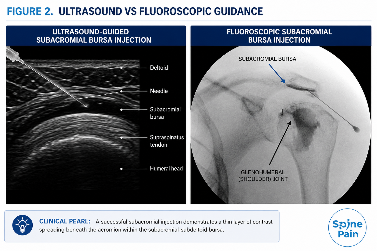
Subacromial bursa procedures may be performed using ultrasound or fluoroscopic guidance. Both techniques provide image-guided needle placement, but they display different anatomical information and may be better suited to different patients.
At SpinePain Solutions, the imaging method is selected according to body habitus, procedural depth, the tissue being treated, and the physician’s judgment. We commonly use ultrasound in thinner patients and when evaluation of the rotator cuff tendons is important. Fluoroscopy may be selected in patients with a larger body habitus or when ultrasound visualization is less dependable.
Ultrasound-Guided Treatment
Ultrasound provides real-time visualization of the subacromial-subdeltoid bursa, rotator cuff tendons, deltoid muscle, fluid collections, calcific deposits, and needle pathway. It does not expose the patient to ionizing radiation and permits dynamic assessment while the shoulder moves.
Ultrasound is particularly useful when treatment is directed toward the supraspinatus or another rotator cuff tendon, when PRP is being considered, or when a calcific deposit requires direct visualization.
Fluoroscopically Guided Treatment
Fluoroscopy uses live X-ray imaging to identify the acromion, humeral head, and other bony landmarks. A small amount of contrast may be used to confirm distribution within the intended subacromial plane before medication is delivered.
Fluoroscopy may provide a reliable alternative when soft-tissue visualization is limited by patient anatomy or when bony landmarks offer the clearest route to the treatment target.
| Consideration | Ultrasound | Fluoroscopy |
|---|---|---|
| Primary visualization | Bursa, rotator cuff tendons, deltoid, fluid, calcification, and needle | Acromion, humeral head, and other bony landmarks |
| Common use in our practice | Often selected in thinner patients or when tendon visualization is important | Often selected in heavier patients or when ultrasound visualization is less favorable |
| Radiation | None | Uses a limited amount of X-ray imaging |
| Contrast | Usually unnecessary | May be used to confirm distribution |
| Technique selected by | Patient anatomy, treatment target, procedural goals, and physician judgment | |
Clinical Perspective
Ultrasound and fluoroscopy are complementary technologies. The best guidance method is the one that provides the clearest and safest route to the intended target for that individual patient.
Interventional Treatment Options
A subacromial procedure is not limited to one medication or technique. Treatment is selected according to whether the primary goal is diagnostic clarification, rapid control of inflammation, biological treatment of chronic tendon disease, removal of degenerative tissue, treatment of calcific deposits, or management of associated muscular pain.
Diagnostic Local Anesthetic Injection
A small amount of local anesthetic may be placed within the subacromial space when temporary numbing would help determine how much of the patient’s pain originates from this region. The response is interpreted together with the history, physical examination, and imaging findings.
Corticosteroid Injection
Corticosteroid medication may be selected when active inflammation within the subacromial bursa is believed to be a major contributor to pain. The medication is delivered under ultrasound or fluoroscopic guidance to reduce inflammation and improve comfort while the patient continues rehabilitation.
Corticosteroid injections may provide relatively rapid relief but do not repair tendon degeneration or reconnect a torn tendon. Repeated steroid exposure near compromised rotator cuff tissue should therefore be considered carefully.
Key Point
A corticosteroid injection may calm inflammation but should not automatically become the default treatment for every painful shoulder. When chronic tendon degeneration, a substantial tear, calcific disease, or another structural problem is present, a different intervention may be more appropriate.
Platelet-Rich Plasma (PRP)
PRP may be considered for selected patients with chronic rotator cuff tendinopathy or qualifying partial-thickness tendon abnormalities. PRP is prepared from the patient’s own blood and delivered under ultrasound guidance to the specific tendon region identified during evaluation.
The objective is not to inject PRP indiscriminately into every inflamed bursa. When tendon degeneration is the primary treatment target, PRP is generally directed toward the abnormal tendon or a combined tendon-bursal region according to the clinical findings.
Clinical evidence for PRP in rotator cuff disease remains mixed. Some studies report short-term improvement in pain and function compared with corticosteroid treatment, while other trials have found no meaningful advantage over placebo or rehabilitation. PRP is therefore offered selectively rather than as a routine replacement for corticosteroid injection.
Dry Needling
Dry needling is used only occasionally in our practice. It is not an injection into the subacromial bursa and does not repair rotator cuff tendon disease. It may be considered when associated muscular or myofascial pain involving the deltoid, trapezius, infraspinatus, or other shoulder-girdle muscles contributes to symptoms.
When Is TenJet® Considered?
TenJet is not a treatment for isolated subacromial bursitis. It is an ultrasound-guided percutaneous tendon procedure that may be considered when persistent shoulder pain is associated with selected chronic degenerative rotator cuff tendon tissue and appropriate conservative care has not provided adequate improvement.
During the procedure, ultrasound is used to identify the abnormal tendon region. Through a small skin opening, the TenJet device uses a controlled stream of fluid and suction to remove selected degenerative tissue while preserving healthier portions of the tendon.
TenJet may be considered when:
- Symptoms persist despite rehabilitation and activity modification.
- Ultrasound or MRI demonstrates selected chronic tendon degeneration.
- The tendon abnormality corresponds with the patient’s examination and symptoms.
- Previous injections provided only temporary or incomplete improvement.
- The tendon remains mechanically suitable for a minimally invasive procedure rather than surgical repair.
TenJet cannot reconnect a complete tendon rupture, repair major retraction, reverse advanced fatty muscle atrophy, or correct significant mechanical failure. Patients with substantial rotator cuff tears may require orthopedic surgical evaluation.
Clinical Perspective
TenJet is reserved for carefully selected tendon pathology. It should not be presented as a treatment for every patient with bursitis or every rotator cuff tear.
Interventional Treatment for Calcific Rotator Cuff Tendinopathy
Calcific rotator cuff tendinopathy occurs when calcium deposits develop within a rotator cuff tendon, most commonly the supraspinatus. The deposit may trigger substantial inflammation within the subacromial bursa and produce severe shoulder pain.
Treatment depends on the deposit’s size, consistency, location, symptoms, and response to conservative care. Options may include:
- Rehabilitation and activity modification.
- Subacromial corticosteroid injection for selected inflammatory presentations.
- Ultrasound-guided needling or lavage of an accessible calcific deposit.
- Extracorporeal shockwave therapy.
- Percutaneous tendon treatment in selected chronic cases.
- Orthopedic surgical evaluation when symptoms remain severe or mechanical limitations persist.
Evidence for ultrasound-guided lavage is conflicting. Earlier studies reported improvement compared with corticosteroid injection alone, while a more recent sham-controlled trial did not demonstrate benefit over sham treatment. The procedure should therefore be offered selectively rather than presented as universally effective.
Key Point
Calcific tendon disease is not simply bursitis. Treatment should be directed toward the calcium deposit, tendon condition, associated inflammation, and overall shoulder mechanics.
Comparing Interventional Treatment Options
| Treatment | Primary Target | Primary Goal | Typical Role |
|---|---|---|---|
| Local anesthetic injection | Subacromial space | Temporary pain reduction and diagnostic clarification | Selected diagnostic procedures |
| Corticosteroid injection | Inflamed subacromial-subdeltoid bursa | Reduce inflammation and provide relatively rapid relief | Active inflammatory pain or recalcitrant flare |
| PRP | Selected degenerative rotator cuff tendon tissue | Support the biological environment of chronically injured tissue | Selected chronic tendon-related cases |
| Dry needling | Shoulder-girdle muscular trigger points | Reduce associated myofascial pain | Occasional adjunct; does not treat the bursa itself |
| TenJet® | Selected chronic degenerative tendon tissue | Remove selected abnormal tissue while preserving healthier tendon | Qualifying refractory tendinopathy |
| Ultrasound-guided calcific deposit needling or lavage | Selected accessible calcium deposit | Disrupt or remove calcium material | Selected symptomatic calcific tendinopathy |
| Surgical treatment | Substantial tendon tear or other correctable structural pathology | Repair structural failure and restore mechanical function | Advanced disease or failure of appropriate non-surgical care |
What Happens During a Subacromial Bursa Procedure?
The exact procedure depends on the treatment being performed. Image-guided local anesthetic and corticosteroid injections are generally brief outpatient procedures. PRP, TenJet, and calcific-deposit procedures require different preparation and recovery instructions.
Preparation and Positioning
The patient’s medications, allergies, medical history, imaging studies, and treatment goals are reviewed. The patient is positioned so the shoulder can be safely accessed using the selected imaging technique.
Image-Guided Needle Placement
The skin is cleansed using sterile technique. Ultrasound or fluoroscopy is used to identify the treatment target and guide the needle along the intended pathway. When fluoroscopy is used, a small amount of contrast may be administered when clinically appropriate.
Delivery of the Selected Treatment
Local anesthetic, corticosteroid, or PRP is delivered according to the treatment plan. TenJet and calcific-deposit procedures are performed under continuous ultrasound visualization.
A small bandage is applied afterward. Most patients return home the same day with procedure-specific instructions.
Clinical Perspective
Accurate target selection, appropriate imaging, sterile technique, thoughtful rehabilitation, and realistic expectations are as important as the medication or device used.
Recovery After Subacromial Treatment
Recovery depends on the intervention performed. A routine corticosteroid injection usually has a much shorter recovery than PRP, TenJet, or a calcific-deposit procedure.
After Local Anesthetic or Corticosteroid Injection
- Mild soreness may occur for one or several days.
- Relative rest is generally recommended for the remainder of the day.
- Light daily activity can usually resume shortly afterward.
- Improvement may begin within several days and continue over one to two weeks.
- Temporary numbness or weakness may occur until the local anesthetic wears off.
After PRP
- A temporary increase in soreness is common.
- Improvement is generally gradual rather than immediate.
- Anti-inflammatory medications may be restricted around the procedure according to physician instructions.
- Activity progression is coordinated with rehabilitation and the tendon treated.
- Benefits, when achieved, may develop over several weeks to months.
After TenJet® or Calcific-Deposit Treatment
- A small bandage covers the percutaneous treatment site.
- A sling may be used briefly in selected patients for comfort rather than prolonged immobilization.
- Shoulder activity is advanced according to the tissue treated.
- Progressive range-of-motion and strengthening exercises are introduced through rehabilitation.
- Recovery is generally measured over weeks to months rather than days.
Key Point
Pain relief should create an opportunity to restore shoulder motion, strength, and function. Continuing or restarting an appropriate rehabilitation program is usually essential for maintaining improvement.
What Results Can Be Expected?
The goal is to reduce pain, improve sleep, restore shoulder motion, support rehabilitation, and allow the patient to resume valued activities. No procedure can guarantee a particular degree or duration of relief.
Corticosteroid injection may provide relatively rapid symptom reduction, but relief may be temporary when tendon degeneration, weakness, tearing, or altered mechanics remain untreated. PRP and TenJet are used for different tissue problems and generally involve slower recovery.
Outcome is influenced by:
- Accuracy of the diagnosis and treatment target.
- Severity and chronicity of the tendon or bursal abnormality.
- Presence of a partial or full-thickness rotator cuff tear.
- Muscle weakness, stiffness, arthritis, or cervical referral.
- Adherence to rehabilitation and activity recommendations.
- General health, smoking, metabolic disease, and other healing factors.
Clinical Perspective
A meaningful outcome is not simply a lower pain score. Successful treatment should help the patient sleep, reach overhead, dress, lift, exercise, participate in therapy, or return to other important activities more comfortably.
What Are the Risks?
Image-guided subacromial bursa and rotator cuff procedures are generally well tolerated, but every intervention has potential risks. The exact risk profile depends on the treatment performed.
- Temporary soreness or bruising.
- Temporary increase in pain.
- Bleeding or hematoma.
- Infection.
- Allergic reaction to medication, antiseptic, adhesive, or contrast.
- Temporary numbness or weakness from local anesthetic.
- Skin thinning or pigment change after corticosteroid exposure.
- Transient elevation of blood glucose following corticosteroid treatment.
- Tendon weakening or injury, particularly with repeated steroid exposure near compromised tendon tissue.
- Incomplete removal of calcium during lavage.
- Failure to obtain adequate or durable relief.
- Need for further treatment or surgical evaluation.
When Should You Contact Your Physician?
Contact your physician promptly for fever, progressive redness, drainage, rapidly increasing swelling, severe or worsening pain, new weakness, persistent numbness, or any unexpected symptoms after the procedure.
Who May Not Be a Candidate?
A particular procedure may be modified, postponed, or avoided when the potential risks outweigh the expected benefit.
- Active infection at the treatment site or systemic infection.
- Uncontrolled bleeding risk or inability to safely manage anticoagulant medication.
- Allergy to required medication or contrast without a suitable alternative.
- A complete or substantially retracted rotator cuff tear requiring surgical evaluation.
- Progressive weakness or another condition requiring more urgent treatment.
- Pain arising primarily from the glenohumeral joint, cervical spine, or another untreated source.
- Inability to participate in the rehabilitation required after the selected procedure.
- Medical conditions that significantly impair healing or increase procedural risk.
Our Approach at SpinePain Solutions
Our approach is built around selecting the appropriate target, imaging method, intervention, and recovery plan for the individual patient.
At SpinePain Solutions, treatment may include:
- Ultrasound guidance when direct soft-tissue visualization is most useful.
- Fluoroscopic guidance when body habitus or procedural depth makes bony landmarks more dependable.
- Corticosteroid injection for selected inflammatory presentations.
- PRP for carefully selected chronic tendon-related cases.
- TenJet for qualifying refractory rotator cuff tendinopathy.
- Dry needling only occasionally when muscular pain is clinically important.
- Ultrasound-guided treatment of selected calcific deposits.
- Rehabilitation integrated into the interventional treatment plan.
- Orthopedic referral when structural tendon failure is better treated surgically.
The goal is not to perform the greatest number of procedures. It is to select the least invasive intervention reasonably likely to improve pain and function while preserving future treatment options.
Key Takeaways
- A subacromial bursa injection is one of several interventions available for shoulder pain.
- Ultrasound and fluoroscopy are selected according to patient anatomy and the treatment target.
- Corticosteroid may reduce active inflammation but does not repair tendon degeneration.
- PRP is offered selectively because results vary among rotator cuff conditions and studies.
- Dry needling treats associated muscular pain rather than the bursa itself.
- TenJet may be considered for qualifying chronic tendon disease but cannot repair major structural tears.
- Calcific tendon disease may require a deposit-directed procedure rather than a routine bursa injection.
- Recovery and rehabilitation depend on the specific intervention performed.
- Substantial tendon failure may require orthopedic surgical evaluation.
Related Services and Resources
Bursitis Treatment
Overview of rehabilitation, aspiration, image-guided injections, PRP, and other treatments for painful bursae.
Subacromial Bursitis
Patient resource covering symptoms, causes, diagnosis, and common sources of subacromial pain.
Rotator Cuff Tendinopathy
Information about degeneration and tearing of the tendons responsible for shoulder movement.
TenJet Percutaneous Tenotomy
How ultrasound-guided removal of selected degenerative tendon tissue may help qualifying chronic tendinopathy.
Joint Preservation
A personalized strategy for maintaining mobility and native-joint function.
Frequently Asked Questions
Is a subacromial bursa injection always performed with ultrasound?
No. Ultrasound is commonly used when the bursa and rotator cuff tendons can be clearly visualized. Fluoroscopy may be selected in heavier patients or when bony landmarks provide a more dependable pathway.
Is ultrasound better than fluoroscopy?
Neither is universally better. Ultrasound visualizes soft tissues without radiation, while fluoroscopy provides excellent bony landmarks and may confirm distribution with contrast.
What medication is used?
A conventional injection commonly contains local anesthetic and corticosteroid. PRP may be used in selected tendon-related cases but is prepared and performed differently.
How quickly does a steroid injection work?
Local anesthetic may provide temporary relief shortly after treatment. Corticosteroid benefit often develops over several days and may continue to improve over one to two weeks.
How long does relief last?
Duration varies. Some patients improve for weeks or months, while others obtain temporary or incomplete relief. Persistent tendon disease, tearing, stiffness, or another pain source can affect the response.
Can the injection be repeated?
Repeat treatment may be considered after reassessing the diagnosis, prior response, tendon condition, and cumulative steroid exposure. Repeated injections should not replace treatment of ongoing tendon or biomechanical abnormalities.
Is PRP better than corticosteroid?
Research is mixed. Some studies report better short-term pain and function after PRP in selected tendon disorders, while others have found no clear benefit over placebo. PRP is therefore offered selectively.
Is PRP injected into the bursa or tendon?
The target depends on the clinical findings. When rotator cuff tendinopathy is the primary problem, PRP is generally directed toward the abnormal tendon or combined tendon-bursal target under ultrasound guidance.
What is TenJet used for?
TenJet may be considered for selected chronic degenerative tendon abnormalities. It does not treat isolated bursitis and cannot repair a complete or substantially retracted tear.
What is barbotage or lavage?
It is an ultrasound-guided procedure in which an accessible calcium deposit is needled and fluid may be used to loosen or remove calcium material. It is reserved for selected symptomatic calcific deposits.
Will I need sedation?
Most routine subacromial injections are performed without intravenous sedation. Anesthesia needs vary for more advanced procedures and are reviewed before treatment.
When can I exercise again?
Activity often resumes quickly after a routine steroid injection. PRP, TenJet, and calcific-deposit procedures require a more gradual, tissue-specific progression.
What if the injection does not help?
The diagnosis and treatment target should be reassessed. Persistent symptoms may reflect rotator cuff tearing, glenohumeral arthritis, adhesive capsulitis, biceps pathology, cervical referral, or another pain generator.
References
- Wu T, et al. Ultrasound-guided versus blind subacromial-subdeltoid bursa corticosteroid injection for shoulder pain: A systematic review and meta-analysis. 2015.
- Deng X, et al. Ultrasound-guided versus anatomic landmark-guided corticosteroid injection for subacromial pain syndrome. 2022.
- Kwong CA, et al. Platelet-rich plasma compared with corticosteroid injection for partial-thickness rotator cuff tears or tendinopathy: A randomized controlled trial. 2021.
- Kesikburun S, et al. Platelet-rich plasma injections in chronic rotator cuff tendinopathy: A randomized controlled trial. 2013.
- Lin MT, et al. Effectiveness of platelet-rich plasma injection in rotator cuff tendinopathy: A systematic review and meta-analysis. 2020.
- Maag L, et al. Effectiveness of percutaneous needle tenotomy for tendinopathy: A systematic review. 2025.
- Jacobson JA, et al. Ultrasound-guided percutaneous needle tenotomy: Rationale and technique. 2016.
- de Witte PB, et al. Calcific tendinitis of the rotator cuff: Ultrasound-guided needling and lavage versus subacromial corticosteroid injection. 2013.
- Moosmayer S, et al. Ultrasound-guided lavage with corticosteroid injection versus sham treatment for calcific rotator cuff tendinopathy. 2023.
- American Academy of Orthopaedic Surgeons. Management of Rotator Cuff Injuries: Clinical Practice Guideline.
About This Guide
This article from Dr. Amit Sharma is provided for educational purposes only and should not replace personalized medical advice, diagnosis, or treatment from a qualified healthcare professional. The appropriate imaging method and intervention depend on the patient’s diagnosis, anatomy, medical history, imaging findings, previous treatment response, and individual goals.