

Arthritis Treatment: A Complete Guide to Non-Surgical Joint Pain Relief
Arthritis Treatment has changed dramatically over the past two decades. Many people still believe their only options are to live with chronic joint pain until it becomes severe enough to require joint replacement surgery. While joint replacement remains an excellent solution for many patients with advanced arthritis, it is no longer the only option. Today, a growing range of evidence-based treatments can reduce pain, improve mobility, preserve function, and in some cases delay or even avoid surgery.
Modern arthritis care begins with understanding that every patient is different. The location of the affected joint, the severity of cartilage damage, activity level, age, overall health, and personal goals all influence which treatments are most appropriate. A competitive athlete with early knee arthritis, for example, may benefit from a very different treatment strategy than an older adult with advanced hip osteoarthritis.
Treatment options may include lifestyle modifications, physical therapy, medications, image-guided joint injections, regenerative medicine techniques such as platelet-rich plasma (PRP), hyaluronic acid (gel) injections, radiofrequency ablation for chronic joint pain, and when necessary, referral for joint replacement surgery. Rather than relying on a single treatment, physicians often combine several approaches into a personalized treatment plan that evolves as symptoms and arthritis progress over time.
This comprehensive guide explains how arthritis develops, why joints become painful, the advantages and limitations of today’s treatment options, and how an individualized treatment strategy can help patients remain active and maintain their quality of life.
Arthritis treatment has expanded far beyond the traditional choice between taking medication and waiting until a joint becomes painful enough to replace. Although joint replacement remains an important and sometimes life-changing treatment for advanced disease, many patients can benefit from a broader plan designed to reduce pain, improve mobility, maintain strength, and preserve function for as long as reasonably possible.
Modern arthritis care may include exercise, physical therapy, weight management, medications, bracing, image-guided joint injections, platelet-rich plasma, radiofrequency ablation, and joint-preservation strategies. The best plan depends on much more than the appearance of an X-ray. The joint involved, the source of pain, the severity of structural damage, activity level, medical history, prior treatment response, and the patient’s goals all matter.
This guide focuses primarily on osteoarthritis and other forms of mechanically driven joint pain treated within musculoskeletal and interventional pain care. Inflammatory diseases such as rheumatoid arthritis, psoriatic arthritis, gout, and infection require a different diagnostic and medical approach, although they may produce some overlapping symptoms.
The goal is not to promote one procedure as the answer for everyone. It is to explain the available options, where each treatment may fit, why results differ, and when surgical evaluation should become part of the conversation.
What Are the Main Treatments for Arthritis?
Quick Answer
Arthritis treatment usually begins with education, appropriate activity, exercise, physical therapy, weight management when relevant, and medications selected according to the patient’s health. If pain continues, image-guided corticosteroid, hyaluronic acid, or platelet-rich plasma injections may be considered. Radiofrequency ablation can reduce pain from selected arthritic joints by treating sensory nerve branches, including in some patients who continue to have pain after joint replacement. Joint-preservation surgery or joint replacement may be appropriate when structural damage, mechanical dysfunction, or persistent disability cannot be adequately managed with less invasive care.
There is no universal sequence that every patient must follow. Some people improve substantially with exercise and modest activity changes. Others need injections or radiofrequency treatment to participate comfortably in rehabilitation. Patients with severe mechanical deformity, instability, fracture risk, or advanced loss of function may need an orthopedic evaluation sooner.
Clinical Perspective
The severity of arthritis seen on imaging does not always match the severity of pain. One patient may have marked cartilage loss but remain active, while another may have moderate imaging changes and substantial pain. This is not because the pain is imaginary or because the X-ray is meaningless. It reflects the fact that arthritic pain may arise from several structures, including the synovium, capsule, subchondral bone, ligaments, tendons, muscles, bursae, and sensory nerves surrounding the joint. Effective treatment begins by identifying which of these structures is most likely driving the patient’s symptoms.
What Is Arthritis?
Arthritis is a broad term describing disorders that affect joints and surrounding tissues. More than one hundred forms of arthritis have been identified. They do not all develop for the same reason, affect the same tissues, or respond to the same treatments.
Within a musculoskeletal practice, the most frequently encountered form is osteoarthritis. Osteoarthritis is not simply the passive wearing away of cartilage. It is a disease of the whole joint in which cartilage, bone, synovium, capsule, ligaments, muscles, and other supporting tissues may change over time.
Commonly affected areas include the knees, hips, shoulders, hands, feet, and spinal joints. Osteoarthritis may involve one joint after an injury or several joints because of age, genetics, body mechanics, occupational exposure, prior surgery, or other contributing factors.
Osteoarthritis
Osteoarthritis develops as the tissues responsible for smooth movement and load distribution become altered. Articular cartilage may thin or become irregular. The bone beneath the cartilage may harden, remodel, develop cysts, or form osteophytes commonly called bone spurs. The joint lining may become inflamed, and surrounding muscles may weaken as pain reduces normal activity.
The condition can range from mild, intermittent stiffness to advanced pain, deformity, instability, and loss of mobility. Progression is not identical in every patient, and symptoms may fluctuate even when the underlying structural changes remain relatively stable.
Post-Traumatic Arthritis
Post-traumatic arthritis can develop after a fracture, cartilage injury, ligament tear, meniscal injury, dislocation, or other significant joint trauma. Even after an injury has healed, changes in alignment, stability, or load distribution may increase stress across part of the joint.
Symptoms may arise years after the original injury. Treatment may resemble treatment for primary osteoarthritis, but structural instability or deformity may require additional orthopedic assessment.
Inflammatory Arthritis
Rheumatoid arthritis, psoriatic arthritis, and related autoimmune disorders occur when immune activity drives inflammation within joints and sometimes other organs. These conditions may cause prolonged morning stiffness, warmth, swelling, fatigue, and symmetrical involvement of multiple joints.
Local pain procedures may occasionally have a supportive role, but they do not replace disease-modifying treatment prescribed by a rheumatologist. Controlling the underlying inflammatory disease is essential to limiting progressive joint damage and systemic complications.
Crystal Arthritis
Gout and calcium pyrophosphate deposition disease can produce sudden episodes of intense joint pain, swelling, redness, and warmth. Joint aspiration may be needed to identify crystals and, importantly, to help exclude infection when the diagnosis is uncertain.
Important: A suddenly hot, red, severely painful joint, especially when accompanied by fever, chills, illness, a wound, recent surgery, or immune suppression, requires prompt medical evaluation. Joint infection can resemble an arthritis flare but needs urgent treatment.
Why Does an Arthritic Joint Hurt?
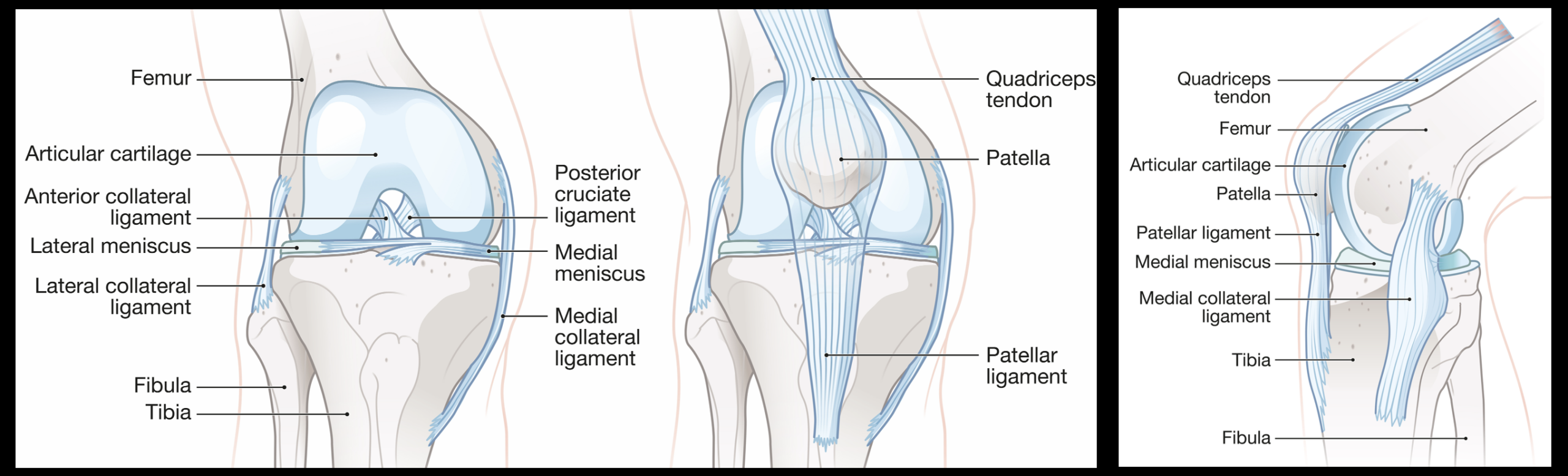
Cartilage deterioration is an important feature of osteoarthritis, but cartilage itself has little or no direct pain sensation. Pain more commonly arises from tissues that contain nerve endings and respond to inflammation, pressure, stress, or abnormal movement.
Synovial Inflammation
The synovium is the lining of a movable joint. It produces fluid that lubricates the joint and supports cartilage. When irritated, the synovium may become inflamed and produce excess fluid, contributing to swelling, stiffness, and pain.
Subchondral Bone Changes
The bone immediately beneath the cartilage is called subchondral bone. It can remodel and become exposed to altered mechanical forces as arthritis progresses. Bone marrow lesions, microstructural stress, cyst formation, and increased pressure within the bone may contribute to pain.
Joint Capsule and Ligaments
The capsule and ligaments help stabilize a joint. Chronic swelling, altered alignment, contracture, or instability can stress these tissues. Patients may experience pain at the limits of motion, a sense of giving way, or discomfort when loading the joint.
Muscles, Tendons, and Bursae
Arthritis changes the way a person moves. Limping, guarding, and reduced activity can weaken muscles and overload tendons or bursae. Hip arthritis may coexist with gluteal tendon pain. Knee arthritis may be accompanied by quadriceps weakness or pes anserine bursitis. Shoulder arthritis may occur alongside rotator cuff disease.
These overlapping conditions matter because an injection placed into the joint may not fully relieve pain arising primarily from a tendon, bursa, muscle, or peripheral nerve.
Sensory Nerves and Pain Sensitization
Sensory nerve branches surrounding a joint transmit pain signals to the nervous system. Persistent pain can also make the nervous system more responsive to stimulation, a process often described as sensitization. This helps explain why pain intensity may not track perfectly with cartilage loss and why some patients continue to hurt even after technically successful joint replacement.
Key Point: Treating arthritis successfully does not always mean treating cartilage alone. The physician must consider the entire joint and the structures surrounding it.
Common Symptoms of Arthritis
Symptoms differ according to the joint involved, the type of arthritis, disease severity, and the patient’s activities. Common complaints include:
- Pain during walking, lifting, reaching, gripping, climbing stairs, or other joint-specific activities
- Stiffness after rest or inactivity
- Reduced range of motion
- Swelling or recurrent joint fluid
- Grinding, cracking, or a rough sensation during movement
- Weakness or a feeling that the joint may give way
- Difficulty getting out of a chair, entering a car, dressing, sleeping, or exercising
- Loss of endurance and reduced participation in work or recreation
- Changes in alignment, such as bowing of the knee
- Pain that persists at rest or interferes with sleep in more advanced cases
Mechanical symptoms such as true locking, repeated catching, marked instability, or sudden loss of motion may indicate an additional structural problem. These symptoms deserve careful evaluation rather than being attributed automatically to arthritis.
How Is Arthritis Evaluated?
The diagnosis usually begins with a detailed history and physical examination. Imaging can confirm structural changes, but the purpose of evaluation is not merely to assign an arthritis grade. It is to determine which condition is producing the patient’s current symptoms and whether another problem is present.
Medical History
Important details include when the pain began, whether an injury occurred, which movements provoke symptoms, the duration of morning stiffness, the presence of swelling, prior surgery, previous treatment response, and whether other joints are involved. Fever, rash, fatigue, unexplained weight change, eye inflammation, bowel disease, psoriasis, or a history of tick exposure may shift the evaluation away from uncomplicated osteoarthritis.
Physical Examination
Examination may assess gait, posture, alignment, swelling, warmth, tenderness, muscle strength, stability, range of motion, and pain during specific maneuvers. The surrounding spine, tendons, bursae, muscles, and nerves may also need to be examined because referred pain can imitate arthritis.
X-Rays
Weight-bearing X-rays are often the most useful initial imaging study for knee, hip, and other load-bearing joint arthritis. They may demonstrate joint-space narrowing, osteophytes, subchondral sclerosis, cysts, alignment changes, or deformity.
A normal or mildly abnormal X-ray does not exclude every meaningful source of joint pain, and severe X-ray findings do not automatically require surgery if symptoms and function remain acceptable.
MRI
MRI can evaluate cartilage, bone marrow, menisci, labrum, tendons, ligaments, and other soft tissues. It may be helpful when symptoms are atypical, a substantial injury occurred, surgery is being considered, or the diagnosis remains uncertain after examination and X-rays.
MRI is not always necessary for straightforward arthritis. It can also reveal age-related abnormalities that are not responsible for the patient’s pain. Findings must therefore be interpreted in clinical context.
Musculoskeletal Ultrasound
Ultrasound can identify joint fluid, synovial thickening, bursitis, tendon abnormalities, cysts, and selected peripheral nerve conditions. It also provides real-time guidance for many aspirations and injections without ionizing radiation.
Laboratory Testing and Joint Aspiration
Blood tests are not required to diagnose typical osteoarthritis, but they may be ordered when inflammatory arthritis, infection, gout, or another systemic condition is suspected. Joint aspiration allows fluid to be analyzed for infection, crystals, inflammatory cells, or bleeding.
Diagnostic Injections and Nerve Blocks
In selected cases, a precisely placed local anesthetic injection can help determine whether pain is arising from the joint or another structure. Before radiofrequency ablation, diagnostic blocks may be used to test whether targeted sensory nerve branches are meaningfully involved in the pain.
What Are the Goals of Arthritis Treatment?
Treatment goals should be specific to the patient. Eliminating every sensation from a structurally altered joint may not always be realistic, but meaningful improvements in pain, movement, sleep, independence, and participation are often possible.
- Reduce pain and inflammatory flares
- Improve range of motion, strength, and joint stability
- Maintain independence and daily function
- Support participation in exercise and rehabilitation
- Reduce avoidable medication exposure
- Address secondary tendon, muscle, bursal, or nerve pain
- Preserve the native joint when clinically reasonable
- Recognize when delaying surgery is no longer beneficial
A useful plan also includes measurable functional targets. Walking for twenty minutes, sleeping without repeated awakening, climbing stairs, returning to golf, or playing with grandchildren may be more meaningful outcomes than a pain score alone.
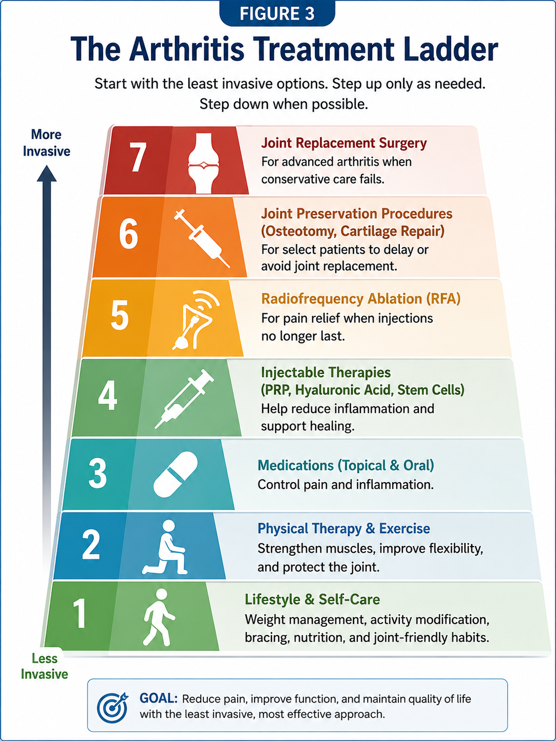
Foundational Non-Surgical Arthritis Treatment
Procedures may provide valuable relief, but they work best within a broader plan. Exercise, muscle conditioning, activity modification, and management of contributing health factors remain central to arthritis care.
Education and Self-Management
Understanding the condition can reduce fear and help patients make sensible decisions about movement. Arthritis does not necessarily mean that all activity is damaging the joint. Avoiding movement entirely often leads to weakness, stiffness, reduced balance, weight gain, and greater disability.
Self-management may include pacing demanding activities, alternating heavier and lighter days, using heat or ice appropriately, planning recovery after activity, and identifying movements that can be modified without abandoning meaningful goals.
Exercise
Appropriately selected exercise can improve pain, strength, endurance, balance, and function. Useful forms may include:
- Progressive strengthening
- Walking or other tolerable weight-bearing activity
- Cycling
- Aquatic exercise
- Range-of-motion work
- Balance and neuromuscular training
- Tai chi or other controlled movement programs
Some temporary soreness can occur when beginning a new program. Sharp pain, major swelling, progressive weakness, instability, or prolonged deterioration should prompt reassessment. The correct dose of exercise is challenging enough to produce adaptation but not so aggressive that each session causes a sustained flare.
Physical Therapy
Physical therapy can address weakness, impaired movement patterns, reduced flexibility, gait changes, and loss of confidence. A therapist may also recommend a home program, assistive device, taping technique, or gradual return-to-activity plan.
Passive treatments alone rarely replace the need for active conditioning. Manual therapy, heat, electrical stimulation, or other modalities may help selected patients, but durable improvement usually depends on restoring strength and useful movement.
Weight Management
For patients with excess body weight and lower-extremity arthritis, gradual weight reduction can decrease the load placed across painful joints and may improve symptoms, mobility, and general health. Even a modest reduction may make exercise easier.
Weight should not be used to dismiss a patient’s pain or deny other appropriate treatment. It is one modifiable contributor among many, and recommendations should be realistic, respectful, and paired with practical support.
Activity Modification
Activity modification does not necessarily mean stopping valued activities. It may mean reducing impact, shortening sessions, adjusting technique, allowing recovery days, changing footwear, improving ergonomics, or substituting a lower-impact activity during a flare.
Braces, Canes, and Other Supports
A properly selected brace may improve confidence or redistribute stress in certain knees, thumbs, wrists, ankles, or other joints. A cane can reduce load and improve balance when used correctly, generally in the hand opposite the painful lower-extremity joint.
Supports that are uncomfortable, poorly fitted, or used without a clear purpose may provide little benefit. They should supplement movement rather than create unnecessary dependence whenever possible.
Treatment Tip: An injection or radiofrequency procedure may create a window of reduced pain. That window is often most valuable when it is used to improve strength, walking tolerance, range of motion, and participation in rehabilitation.
Medications for Arthritis Pain
Medication selection depends on the joint involved, symptom pattern, age, kidney function, cardiovascular health, gastrointestinal history, liver function, anticoagulant use, and other medications. No medication is harmless simply because it is available without a prescription.
Topical Medications
Topical nonsteroidal anti-inflammatory drugs may help pain from accessible joints such as the knee or hand while producing less systemic exposure than oral medication. Topical capsaicin or other preparations may help selected patients, although skin irritation can occur.
Oral Anti-Inflammatory Medication
Oral nonsteroidal anti-inflammatory drugs can reduce pain and inflammation, but they may increase the risk of gastrointestinal bleeding, kidney injury, fluid retention, blood-pressure elevation, or cardiovascular complications. The lowest effective dose for the shortest appropriate duration is generally preferred.
Acetaminophen
Acetaminophen may provide limited relief for some patients who cannot take anti-inflammatory medication. Total daily intake must account for acetaminophen contained in combination cold, sleep, and prescription products. Excessive dosing can injure the liver.
Duloxetine and Other Selected Medications
Duloxetine may be considered for selected patients with chronic osteoarthritis pain, particularly when widespread pain, altered pain processing, anxiety, or depression is also present. Side effects, drug interactions, and the need for gradual tapering should be discussed.
Opioid Medication
Opioids generally have a limited role in chronic arthritis because of tolerance, sedation, constipation, falls, dependence, overdose risk, and uncertain long-term functional benefit. They do not treat the underlying joint disease. Exceptional circumstances require careful individualized assessment and monitoring.
Important: Patients with kidney disease, cardiovascular disease, a history of ulcers or gastrointestinal bleeding, liver disease, anticoagulant use, uncontrolled hypertension, or multiple medications should speak with a qualified clinician before beginning or increasing arthritis medication.
Image-Guided Joint Procedures
When foundational treatment does not provide adequate relief, an image-guided procedure may be considered. Ultrasound and fluoroscopy allow the physician to visualize anatomy or bony landmarks, confirm needle position, avoid selected nearby structures, and document accurate placement.
The preferred guidance method depends on the joint and procedure. Ultrasound provides real-time soft-tissue visualization without radiation. Fluoroscopy provides detailed X-ray guidance and can confirm intra-articular placement with a small amount of contrast when appropriate.
Accuracy is important, but accurate placement does not guarantee clinical success. A technically perfect injection will not fully relieve pain if the treated structure is not the main pain generator or if advanced mechanical dysfunction remains dominant.
A broader guide to these procedures will be available on the planned
Joint Injections
service page.
Corticosteroid Injections for Arthritis
Corticosteroid injections place anti-inflammatory medication directly into an affected joint. They are commonly considered when inflammation, swelling, or a painful flare limits activity and more conservative treatment has not been sufficient.
Relief may begin within several days, although the timing and duration vary. Some patients obtain meaningful improvement for weeks or months, while others experience little benefit. Corticosteroids do not restore lost cartilage and should not be described as a cure for arthritis.
Potential Advantages
- May reduce inflammation and pain relatively quickly
- Can help calm an acute flare
- May improve participation in exercise or physical therapy
- Can sometimes provide diagnostic information when accurately placed
- Requires little downtime for most patients
Limitations and Precautions
Possible effects include temporary post-injection pain, facial flushing, skin or fat changes near the injection site, transient blood-sugar elevation, bleeding, allergic reaction, and rare infection. The timing and frequency of repeat injections should be individualized, particularly in weight-bearing joints or when joint replacement is being planned.
Learn more in the complete guide to
corticosteroid joint injections for arthritis.
Hyaluronic Acid Injections, or Gel Shots
Hyaluronic acid is a naturally occurring component of joint fluid and other connective tissues. Injectable hyaluronic acid products, often called viscosupplements or gel shots, are used primarily for symptomatic knee osteoarthritis after simpler treatments have not provided adequate relief.
These products are intended to improve the joint environment and reduce pain rather than rebuild substantial areas of missing cartilage. Some patients improve, while others do not. Benefits, when present, may develop more gradually than after corticosteroid injection.
Professional guidelines differ in how they interpret the average benefit of hyaluronic acid. This disagreement reflects variation in trial quality, product formulation, injection schedules, patient characteristics, and the difference between an average study result and the response of an individual patient.
Who May Consider Gel Injections?
- Patients with symptomatic knee osteoarthritis
- Patients who have not obtained adequate relief from exercise, therapy, or medication
- Patients seeking an option that does not contain corticosteroid
- Patients who are not ready for or cannot undergo knee replacement
- Selected patients who previously responded favorably to viscosupplementation
Temporary soreness or swelling can occur. A more intense inflammatory reaction is uncommon but possible. Infection and bleeding are rare procedural risks.
Read the detailed guide to
hyaluronic acid joint injections and gel shots.
Platelet-Rich Plasma for Arthritis
Platelet-rich plasma, commonly abbreviated PRP, is prepared from a sample of the patient’s own blood. The blood is processed to concentrate platelets within plasma, and the resulting preparation is injected into the selected joint under image guidance.
Platelets contain signaling proteins involved in tissue response and inflammation. In osteoarthritis care, PRP is intended to modify the painful joint environment and improve symptoms. It should not be represented as reliably regrowing an advanced joint or reversing all structural damage.
Evidence is most developed for knee osteoarthritis. Some studies suggest that appropriately prepared PRP can improve pain and function in selected patients, but results vary and protocols are not uniform. Patient age, arthritis severity, alignment, metabolic health, platelet concentration, leukocyte content, activation method, injection schedule, and rehabilitation may influence outcome.
Potential Advantages
- Uses an autologous preparation derived from the patient’s blood
- Avoids exposure to corticosteroid
- May provide longer-lasting improvement than some other injections in selected patients
- Can be combined with a broader joint-preservation and rehabilitation plan
Limitations
- Not every patient responds
- Improvement may take several weeks
- Temporary post-procedure soreness is common
- Insurance coverage is often limited
- Preparation systems and treatment protocols differ
- PRP cannot reliably correct severe deformity, instability, or mechanical joint failure
Explore
PRP for arthritis
or the broader
platelet-rich plasma therapy
service page.
Important: PRP should not be confused with unapproved products marketed broadly as stem cells, exosomes, amniotic cells, or secretome therapy. Patients should ask exactly what material is being injected, how it is obtained, what regulatory status applies, and what evidence supports its use.
Cortisone vs Gel Shots vs PRP
These injections are not interchangeable. They contain different materials, work through different biological mechanisms, and have different advantages, limitations, costs, and expected timelines.
| Treatment | Primary Purpose | Typical Onset | Potential Strength | Important Limitation |
|---|---|---|---|---|
| Corticosteroid | Reduce inflammation and pain | Often several days | May act relatively quickly during a painful inflammatory flare | Relief may be temporary; repeat exposure requires clinical judgment |
| Hyaluronic Acid | Improve symptoms in selected knee osteoarthritis patients | Usually gradual | Non-steroid option with potential multi-month relief | Average benefit is debated and response is inconsistent |
| PRP | Modify the painful joint environment using an autologous blood product | Often several weeks | May provide sustained improvement in selected patients | Protocols vary; cost and insurance coverage may be limiting |
A more detailed comparison is available in
PRP vs hyaluronic acid vs cortisone injections.
Joint Radiofrequency Ablation
Radiofrequency ablation, commonly called RFA, is a minimally invasive procedure that treats selected sensory nerve branches carrying pain signals from an arthritic joint. It does not remove the joint, repair cartilage, or weaken the primary motor nerves controlling the limb.
The physician places specialized needles near carefully selected nerve targets using fluoroscopic or ultrasound guidance. Radiofrequency energy creates a controlled treatment zone intended to interrupt pain transmission. Because small sensory nerves can recover over time, relief is not necessarily permanent, and the procedure may sometimes be repeated.
RFA may be considered when arthritis pain remains functionally limiting despite rehabilitation, medication, or injections. It can be especially useful for patients who are not ready for joint replacement, are medically unable to undergo major surgery, or continue to experience selected joint-mediated pain after replacement surgery.
Key Point: Radiofrequency ablation treats pain transmission. It does not correct instability, severe deformity, loosening of a prosthesis, infection, fracture, or another mechanical complication. Persistent pain after joint replacement should be evaluated before nerve treatment is considered.
Knee Radiofrequency Ablation
Knee RFA commonly targets genicular sensory nerve branches. It may be considered for chronic knee osteoarthritis pain and for selected patients with persistent pain after knee replacement when infection, loosening, fracture, instability, and other correctable causes have been addressed.
Learn more about knee radiofrequency ablation.
Shoulder Radiofrequency Ablation
Shoulder RFA may target sensory articular branches associated with the suprascapular, axillary, or lateral pectoral nerves. It may be considered for selected patients with glenohumeral arthritis, severe chronic shoulder pain, or persistent pain after shoulder replacement when no surgically correctable complication is identified.
A dedicated shoulder radiofrequency ablation guide is planned.
Hip Radiofrequency Ablation
Hip RFA generally focuses on sensory articular branches arising from the femoral and obturator nerves. It may be considered for chronic hip arthritis pain in carefully selected patients, including some who are not candidates for hip replacement or who have persistent pain after arthroplasty without a remediable surgical cause.
A complete hip radiofrequency ablation guide is planned.
Diagnostic Blocks Before RFA
A diagnostic nerve block may be performed before ablation. A small amount of local anesthetic is placed near the intended sensory nerve targets, and the patient evaluates whether meaningful temporary pain reduction occurs during normally painful activity.
A favorable block can support proceeding with RFA, but it does not guarantee the duration or degree of relief. Selection protocols differ according to the joint, clinical circumstances, and payer requirements.
The planned Joint Radiofrequency Ablation service page will provide an overview of knee, shoulder, and hip applications.
Joint Preservation
Joint preservation is an overall treatment strategy rather than a single procedure. Its purpose is to maintain useful function of the native joint, address modifiable sources of stress, and postpone joint replacement when doing so remains medically appropriate and consistent with the patient’s goals.
A joint-preservation plan may include:
- Strength and movement restoration
- Weight and metabolic health management
- Correction of modifiable biomechanics
- Bracing or unloading strategies
- Image-guided injection therapy
- PRP or selected orthobiologic treatment
- Treatment of associated tendon or muscle dysfunction
- Radiofrequency ablation for pain control
- Arthroscopic or reconstructive surgery in carefully selected conditions
- Ongoing reassessment of whether preservation remains beneficial
Joint preservation should not become a promise to avoid replacement at any cost. Repeated temporary procedures may be counterproductive if a patient has progressive deformity, substantial instability, severe loss of motion, or unacceptable disability that is likely to respond better to surgery.
The planned Joint Preservation service page will examine this approach in greater detail.
When the Pain Is Not Coming Only From Arthritis
A patient may have arthritis on imaging and still have pain predominantly arising from a tendon, bursa, muscle, peripheral nerve, or the spine. Treating the arthritic joint alone may therefore produce incomplete relief.
Tendon Disease
Chronic tendinopathy can coexist with arthritis and may require progressive loading, physical therapy, PRP, or an image-guided tendon procedure. For selected chronic tendon disorders that have not improved with conservative care, percutaneous needle tenotomy may be considered.
The practice also offers TenJet, an ultrasound-guided technology used for selected areas of diseased tendon tissue.
Myofascial Pain
Protective muscle guarding and altered movement can generate painful trigger points around an arthritic region. Exercise, posture correction, manual treatment, and trigger point injections may be considered when myofascial pain is a meaningful part of the clinical picture.
Peripheral Nerve Entrapment
Numbness, burning, tingling, or electric pain may indicate nerve involvement rather than joint pain alone. Selected nerve entrapments can be treated with ultrasound-guided hydrodissection.
Carpal tunnel syndrome may also be evaluated separately and treated with an image-guided carpal tunnel injection when appropriate.
Muscle Overactivity and Spasm
Persistent muscle overactivity may sometimes require a different treatment pathway. The planned Botulinum Toxin Injections service page will discuss treatment using products such as Botox® and Dysport® for selected muscle-spasm and pain conditions.
How Is the Right Arthritis Treatment Selected?
Treatment selection is not based on a single X-ray finding or a rigid ladder. The following factors help shape a reasonable plan:
- The joint involved
- The type and severity of arthritis
- The structures most likely producing pain
- Mechanical alignment and stability
- Age and activity goals
- Body weight and metabolic health
- Strength, balance, and rehabilitation potential
- Previous response to medication, therapy, and injections
- Diabetes, kidney disease, cardiovascular risk, bleeding risk, or immune suppression
- Upcoming surgery or recent joint replacement
- Insurance coverage and out-of-pocket cost
- The patient’s preferences and tolerance for uncertainty
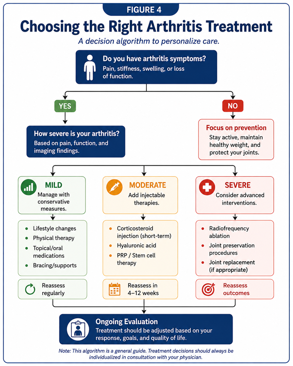
| Clinical Situation | Options That May Be Considered | Important Considerations |
|---|---|---|
| Early or mild symptomatic arthritis | Education, exercise, physical therapy, activity modification, topical medication | Look for reversible weakness, poor mechanics, tendon disease, or overuse |
| Painful inflammatory flare | Medication adjustment, aspiration when indicated, corticosteroid injection | Exclude infection or crystal disease when presentation is atypical |
| Persistent knee osteoarthritis pain | Corticosteroid, hyaluronic acid, PRP, diagnostic genicular blocks, knee RFA | Choice depends on inflammation, severity, goals, prior response, evidence, and coverage |
| Severe arthritis but poor surgical candidacy | Rehabilitation, medication optimization, assistive devices, injections, joint RFA | Treatment may focus on pain reduction and maintaining independence |
| Persistent pain after joint replacement | Diagnostic evaluation, rehabilitation, selected nerve blocks or RFA | First assess infection, loosening, fracture, instability, component problems, and referred pain |
| Progressive deformity or major functional loss | Orthopedic consultation for preservation surgery or joint replacement | Repeated temporary procedures should not postpone appropriate surgery indefinitely |
When Is Surgery Appropriate?
Non-surgical treatment is not automatically preferable simply because it is less invasive. Surgery may offer the most predictable functional improvement when arthritis has caused severe structural damage, deformity, instability, loss of motion, or persistent disability.
An orthopedic consultation may be appropriate when:
- Pain substantially limits basic daily activities despite reasonable non-surgical treatment
- The joint has progressive deformity or instability
- Walking, sleep, work, or personal care remains significantly impaired
- Imaging demonstrates advanced disease that matches the clinical symptoms
- Repeated injections provide inadequate or very short-lived relief
- The patient is medically suitable for surgery and understands the expected recovery
- A correctable structural problem is unlikely to respond to pain procedures alone
Joint replacement does not need to be delayed until a patient is completely disabled. Conversely, an abnormal X-ray alone does not mandate immediate surgery. Timing should reflect symptom severity, function, medical readiness, and informed patient preference.
What If Pain Continues After Joint Replacement?
Most patients undergo joint replacement to improve pain and function, but a minority continue to experience significant discomfort. Persistent pain should not be assumed to represent ordinary arthritis because the native joint surfaces have been replaced.
Potential causes may include infection, component loosening, fracture, instability, malalignment, tendon or bursal pain, scar sensitivity, nerve injury, complex regional pain syndrome, referred spinal pain, or persistent sensory nerve signaling.
Evaluation by the operating surgeon or another qualified orthopedic specialist may be required. When no correctable mechanical or infectious cause is identified, targeted nerve blocks and radiofrequency ablation may be considered for selected knee, shoulder, or hip replacement patients.
Preparing for an Arthritis Injection or Radiofrequency Procedure
Preparation varies according to the treatment. Patients should provide an accurate medication list and disclose anticoagulants, antiplatelet medication, diabetes, allergies, infection symptoms, recent antibiotic use, immune suppression, pregnancy, and any planned surgery.
- Do not stop prescribed blood-thinning medication unless instructed by the prescribing and procedural clinicians.
- Report fever, active infection, skin wounds, or antibiotics before the procedure.
- Discuss blood-sugar planning when corticosteroid will be used.
- Ask whether driving restrictions or transportation arrangements apply.
- Wear clothing that allows access to the treatment area.
- Clarify whether anti-inflammatory medication should be adjusted around PRP.
- Discuss the timing of injections when joint replacement is being considered.
Patients should receive procedure-specific instructions rather than relying solely on general information from an article.
Recovery After Arthritis Procedures
Most joint injections and diagnostic nerve blocks are outpatient procedures. Patients usually return home the same day. Recovery depends on the medication or procedure performed, the treated joint, and the patient’s baseline condition.
| Treatment | Early Recovery | When Improvement May Be Noticed |
|---|---|---|
| Corticosteroid Injection | Relative rest for the remainder of the day; temporary soreness possible | Often within several days |
| Hyaluronic Acid | Temporary fullness or soreness may occur | Often gradual over several weeks |
| PRP | A temporary inflammatory soreness is common; activity is advanced gradually | Often several weeks, with continued change over subsequent months |
| Diagnostic Nerve Block | Temporary numbness or soreness may occur | Assessed during the anesthetic window, often the same day |
| Joint RFA | Localized soreness, bruising, or temporary nerve irritation may occur | Often develops over several weeks |
Patients should follow the instructions given for their particular procedure. Increasing redness, drainage, fever, rapidly worsening swelling, new major weakness, loss of bowel or bladder control, chest pain, or shortness of breath requires prompt medical attention.
Risks, Limitations, and Reasons Treatment May Not Work
Every treatment has limitations. Even when the diagnosis and technical performance are appropriate, results vary.
General Procedural Risks
- Temporary pain or swelling
- Bruising or bleeding
- Infection
- Allergic or medication reaction
- Injury to a nearby nerve, vessel, tendon, or other structure
- Temporary numbness or weakness
- Failure to improve
- Recurrence of pain
Why Results Differ
- The treated structure may not be the main pain generator.
- Several pain generators may be present at the same time.
- Advanced deformity or instability may overwhelm the benefit of pain treatment.
- Muscle weakness and impaired movement may remain unaddressed.
- Inflammatory or neuropathic pain may coexist with mechanical pain.
- The patient may have a systemic condition affecting recovery.
- The biological preparation or technique may differ from that used in published studies.
- The patient’s activity after treatment may be either too limited or too aggressive.
- Expectations may exceed what the procedure can reasonably accomplish.
A treatment should be judged by meaningful improvement in function and quality of life, not by whether all pain disappears permanently.
An Individualized Approach to Arthritis Care
The same diagnosis can require different treatment in different people. A younger patient with focal post-traumatic cartilage injury may prioritize joint preservation and return to demanding activity. An older patient with advanced arthritis and significant medical risk may prioritize walking, sleep, and avoiding major surgery. A patient with persistent pain after joint replacement needs a careful search for prosthetic, mechanical, infectious, neurologic, and soft-tissue causes before additional treatment.
A thoughtful plan often combines several elements:
- A clear working diagnosis
- Realistic functional goals
- A sustainable exercise and strength program
- Medication selected around individual medical risk
- Precise procedures when a reasonable target exists
- Reassessment based on actual response
- Timely referral when surgical treatment offers a better path
Clinical Perspective
The most sophisticated treatment plan is not necessarily the one containing the greatest number of procedures. It is the plan that identifies the major pain generator, uses the least burdensome treatment likely to help, measures whether function improves, and changes direction when the evidence from the patient’s response says it should.
Frequently Asked Questions About Arthritis Treatment
Can arthritis be cured?
Most established osteoarthritis cannot currently be cured or reliably reversed. Treatment can nevertheless reduce pain, improve strength and mobility, control flares, and help patients remain active. Some contributing conditions, such as weakness, poor movement patterns, or associated tendon pain, may be substantially improved even when structural arthritis remains.
Does bone-on-bone arthritis always require joint replacement?
No. Treatment is based on symptoms, function, examination findings, imaging, health, and patient preference. Some patients with advanced imaging changes function reasonably well with non-surgical care. Others have severe disability and are more likely to benefit from replacement. “Bone-on-bone” describes imaging but does not determine treatment by itself.
Can exercise make arthritis worse?
Appropriately dosed exercise generally supports strength and function rather than accelerating routine osteoarthritis. The program may need modification for severe pain, instability, acute swelling, fracture, major tendon injury, or other specific problems. A gradual progression is usually better tolerated than sudden high-intensity activity.
Which arthritis injection is best?
There is no best injection for every patient. Corticosteroid may be useful when rapid anti-inflammatory treatment is needed. Hyaluronic acid is a non-steroid option used primarily for knee osteoarthritis. PRP may be considered for selected patients seeking an autologous biologic treatment. The joint, severity, previous response, medical risks, goals, cost, and available evidence all matter.
How long do arthritis injections last?
Duration varies widely. Some patients obtain little relief, while others improve for weeks or months. The type of injection, joint treated, arthritis severity, activity, and individual response influence the result. Prior response can be informative but does not guarantee the same outcome after a future injection.
How often can cortisone injections be repeated?
There is no single repeat interval appropriate for every joint and patient. The decision should consider the dose, joint, degree and duration of prior benefit, diabetes, tissue quality, surgical plans, and whether repeated injections are merely postponing a treatment more likely to restore function.
Does PRP regrow cartilage?
PRP should not be promised to regrow large areas of lost cartilage or restore an advanced joint to normal. Its practical role is to improve pain and function in appropriately selected patients by influencing the joint environment. Structural regeneration remains an area of continuing investigation.
Are gel shots the same as steroids?
No. Hyaluronic acid products do not contain corticosteroid. They have a different composition, mechanism, expected onset, and side-effect profile.
Does radiofrequency ablation damage the joint?
Joint RFA targets selected sensory nerve branches outside the primary joint surfaces. It does not remove cartilage or alter a prosthetic implant. It also does not repair the underlying arthritis. Accurate targeting and appropriate patient selection are important.
Will joint RFA make the leg or arm weak?
The intended targets are sensory articular branches rather than the major motor nerves controlling the limb. Temporary numbness, soreness, or unusual sensations can occur. Major weakness is not the desired effect and should be evaluated promptly if it develops.
Can RFA help after joint replacement?
It may help selected patients with persistent knee, shoulder, or hip pain after replacement. Before considering RFA, the joint should be evaluated for infection, implant loosening, fracture, instability, malalignment, tendon problems, and other potentially correctable causes.
Can I still have joint replacement after injections or RFA?
In most cases, these treatments do not eliminate the future option of joint replacement. Injection timing may matter because surgeons may require an interval between an intra-articular injection and elective surgery. Patients considering replacement should coordinate treatment with the orthopedic surgeon.
Can several arthritic joints be treated?
Yes, but the plan should remain practical and medically appropriate. Treating every abnormal joint simultaneously may make it difficult to evaluate response and may increase medication exposure, procedural burden, or cost. Prioritizing the joint causing the greatest functional limitation is often sensible.
When should I see a rheumatologist?
Rheumatology evaluation may be appropriate when multiple joints are inflamed, morning stiffness is prolonged, swelling is persistent, symptoms are symmetrical, inflammatory blood tests are abnormal, or there are associated features such as psoriasis, rash, eye inflammation, fever, or systemic illness.
When should I see an orthopedic surgeon?
An orthopedic consultation is reasonable when arthritis causes major functional impairment, progressive deformity, instability, severe loss of motion, or persistent pain despite appropriate non-surgical care. Consultation does not obligate the patient to undergo surgery; it provides information about whether a surgical option is suitable.
Key Takeaways
- Arthritis treatment should be based on symptoms, function, examination findings, imaging, medical health, and personal goals.
- Exercise, strength, weight management when relevant, and physical therapy remain foundational components of care.
- Corticosteroid, hyaluronic acid, and PRP injections have different purposes and should not be treated as interchangeable products.
- Joint radiofrequency ablation may reduce knee, shoulder, or hip pain by treating selected sensory nerve branches.
- RFA may also be considered for carefully selected patients with persistent pain after joint replacement once correctable causes have been evaluated.
- Joint preservation is a coordinated strategy, not a guarantee that replacement can or should always be avoided.
- Sometimes joint replacement is the most appropriate treatment, particularly when structural failure and disability outweigh the likely benefit of repeated temporary care.
Related Joint and Muscle Care Services
Joint Injections
An overview of image-guided injection options for painful arthritic joints.
Corticosteroid Joint Injections
How cortisone shots reduce inflammation, what patients may expect, and how repeat treatment is considered.
Hyaluronic Acid Joint Injections
A patient guide to viscosupplementation and gel shots for knee osteoarthritis.
PRP for Arthritis
How platelet-rich plasma is prepared and used for selected arthritic joints.
Joint Radiofrequency Ablation
An overview of sensory nerve treatment for chronic knee, shoulder, and hip joint pain.
Joint Preservation
Strategies intended to maintain mobility and native-joint function when clinically appropriate.
Percutaneous Needle Tenotomy
A minimally invasive approach for selected chronic tendon conditions that contribute to musculoskeletal pain.
Hydrodissection
An ultrasound-guided treatment for selected peripheral nerve entrapment conditions.
Botulinum Toxin Injections
Treatment using Botox®, Dysport®, and other selected formulations for appropriate muscle-spasm and pain conditions.
References
- Kolasinski SL, Neogi T, Hochberg MC, et al. 2019 American College of Rheumatology/Arthritis Foundation guideline for the management of osteoarthritis of the hand, hip, and knee. Arthritis Care & Research. 2020;72(2):149-162.
- American Academy of Orthopaedic Surgeons. Management of Osteoarthritis of the Knee (Non-Arthroplasty), Third Edition: Clinical Practice Guideline. Published August 31, 2021.
- Bannuru RR, Osani MC, Vaysbrot EE, et al. OARSI guidelines for the non-surgical management of knee, hip, and polyarticular osteoarthritis. Osteoarthritis and Cartilage. 2019;27(11):1578-1589.
- National Institute of Arthritis and Musculoskeletal and Skin Diseases. Osteoarthritis: overview, symptoms, causes, diagnosis, and treatment. National Institutes of Health.
- World Health Organization. Osteoarthritis fact sheet. Published July 14, 2023.
- American Academy of Orthopaedic Surgeons. Platelet-Rich Plasma for Knee Osteoarthritis Technology Overview.
- U.S. Food and Drug Administration. Important patient and consumer information about regenerative medicine therapies. Updated June 3, 2021.
- National Institute for Health and Care Excellence. Osteoarthritis in Over 16s: Diagnosis and Management. NICE Guideline NG226. Published October 2022.
- Hunter DJ, Bierma-Zeinstra S. Osteoarthritis. The Lancet. 2019;393(10182):1745-1759.
- Katz JN, Arant KR, Loeser RF. Diagnosis and treatment of hip and knee osteoarthritis: a review. JAMA. 2021;325(6):568-578.
- Chou R, McDonagh MS, Nakamoto E, Griffin J. Analgesics for osteoarthritis: an update of the 2006 comparative effectiveness review. Agency for Healthcare Research and Quality.
- Chen AF, Khalouf F, Zora K, et al. Cooled radiofrequency ablation provides extended clinical utility in the management of knee osteoarthritis: 12-month results from a prospective, multi-center, randomized, cross-over trial. BMC Musculoskeletal Disorders. 2020;21:363.
About This Guide
This resource is provided for educational purposes and should not replace personalized medical advice, diagnosis, or treatment from a qualified healthcare professional. Recommendations vary according to the joint involved, diagnosis, medical history, examination findings, imaging, and individual treatment goals.



