
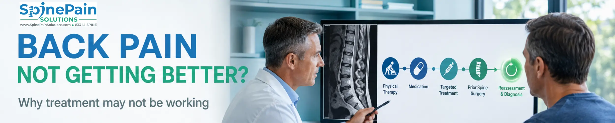
Why Is My Back Pain Not Getting Better?
Persistent pain after physical therapy, medication, an injection or spine surgery can be deeply frustrating. Patients may begin to wonder whether the MRI missed something, whether they performed therapy incorrectly or whether they simply have to live with the symptoms.
The more useful question is not only, “Why did the treatment fail?” It is:
What was the treatment intended to accomplish, what actually changed and does the current diagnosis still explain the symptoms?
A treatment may be technically successful yet fail to address the dominant pain generator. It may improve one component while another remains. Pain can also persist because nerve recovery is incomplete, rehabilitation is mismatched or the nervous system has become more sensitive over time.
What the Pattern of Persistent Pain May Suggest
| What happened? | Question to reconsider | Possible next step |
|---|---|---|
| Pain never changed with treatment | Was the correct structure or mechanism treated? | Reassess the diagnosis, examination and original imaging |
| Pain improved briefly and returned | Was the treatment temporary by design, or is the underlying process still active? | Use the duration and location of relief as diagnostic information |
| Back pain improved but leg pain remained | Is nerve compression or nerve injury still present? | Neurological examination, MRI review and possibly EMG |
| Leg pain improved but axial back pain remained | Was a separate facet, disc, endplate, SI-joint or muscular source present? | Evaluate the remaining pain as a distinct problem |
| Pain changed location or character | Has a new condition developed? | Repeat examination and selective updated testing |
| New weakness, balance difficulty or bowel/bladder symptoms | Is neurological function threatened? | Urgent or emergency evaluation |
Nine Reasons Back Pain May Not Be Getting Better
1. The Original Diagnosis Was Incorrect or Incomplete
“Back pain” describes a symptom, not one disease. Pain may arise from a spinal nerve, facet joint, intervertebral disc, vertebral endplate, sacroiliac joint, muscle, ligament, compression fracture or another structure.
Hip arthritis, peripheral neuropathy, cluneal-nerve entrapment, vascular disease and other conditions can also resemble spinal pain.
Treatment aimed at the wrong structure may produce no improvement even when it is performed correctly. Read how doctors approach diagnosing back pain and identifying the pain generator.
2. An Incidental MRI Finding Was Treated
Disc bulges, degeneration, facet arthritis and even stenosis are common in people who have no pain. An MRI abnormality becomes clinically persuasive only when it matches:
- The location and character of the symptoms
- The correct side and spinal level
- The neurological or mechanical examination
- The patient’s functional limitation
- The response to appropriately selected treatment
The most dramatic phrase in a radiology report is not always the pain generator. Conversely, an MRI may appear relatively unimpressive while the patient has meaningful pain arising from vertebral endplates, a peripheral nerve, the SI joint or a process that routine imaging cannot fully characterize.
Learn more about why pain and MRI findings do not always match.
3. Physical Therapy Did Not Match the Pain Generator
Physical therapy is not one standardized treatment. A program for disc-related radicular pain should not necessarily look like rehabilitation for spinal stenosis, facet pain, instability, osteoporosis or postsurgical deconditioning.
Therapy may provide limited benefit when:
- The exercises repeatedly provoke the dominant pain mechanism
- The program is too generic for the patient’s functional demands
- The initial load or progression is excessive
- The program never progresses enough to restore strength and tolerance
- Hip, gait, balance or peripheral-nerve problems are overlooked
- Severe structural compression prevents meaningful progress
- Pain causes the patient to avoid enough movement that rehabilitation cannot advance
Temporary muscular soreness after beginning or advancing exercise is not the same as worsening neurological pain. New radiating pain, progressive weakness or steadily declining function should prompt reassessment rather than automatic continuation of the same program.
4. Medication Was Not Directed at the Correct Pain Mechanism
Different medications address different processes. An anti-inflammatory medication, muscle relaxant and neuropathic-pain medication are not interchangeable.
Medication may reduce symptoms enough to support sleep, movement and rehabilitation, but it cannot:
- Remove a recurrent disc fragment
- Mechanically enlarge a severely narrowed spinal canal
- Stabilize an unstable spinal segment
- Reverse every form of advanced nerve injury
- Determine which of several MRI abnormalities is painful
A lack of response to one medication does not establish that the pain is untreatable. It may mean that the medication, dose, duration or presumed pain mechanism was incorrect—or that medication alone was never capable of correcting the underlying problem.
5. Nerve Compression or Injury Remains Active
A spinal nerve may remain compressed by a disc herniation, foraminal narrowing, lateral-recess stenosis, a synovial cyst or another structural lesion. Inflammation may improve while fixed mechanical compression remains.
Longstanding compression can also injure a nerve. Removing the compression does not make an injured nerve recover instantly. Pain may improve before numbness or weakness, and some neurological deficits may recover incompletely.
Persistent or progressive weakness, loss of coordination, worsening walking tolerance or increasing numbness deserves a repeat neurological examination.
6. More Than One Pain Generator Is Present
A patient may simultaneously have:
- Spinal stenosis causing leg heaviness while walking
- Facet arthritis causing extension-related back pain
- Vertebrogenic or disc-related axial pain
- Sacroiliac-joint pain
- Hip arthritis
- Peripheral neuropathy
- Muscular deconditioning or myofascial pain
Treating one problem may produce a real but incomplete improvement. This should not automatically be called failure. The residual symptoms should be mapped again rather than assuming that repeating the same treatment will address everything.
7. Pain Signaling Has Become More Sensitive
Persistent nerve or tissue irritation can alter pain processing. Neuropathic pain may continue after the original injury because the affected nerve remains damaged or abnormally excitable. In some patients, central pain amplification may also contribute.
Features that can raise this possibility include:
- Burning, electrical or shock-like pain
- Pain from light touch or normally nonpainful movement
- Symptoms spreading beyond one clear anatomical structure
- Disproportionate and prolonged flares after modest activity
- Sleep disruption, fatigue and reduced activity tolerance accompanying persistent pain
Central sensitization does not mean the pain is imaginary. It also should not become a label applied whenever imaging is inconclusive. Structural, neurological, inflammatory and non-spinal causes still require appropriate evaluation.
Management may need to combine graded rehabilitation, sleep and activity restoration, pain education, psychological strategies and appropriately selected medication or procedures rather than repeatedly treating a single structure.
8. The Treatment May Need More Time
Some treatments work quickly; others do not. Judgment should depend on the procedure, diagnosis and expected biological recovery.
| Treatment or recovery process | Why improvement may not be immediate | What should be monitored |
|---|---|---|
| Physical therapy | Strength, movement tolerance and confidence develop progressively | Function, activity tolerance and direction of change—not soreness alone |
| Epidural injection | The local anesthetic and anti-inflammatory medication have different timelines | Change in radiating pain, sleep, walking and rehabilitation tolerance |
| Radiofrequency ablation | Temporary post-procedure soreness or neuritis may precede benefit | Whether the familiar facet-related pain gradually declines |
| Nerve decompression | An injured nerve may recover slowly after pressure is relieved | Pain, sensation, strength and neurological trajectory |
| Fusion or major reconstruction | Bone healing, muscular recovery and conditioning require time | Alignment, stability, neurological function and progressive rehabilitation |
Time should not be used as an excuse to ignore deterioration. A patient who is gradually increasing activity is different from a patient developing progressive weakness or steadily worsening pain.
9. A Persistent, Recurrent or New Problem Developed After Surgery
Pain after spine surgery is sometimes called failed back surgery syndrome. The more precise term is persistent spinal pain syndrome type 2. It avoids assuming that the operation itself “failed” before the cause of the current symptoms is understood.
Potential explanations include:
- The original pain generator was not fully identified before surgery
- The operation corrected one problem but not another simultaneous source
- Nerve compression was incompletely relieved
- A nerve sustained advanced injury before decompression
- A disc herniated again at the operated level
- Epidural fibrosis developed around a nerve root
- An adjacent spinal level became symptomatic
- Instability, pseudarthrosis or a hardware-related problem developed
- Hip, SI-joint, peripheral-nerve or muscular pain was mistaken for recurrent spinal disease
Recurrent Disc Herniation
A disc can herniate again after discectomy. Recurrent radiating leg pain after an interval of meaningful improvement raises a different question from pain that never changed after surgery. Updated MRI—sometimes with contrast depending on the timing and clinical question—may help distinguish recurrent disc material from postoperative scar.
Epidural Fibrosis
Scar formation is part of normal healing after surgery. Its presence on imaging does not automatically prove that it causes pain.
Epidural fibrosis becomes more clinically relevant when scar surrounds or tethers a nerve root and the location matches persistent radicular symptoms. Even then, the complete pattern—not the word “scar” in isolation—should guide treatment.
Adjacent-Segment Degeneration Versus Disease
After fusion, the levels above or below may show increasing degeneration over time. However, adjacent-segment degeneration is an imaging finding, while adjacent-segment disease means the new degeneration is producing corresponding symptoms or neurological effects.
This distinction matters because not every worn adjacent disc requires another operation.
Instability, Pseudarthrosis or Hardware Problems
Persistent mechanical pain after fusion may occasionally relate to failure of solid fusion, abnormal movement, hardware loosening or another alignment problem. CT, standing radiographs or flexion-extension imaging may answer questions that a routine supine MRI does not.
Review the broader guide to conditions that can occur after spine surgery.
When Should the Diagnosis Be Reconsidered?
Reassessment is reasonable when:
- Appropriately selected treatment produced no meaningful change
- The response was opposite to what the diagnosis predicted
- Symptoms changed location, character or severity
- New numbness, weakness or gait difficulty developed
- The MRI abnormality does not match the side or pattern of pain
- Several treatments aimed at the same structure repeatedly failed
- Pain persists after surgery without a clear explanation
- Hip, SI-joint, peripheral-nerve or systemic disease has not been considered
Reconsidering the diagnosis does not mean starting from zero. The response—or lack of response—to previous care is itself useful evidence.
When Additional Testing May Help
Testing should answer a focused question. Repeating every test without a clear decision attached to the result can create more incidental findings without clarifying the diagnosis.
Repeat MRI or Other Imaging
Repeat imaging may be appropriate when:
- Symptoms or neurological findings have materially changed
- Weakness or walking ability is worsening
- A new injury occurred
- Infection, fracture, tumor or another serious process is suspected
- Prior surgery has changed the anatomy
- A procedure or surgical decision requires updated information
MRI is not always the only useful study. CT may better evaluate bone, fusion and hardware. Standing or flexion-extension X-rays may reveal alignment or instability that is not apparent while lying down.
EMG and Nerve-Conduction Testing
Electrodiagnostic testing may help distinguish:
- Radiculopathy from peripheral neuropathy
- Lumbar nerve-root disease from peroneal neuropathy
- Cervical radiculopathy from carpal-tunnel or ulnar neuropathy
- Active nerve injury from certain chronic or non-neurological symptoms
EMG complements imaging; it does not replace it. It may be normal in early or sensory-predominant radiculopathy and does not diagnose most disc, facet, SI-joint or muscular pain.
Diagnostic Injections
A selective nerve-root block, medial branch block, sacroiliac-joint injection or peripheral-joint injection may provide additional evidence when several sources remain plausible.
The procedure should be chosen to answer a specific question, performed with appropriate imaging guidance and interpreted alongside the entire clinical picture. Review the limitations of diagnostic injections for identifying a pain generator.
Laboratory or Bone-Health Evaluation
Laboratory testing may be appropriate when infection, inflammatory disease, metabolic bone disease or another systemic process is possible. Persistent pain after a compression fracture should also trigger attention to osteoporosis and future-fracture prevention rather than focusing only on the painful vertebra.
When Another Procedure May Be Appropriate
Another procedure should not be selected merely because the previous one failed. It becomes reasonable when reassessment identifies a better-supported target.
Depending on the diagnosis, options may include:
- A targeted epidural injection for matching radicular inflammation
- Medial branch blocks and radiofrequency ablation for confirmed facet pain
- Basivertebral nerve ablation for selected vertebrogenic pain with qualifying clinical and MRI findings
- Minimally invasive or endoscopic decompression for selected structural nerve compression
- SI-joint treatment for appropriately confirmed SI-joint pain
- Neuromodulation for selected persistent neuropathic or postsurgical pain
- Revision surgery when a surgically correctable structural problem is present
A more invasive treatment is not automatically more effective. The value of the next procedure depends on whether it addresses the reason the previous treatment did not work.
Read the complete guide to treatment options before spine surgery.
When a Surgical Opinion Is Appropriate
A surgical opinion does not commit a patient to surgery. It may clarify whether continued nonsurgical care is reasonable or whether delay could compromise neurological recovery.
A consultation becomes particularly important when:
- Weakness is progressive
- Foot drop or declining walking ability develops
- Spinal-cord or cauda-equina symptoms are suspected
- Severe nerve compression matches the neurological deficit
- Instability, deformity, pseudarthrosis or hardware failure is suspected
- Disabling radicular pain persists despite reasonable nonsurgical treatment
- A recurrent disc herniation or adjacent-segment disease is clinically significant
Another opinion is also reasonable before a major fusion when the dominant pain generator remains uncertain or the proposed operation does not clearly match the patient’s principal symptoms.
What to Bring for a Second Spine Evaluation
- Actual MRI, CT and X-ray images—not only the written reports
- Prior operative and procedure reports
- A timeline of symptoms before and after each treatment
- Physical-therapy programs and exercises attempted
- Medication history, including benefits and side effects
- Injection records and the duration and location of relief
- EMG and nerve-conduction results
- A description of which activities remain limited
The pattern of response often provides more diagnostic information than a simple list stating that treatment “failed.”
Frequently Asked Questions
Why is my back pain not getting better after physical therapy?
Physical therapy may not help when the rehabilitation program does not match the pain generator, progression or exercise dose is inappropriate, nerve compression remains active, several pain sources are present or the original diagnosis needs reconsideration. It may also be too early to judge a properly designed program.
How long should physical therapy take to help back pain?
There is no universal timetable. Some patients notice functional improvement within several weeks, while chronic, postsurgical or neurologically complicated conditions may require longer. The more important question is whether pain, mobility, strength or activity tolerance is showing a measurable trend in the right direction.
Why does back pain continue despite medication?
Medication may reduce inflammation, muscle spasm or pain signaling, but it cannot mechanically decompress a severely compressed nerve, stabilize an unstable segment or correct every structural pain source. An ineffective medication may also have been directed at the wrong pain mechanism.
Can an MRI miss the cause of persistent back pain?
Yes. A routine MRI may not fully demonstrate dynamic instability, certain peripheral-nerve disorders, muscular or connective-tissue pain, some SI-joint or hip conditions, central sensitization or which visible abnormality is actually painful. Imaging must be interpreted with the symptoms and examination.
Why can back or leg pain persist after spine surgery?
Possible explanations include incomplete or recurrent nerve compression, longstanding nerve injury, recurrent disc herniation, epidural fibrosis, adjacent-segment disease, instability, pseudarthrosis, hardware problems, another untreated pain generator or altered pain processing. Persistent pain does not necessarily mean that the operation was performed incorrectly.
When should an MRI be repeated for persistent back pain?
Repeat imaging becomes more useful when symptoms have materially changed, weakness or other neurological findings are progressing, significant trauma has occurred, infection or tumor is suspected, prior surgery changes the diagnostic question or the result would guide a procedure or surgical decision.
Can EMG identify why back pain is not improving?
EMG and nerve-conduction testing may help identify radiculopathy, peripheral neuropathy or a peripheral-nerve entrapment when symptoms include weakness, numbness or radiating limb pain. EMG is not a general test for facet, disc, sacroiliac or muscular back pain and can be normal in sensory-predominant radiculopathy.
Does persistent pain mean that treatment failed?
Not always. A treatment may have improved one component while another pain generator remains, or healing and nerve recovery may still be occurring. The response should be evaluated by changes in function, pain pattern, neurological status and duration rather than by pain intensity alone.
When should I obtain another spine or surgical opinion?
Another opinion is reasonable when the diagnosis remains uncertain, symptoms and imaging do not match, appropriately selected care has not produced meaningful progress, pain recurs after surgery or a major operation is being proposed. Progressive weakness, bowel or bladder changes and spinal-cord symptoms require urgent evaluation.
Evaluation for Persistent Back Pain on Long Island
Amit Sharma, MD evaluates persistent back, neck, joint and nerve pain after physical therapy, medication, procedures or previous spine surgery.
The objective is not to repeat treatments automatically. It is to determine what the previous response reveals, whether the original diagnosis remains persuasive and whether rehabilitation, a targeted procedure, minimally invasive treatment or surgical consultation is now appropriate.
Learn more about evaluation by an interventional spine specialist on Long Island or review our locations across Nassau and Suffolk Counties.
References
- American College of Radiology. ACR Appropriateness Criteria®: Low Back Pain. Read source →
- Brinjikji W, et al. Systematic Literature Review of Imaging Features of Spinal Degeneration in Asymptomatic Populations. AJNR Am J Neuroradiol. 2015;36(4):811-816. Read source →
- Hayden JA, et al. Exercise Therapy for Chronic Low Back Pain. Cochrane Database Syst Rev. 2021;9:CD009790. Read source →
- Briggs AM, et al. The World Health Organization Guideline for Nonsurgical Management of Chronic Primary Low Back Pain in Adults. Read source →
- Schuttert I, et al. The Definition, Assessment and Prevalence of Human-Assumed Central Sensitisation in Patients With Chronic Low Back Pain: A Systematic Review. J Clin Med. 2021;10(24):5931. Read source →
- Dillingham TR, et al. Evaluation of Persons With Suspected Lumbosacral and Cervical Radiculopathy: Electrodiagnostic Assessment. Muscle Nerve. 2020. Read source →
- Christelis N, et al. Persistent Spinal Pain Syndrome: A Proposal for Failed Back Surgery Syndrome and ICD-11. Pain Med. 2021;22(4):807-818. Read source →
- Wu Q, et al. Chronic Pain After Spine Surgery: Insights Into Pathogenesis, New Treatment and Preventive Therapy. J Orthop Translat. 2023. Read source →
- Cannizzaro D, et al. Lumbar Adjacent-Segment Degeneration After Spinal Fusion Surgery: A Systematic Review and Meta-Analysis. J Neurosurg Sci. 2023;67(6):740-749. Read source →
Medical content reviewed by Amit Sharma, MD. This page is educational and does not replace an individualized medical evaluation. New or rapidly progressive neurological symptoms require urgent medical attention.
Dr. Amit Sharma & our minimally invasive pain & spine team.



