
Knee Radiofrequency Ablation (Genicular Nerve Ablation)
Chronic knee pain can make ordinary activities such as walking, climbing stairs, standing from a chair, or sleeping comfortably increasingly difficult. For many people, the pain is caused by knee osteoarthritis. Others continue to experience pain after knee replacement surgery or despite medications, physical therapy, activity modification, and injections.
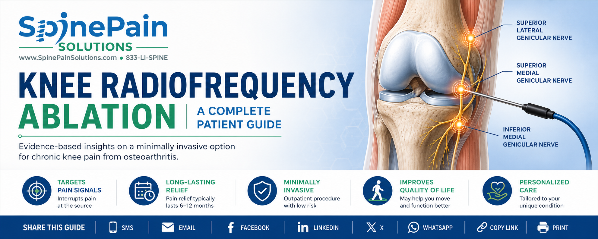
When these treatments no longer provide adequate relief, knee radiofrequency ablation may offer another option. Knee radiofrequency ablation, also called genicular nerve radiofrequency ablation or genicular nerve ablation, is a minimally invasive procedure designed to reduce pain signals traveling from the knee to the brain.
The procedure does not remove arthritis, rebuild cartilage, or repair the knee joint. Instead, it targets selected sensory nerves around the knee that carry pain information. For appropriately selected patients, reducing these pain signals may improve comfort, mobility, and the ability to participate in everyday activities.
This guide explains why chronic knee pain develops, how the genicular nerves contribute to pain, how diagnostic nerve blocks help determine whether radiofrequency ablation may be appropriate, what happens during the procedure, and what patients should realistically expect afterward.
Clinical Perspective
Radiofrequency ablation is not performed to “burn the knee.” The treatment is directed toward small sensory nerves outside the joint that transmit pain signals. It does not remove arthritis or damage the knee joint. Understanding this distinction is important when deciding whether the procedure fits a patient’s diagnosis and treatment goals.
Quick Answer
Knee radiofrequency ablation is a minimally invasive treatment for selected patients with chronic knee pain. It uses controlled radiofrequency energy to reduce pain signals carried by small sensory nerves around the knee, commonly called the genicular nerves.
Who may benefit?
- Patients with chronic knee pain from osteoarthritis
- Patients trying to delay or avoid knee replacement surgery
- Patients who may not be good surgical candidates
- Patients with persistent pain after knee replacement
- Patients whose pain temporarily improves after diagnostic genicular nerve blocks
What are the potential advantages?
- Performed as an outpatient procedure
- No large surgical incision
- Usually requires only a brief recovery period
- May provide longer relief than a diagnostic nerve block
- May be repeated when clinically appropriate
- Does not prevent a future knee replacement if surgery later becomes necessary
Important: Knee radiofrequency ablation treats pain signals. It does not regrow cartilage, reverse arthritis, repair a torn meniscus, correct joint instability, or replace the need for surgery when a major structural problem requires surgical treatment.
Understanding Chronic Knee Pain
The knee is the largest joint in the body and plays a vital role in nearly every movement involving the legs. Walking, climbing stairs, standing from a chair, kneeling, and maintaining balance all depend on healthy knee function. Because the knee bears much of the body’s weight while allowing a wide range of motion, it is particularly vulnerable to injury, arthritis, and age-related wear.
Many people assume knee pain simply means “bone rubbing on bone,” but the reality is more complex. The knee contains cartilage, bone, ligaments, tendons, the joint lining (synovium), and numerous small sensory nerves. Pain may arise from one or several of these structures, which is why two people with similar X-rays can experience very different levels of discomfort.
Clinical Perspective
One of the most surprising things I explain to patients is that an X-ray cannot measure pain. Some people have severe arthritis with relatively little discomfort, while others experience significant pain despite only mild arthritic changes. Successful treatment depends on identifying the true source of pain rather than treating an image alone.
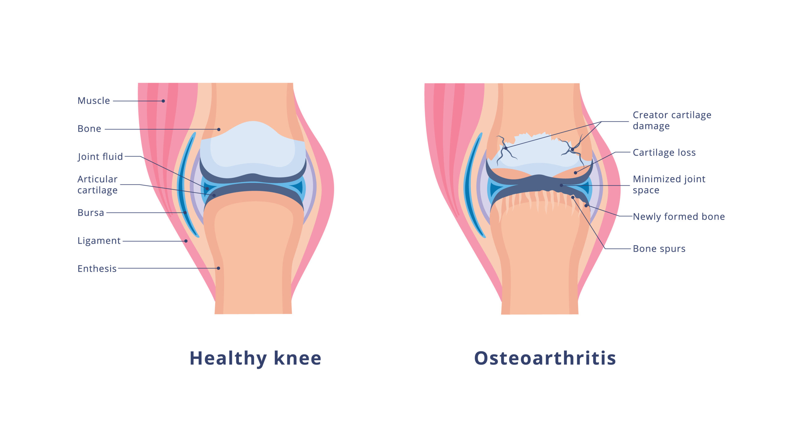
A healthy knee contains smooth articular cartilage that cushions the ends of the bones and allows the joint to move with minimal friction. As osteoarthritis progresses, cartilage gradually wears away, the joint space narrows, bone spurs may develop, and inflammation of surrounding tissues contributes to pain, stiffness, and reduced mobility. Although these structural changes occur within the joint, much of the pain is transmitted by small sensory nerves surrounding the knee that become the target of knee radiofrequency ablation.
Common Causes of Chronic Knee Pain
Several conditions may contribute to chronic knee pain, including:
- Osteoarthritis – the most common cause of chronic knee pain, resulting from gradual cartilage degeneration.
- Persistent pain after knee replacement – discomfort that continues despite technically successful joint replacement surgery.
- Synovitis – inflammation of the joint lining.
- Degenerative meniscal tears, particularly in middle-aged and older adults.
- Tendon disorders, including quadriceps and patellar tendinopathy.
- Referred pain originating from the hip or lumbar spine.
Key Point: Cartilage itself contains very few pain fibers. Much of the pain associated with knee osteoarthritis arises from inflammation and irritation of the tissues surrounding the joint. These tissues communicate with the brain through small sensory nerves called the genicular nerves, which are the primary targets of knee radiofrequency ablation.
How Knee Osteoarthritis Causes Pain
Many people believe that arthritis pain comes directly from worn-out cartilage. Surprisingly, cartilage itself contains very few pain fibers. Instead, the discomfort associated with knee osteoarthritis arises from inflammation and irritation of the tissues surrounding the joint.
As cartilage gradually breaks down, the body responds with inflammation. The joint lining (synovium) may become irritated, the bone beneath the cartilage experiences increased stress, and the joint capsule, ligaments, and surrounding soft tissues become increasingly sensitive. Together, these changes activate small sensory nerves that constantly send pain signals to the brain.
These nerves are known as the genicular nerves. They wrap around the knee joint like a network of telephone wires, carrying pain information from the arthritic knee to the spinal cord and ultimately the brain. Although they do not cause arthritis, they are responsible for transmitting much of the pain associated with it.
Clinical Perspective
One of the most common questions I hear is, “If my arthritis is permanent, why would treating a nerve help?” The answer is that radiofrequency ablation does not repair the arthritic joint. Instead, it interrupts the pain signals traveling through the genicular nerves, allowing many patients to move more comfortably despite the underlying arthritis remaining present.
Key Point: Knee radiofrequency ablation targets the pain messenger, not the arthritis itself. The goal is to reduce pain and improve function while preserving future treatment options such as knee replacement if they become necessary.
What Is Knee Radiofrequency Ablation?
Knee radiofrequency ablation, also called genicular nerve radiofrequency ablation or genicular nerve ablation, is a minimally invasive treatment for certain types of chronic knee pain. The procedure uses controlled radiofrequency energy to reduce the ability of selected sensory nerves around the knee to transmit pain signals.
The treatment does not remove the nerves, repair damaged cartilage, or reverse osteoarthritis. Instead, it creates a small and carefully controlled area of thermal injury along the targeted nerve. This temporarily disrupts pain transmission while preserving the muscles, ligaments, and major nerves responsible for strength and movement.
Because genicular nerves are primarily sensory, treating them does not normally weaken the knee. Patients remain able to walk, bend the knee, and participate in rehabilitation or exercise according to their underlying joint condition.
Key Point: Knee radiofrequency ablation is a treatment for chronic pain arising from the knee. It does not replace cartilage or cure arthritis. Its purpose is to reduce pain sufficiently to improve walking, sleep, exercise tolerance, and participation in physical therapy.
How the Treatment Works
During the procedure, the physician places specialized radiofrequency needles near selected genicular nerve targets. Live X-ray guidance, called fluoroscopy, is commonly used to identify the bony landmarks near which these nerves travel.
Once the needle position has been confirmed, sensory and sometimes motor stimulation may be performed. This helps confirm that the needle is appropriately positioned near a pain-carrying sensory nerve and away from a nerve responsible for muscle movement.
Local anesthetic is then administered, and controlled radiofrequency energy is applied. The energy heats a small area around the tip of the needle, creating a treatment zone along the targeted nerve. Pain relief is not always immediate because the treated nerve may remain irritated for several days before its ability to transmit pain gradually decreases.
Clinical Perspective
Patients sometimes imagine that radiofrequency ablation burns a large area of the knee. In reality, the treatment is focused around the tip of a carefully positioned needle. The objective is to create a controlled treatment zone along a small sensory nerve while avoiding the structures responsible for strength, balance, and joint stability.
Types of Radiofrequency Treatment
Several radiofrequency techniques may be used to treat genicular nerves. The underlying goal is similar, but the shape, size, and temperature characteristics of the treatment zone may differ.
- Conventional radiofrequency ablation creates a focused thermal lesion near the active tip of the radiofrequency needle.
- Cooled radiofrequency ablation circulates fluid through the probe, allowing the system to create a larger treatment zone while controlling tissue temperature near the electrode.
- Bipolar or multipolar radiofrequency techniques use more than one electrode to create a treatment zone between or around the needles.
No single technology is automatically best for every patient. Outcomes depend on several factors, including correct diagnosis, appropriate candidate selection, the nerves chosen for treatment, needle placement, lesion size, and the patient’s underlying knee condition.
Treatment Tip: Patients should focus less on a particular brand name and more on whether the physician has identified the correct pain source, performed an appropriate diagnostic evaluation, and uses careful imaging-guided technique.
Who May Be a Candidate for Knee Radiofrequency Ablation?
Knee radiofrequency ablation is generally considered for people with chronic knee pain that has not improved sufficiently with appropriate conservative treatment. It is not intended for every type of knee pain, and the presence of arthritis on an X-ray alone does not establish that the procedure will help.
The evaluation begins by determining whether the pain is actually arising from the knee joint and whether sensory nerves around the knee are carrying a meaningful portion of that pain. Symptoms, physical examination findings, imaging studies, previous treatments, and the patient’s overall health must all be considered together.
Common Reasons to Consider Knee RFA
A patient may be evaluated for knee radiofrequency ablation when one or more of the following circumstances are present:
- Chronic knee osteoarthritis pain that continues despite treatments such as activity modification, physical therapy, weight management, medications, bracing, or joint injections.
- Persistent pain after knee replacement when the prosthetic joint has been appropriately evaluated and no correctable surgical or mechanical problem has been identified.
- Medical conditions that increase surgical risk or make knee replacement unsuitable.
- A desire to postpone knee replacement while continuing nonsurgical treatment.
- Continued pain while awaiting surgery when temporary improvement in comfort and function may be helpful.
- Previous benefit from a diagnostic genicular nerve block, suggesting that the targeted sensory nerves contribute to the patient’s pain.
Clinical Perspective
The best candidate is not simply the person with the worst-looking X-ray. I look for a logical relationship among the patient’s symptoms, examination, imaging, previous treatment response, and diagnostic block. Radiofrequency ablation is most useful when we have reasonable evidence that the targeted nerves are carrying pain from the knee.
Persistent Pain After Knee Replacement
Most patients undergo knee replacement to reduce severe arthritic pain and restore function. However, some continue to experience pain after surgery. Radiofrequency ablation may be considered in selected patients, but only after appropriate evaluation of the replaced joint.
Possible causes such as infection, fracture, implant loosening, instability, malalignment, tendon problems, or another surgically correctable condition must first be considered. Genicular nerve ablation may reduce pain transmission, but it cannot correct a loose implant, infection, or mechanical failure.
Important: New or worsening pain after knee replacement should not automatically be treated as nerve pain. Increasing swelling, redness, warmth, drainage, fever, instability, or a sudden change in function requires prompt medical evaluation.
When Knee RFA May Not Be Appropriate
Knee radiofrequency ablation may not be appropriate when the diagnosis is uncertain, the pain is primarily coming from another structure, or the risks of the procedure outweigh the expected benefit.
Examples include:
- An active infection involving the skin, soft tissues, bloodstream, or knee joint.
- An untreated bleeding disorder or use of certain blood-thinning medications that cannot be safely managed around the procedure.
- A fracture, unstable joint, or mechanical problem requiring orthopedic treatment.
- Pain arising primarily from the hip, lumbar spine, peripheral nerves, tendons, or another condition outside the knee joint.
- Severe uncontrolled medical illness that makes an elective procedure unsafe.
- A diagnostic block that does not produce meaningful temporary improvement when a prognostic block is used as part of the selection process.
Key Point: Radiofrequency ablation should not be used to conceal pain from an untreated infection, fracture, unstable joint, or failed knee implant. The underlying diagnosis must come first.
The Diagnostic Genicular Nerve Block
Before radiofrequency ablation, many physicians perform a diagnostic or prognostic genicular nerve block. A small amount of local anesthetic is placed near selected genicular nerves using fluoroscopic or ultrasound guidance.
The purpose of the block is not to provide permanent treatment. It temporarily numbs the targeted sensory nerves so the patient and physician can evaluate whether those nerves appear to be carrying a meaningful portion of the knee pain.
What Happens During the Block?
- The patient identifies the usual pain level and which activities normally reproduce the pain.
- The skin is cleaned, and a small amount of local anesthetic may be used to numb the needle-entry sites.
- Using imaging guidance, thin needles are positioned near the selected genicular nerve targets.
- A small volume of local anesthetic is injected around each target.
- Afterward, the patient performs safe activities that would ordinarily provoke the knee pain, such as walking, standing, or climbing a few steps.
- The patient records the amount and duration of pain relief as the anesthetic takes effect and then wears off.
Treatment Tip: Do not spend the entire diagnostic period resting if rest is not what normally triggers the pain. Within the physician’s safety instructions, gently test the activities that usually hurt so the response to the block can be meaningfully assessed.
What Is Considered a Positive Block?
A block is considered helpful when it produces a clear and temporary reduction in the patient’s usual knee pain and improves the ability to perform activities that were painful before the injection.
The exact percentage of relief required before proceeding to radiofrequency ablation varies among physicians, research protocols, and insurance plans. Some use a threshold of at least 50 percent relief, while others look for a more substantial response. The quality of improvement is also important: walking farther, standing more comfortably, or climbing stairs with less pain may be more informative than a number recorded while resting.
Clinical Perspective
I do not evaluate a diagnostic block by asking only, “What percentage better are you?” I also want to know what the patient was able to do differently. A meaningful response should make sense in the context of the patient’s usual pain pattern and functional limitations.
Does a Successful Block Guarantee RFA Relief?
No. A diagnostic block is a screening tool, not a promise. The temporary anesthetic injection and radiofrequency ablation are different procedures, and several factors influence the eventual result.
The anesthetic may spread beyond the intended nerve, individual nerve anatomy varies, and not every painful structure within the knee is supplied by the same nerve branches. Conversely, an imperfect block does not always mean the nerves are irrelevant. The result must be interpreted together with the complete clinical evaluation.
Key Point: The diagnostic block improves patient selection, but it cannot predict the result with certainty. Knee radiofrequency ablation remains a treatment trial based on the best available clinical evidence for that individual patient.
How Knee Radiofrequency Ablation Is Performed
Knee radiofrequency ablation is typically performed as an outpatient procedure. The patient is positioned so the physician can obtain clear imaging of the knee and safely reach the selected genicular nerve targets.
The exact number and location of the nerves treated may vary. Traditional techniques commonly target three genicular nerves, while expanded approaches may include additional sensory branches based on the patient’s pain pattern, anatomy, previous surgery, and the physician’s treatment strategy.
Before the Procedure
Before treatment, the physician reviews the patient’s medical history, medications, allergies, imaging studies, response to previous treatment, and the results of any diagnostic genicular nerve block.
Patients should provide a complete list of medications and supplements, particularly blood thinners, antiplatelet medications, diabetes medications, and drugs that may affect sedation. Medication changes should be made only under the direction of the prescribing physician and the procedural team.
Important: Do not stop a prescribed blood thinner on your own. The risk of bleeding must be balanced against the medical reason the medication was prescribed. Instructions should be individualized by the physicians managing your care.
Positioning and Imaging Guidance
The patient usually lies on the back with the treated knee supported in a comfortable position. The skin is cleaned with antiseptic solution, and sterile drapes are placed around the procedure area.
Fluoroscopy provides live X-ray images of the knee from more than one direction. These views allow the physician to identify the femur, tibia, joint line, and other bony landmarks near which the targeted sensory nerves travel.
Because the nerves themselves are usually too small to be seen directly on an X-ray, the needles are positioned using their known relationship to these anatomical landmarks. A second view is then obtained to confirm the depth of each needle.
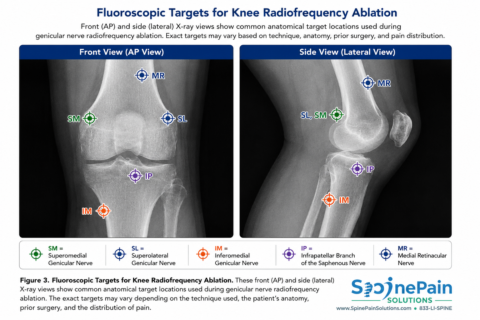
Front and side X-ray views help the physician position each radiofrequency needle near selected bony landmarks where sensory nerves travel. The exact targets may vary depending on the technique used, the patient’s anatomy, prior surgery, and the distribution of pain.
Placing the Radiofrequency Needles
A small amount of local anesthetic is used to numb the skin and deeper tissues at each needle-entry site. Specialized radiofrequency needles are then advanced toward the selected targets under continuous or intermittent imaging guidance.
The physician adjusts each needle until its position is satisfactory in both the front and side imaging views. This two-view confirmation is important because an apparently correct position in one view may still be too shallow, too deep, or displaced when viewed from another angle.
Clinical Perspective
The fluoroscopic landmarks are a map, not the nerve itself. Genicular nerve anatomy varies from person to person, which is one reason careful positioning, appropriate treatment-zone size, and thoughtful target selection matter as much as the radiofrequency technology being used.
Sensory and Motor Testing
After the needles are positioned, electrical stimulation may be used to further evaluate their relationship to nearby nerves.
- Sensory stimulation may produce tingling, pressure, buzzing, or a familiar sensation near the knee. This can help support that the needle is near a sensory target.
- Motor stimulation is used to look for unintended muscle contraction. The absence of concerning motor activity helps reduce the likelihood that the needle is positioned near a major motor nerve.
Testing protocols differ among physicians and radiofrequency techniques. Imaging position, anatomical knowledge, stimulation findings, and the complete clinical picture are considered together.
Creating the Radiofrequency Treatment Zone
Once the needle positions are confirmed, local anesthetic is administered through the radiofrequency needles. The radiofrequency generator then delivers controlled energy through the active portion of each probe.
This energy creates a small treatment zone around the needle tip. The goal is to reduce the targeted nerve’s ability to transmit pain while limiting the effect on surrounding tissue.
Treatment temperature, duration, needle configuration, probe type, and lesion size depend on the radiofrequency technique being used. Conventional, cooled, bipolar, and other approaches create treatment zones with different shapes and dimensions.
Key Point: The procedure does not remove the nerve or place an implant inside the knee. It creates a controlled treatment zone along selected sensory nerve branches to reduce pain transmission.
What Does the Procedure Feel Like?
Patients commonly feel brief pressure or a pinching sensation when the skin is numbed. During needle positioning, there may be pressure around the knee. Sensory testing can cause temporary tingling, buzzing, or a sensation similar to the patient’s usual pain.
Local anesthetic is given before radiofrequency energy is applied. Some patients still notice warmth, pressure, or temporary discomfort during treatment. The procedural team should be told immediately if the sensation becomes unexpectedly sharp, severe, or travels into a different area.
Is Sedation Required?
Knee radiofrequency ablation can often be performed using local anesthetic alone. Light sedation may be offered in some settings depending on patient anxiety, procedural complexity, medical history, and physician preference.
Deep sedation is not always desirable because communication during positioning and sensory testing can provide useful information. When sedation is used, the goal is generally to keep the patient comfortable while preserving safety and appropriate responsiveness.
Treatment Tip: Ask in advance whether sedation is planned and whether you will need to fast or arrange transportation. Instructions vary according to the facility, medications used, and the patient’s medical condition.
How Long Does the Procedure Take?
The radiofrequency treatment itself is usually completed during a relatively brief outpatient visit, although the total time at the facility is longer. Registration, preparation, imaging, sterile setup, treatment of multiple targets, and post-procedure observation all contribute to the overall visit.
The amount of time required varies according to the number of nerves treated, whether one or both knees are addressed, the imaging technique, the radiofrequency method, and the individual patient’s anatomy.
What to Expect After Knee Radiofrequency Ablation
Most patients return home within 30 to 60 minutes after knee radiofrequency ablation. Because the procedure is minimally invasive and performed through small needles rather than surgical incisions, recovery is generally much easier than knee surgery. Many people are able to walk immediately after the procedure, although someone else should drive if sedation was used.
It is common to experience temporary soreness at the needle insertion sites or a mild increase in knee discomfort during the first several days. This occurs because the treated nerves and surrounding tissues become temporarily inflamed before healing begins. Applying ice packs, limiting strenuous activity for 24 hours, and using over-the-counter pain medication if approved by your physician are usually sufficient to manage these symptoms.
Treatment Tip
Walking is encouraged after the procedure, but avoid strenuous exercise, heavy lifting, or high-impact activities for the first 24 to 48 hours unless your physician advises otherwise.
When Will I Notice Pain Relief?
Pain relief is not immediate. Although the diagnostic nerve block produces temporary numbness within minutes, radiofrequency ablation works differently. The controlled heat created during the procedure gradually disrupts the nerve’s ability to transmit pain signals. Most patients begin noticing improvement within two to four weeks, although some experience meaningful relief sooner and others require six to eight weeks for maximum benefit.
During this healing period, patients should continue physical therapy, home exercises, weight management, and other treatments recommended by their physician. Improving strength and joint function while pain is reduced often leads to better long-term outcomes.
Key Point
Radiofrequency ablation does not repair arthritis or regenerate cartilage. Instead, it reduces pain by interrupting the nerves that carry pain signals from the arthritic knee, allowing many patients to become more active and participate more comfortably in rehabilitation.
How Long Does Knee Radiofrequency Ablation Last?
Pain relief commonly lasts between 6 and 12 months, although individual results vary. Some patients experience relief for well over a year, while others notice the pain gradually returning sooner as the treated nerves regenerate. Because the procedure does not permanently destroy the nerves, regeneration is expected and is one reason pain may eventually recur.
If knee pain returns after an initially successful treatment, the procedure can often be repeated. Many patients undergo repeat radiofrequency ablation after their pain gradually returns and continue to achieve meaningful improvement in function and quality of life.
Clinical Perspective
The duration of pain relief depends on several factors, including the severity of arthritis, patient activity level, the specific nerves treated, and individual healing characteristics. A successful diagnostic nerve block before the procedure is associated with a greater likelihood of meaningful pain relief following radiofrequency ablation.
How Knee Radiofrequency Ablation Reduces Pain
Radiofrequency ablation (RFA) relieves pain by targeting the small sensory nerves that carry pain signals from the arthritic knee to the brain. The treatment does not repair worn cartilage or reverse arthritis. Instead, it creates a small, controlled heat lesion that reduces the nerve’s ability to transmit pain while preserving the normal structure and stability of the knee. Studies have shown that genicular nerve radiofrequency ablation can significantly reduce pain and improve function in appropriately selected patients.

Learn More
Would you like a deeper explanation of how radiofrequency ablation works? Our companion article explains how pain signals travel from the knee to the brain, why only the pain-carrying sensory nerves are treated, and why muscle strength and normal knee movement are preserved after the procedure.
→ Read our in-depth guide: How Knee Radiofrequency Ablation Reduces Pain
How Effective Is Knee Radiofrequency Ablation?
Multiple clinical studies have shown that knee radiofrequency ablation can provide meaningful pain relief and improve function for appropriately selected patients with chronic knee pain, particularly those with osteoarthritis who have responded positively to diagnostic genicular nerve blocks. While individual results vary, many patients experience a noticeable reduction in pain that allows them to walk farther, participate more comfortably in physical therapy, and return to activities that had become difficult because of knee pain.
It is important to remember that radiofrequency ablation is designed to reduce pain, not cure arthritis. The underlying joint degeneration remains, but reducing pain often enables patients to become more active, strengthen the muscles supporting the knee, and improve their overall quality of life.
Key Point
The best results are typically seen in patients who experienced significant temporary pain relief after a diagnostic genicular nerve block, as this confirms that the targeted nerves are an important source of their knee pain.
What Do Clinical Studies Show?
Systematic reviews and randomized clinical trials have consistently demonstrated that genicular nerve radiofrequency ablation can reduce pain and improve physical function in patients with chronic knee osteoarthritis. Many patients continue to experience meaningful improvement for six months to one year, with some maintaining benefit even longer before the treated nerves gradually regenerate.
Research continues to evolve as physicians study newer targeting strategies, additional sensory nerve branches, cooled radiofrequency systems, bipolar techniques, and larger treatment lesions. Although procedural techniques differ among practitioners, the overall evidence supports radiofrequency ablation as an effective non-surgical treatment option for carefully selected patients with chronic knee pain.
An Individualized Approach to Knee Radiofrequency Ablation
The anatomy of the nerves supplying the knee is more complex than originally believed. While traditional knee radiofrequency ablation has historically focused on three genicular nerves, ongoing anatomical research has demonstrated that additional sensory nerve branches may also contribute to chronic knee pain. As our understanding of knee innervation continues to evolve, many physicians now individualize treatment rather than applying the same nerve targets to every patient.
At SpinePain Solutions, treatment plans are tailored to each patient’s symptoms, physical examination, imaging findings, previous surgeries, and response to diagnostic nerve blocks. Rather than relying solely on a traditional three-nerve approach, additional sensory branches may be considered when clinically appropriate in an effort to more completely address the patient’s pattern of pain.
Clinical Perspective
No single radiofrequency ablation technique is appropriate for every patient. The exact nerves selected for treatment should reflect each patient’s anatomy, pain distribution, previous surgery, and clinical findings rather than a fixed procedural template.
Expanding Beyond the Traditional Three-Nerve Technique
The classic technique for genicular nerve radiofrequency ablation targets the superomedial, superolateral, and inferomedial genicular nerves. In selected patients, however, additional sensory nerves may contribute significantly to persistent knee pain. One such nerve is the recurrent branch of the fibular nerve, which provides sensation to portions of the lateral knee and may be an important pain generator in some individuals.
When clinically appropriate, Dr. Sharma frequently performs radiofrequency ablation of four sensory nerve targets by including the recurrent branch of the fibular nerve in addition to the traditional genicular nerves. The objective is not to treat more nerves indiscriminately, but to target the sensory pathways most likely to be responsible for each patient’s pain while preserving normal muscle strength and knee function.
Treatment Philosophy
Every knee is different. Rather than performing an identical procedure on every patient, treatment should be individualized using the patient’s history, physical examination, imaging studies, diagnostic nerve block results, and current understanding of knee sensory anatomy.
Recovery After Knee Radiofrequency Ablation
One of the advantages of knee radiofrequency ablation is that it is performed as an outpatient procedure with little recovery time. Most patients are able to walk immediately after the procedure and return home the same day. Although the procedure itself is minimally invasive, the treated nerves require time to undergo the biological changes responsible for pain relief.
What Should I Expect After the Procedure?
It is common to experience mild soreness, bruising, or a temporary increase in knee discomfort for several days after treatment. This occurs because the needle and radiofrequency lesion create temporary irritation before the nerve gradually stops transmitting pain signals.
Key Point
Pain relief is rarely immediate. Most patients begin noticing improvement within two to six weeks as the treated sensory nerves gradually lose their ability to transmit pain.
Typical Recovery Timeline
| Time After Procedure | What to Expect |
|---|---|
| First 24 Hours | Mild soreness, bruising, or temporary discomfort. Walking is encouraged. |
| 2-7 Days | Injection soreness gradually improves. Most patients resume normal daily activities. |
| 2-6 Weeks | Pain relief gradually develops as treated nerves stop transmitting pain signals. |
| Several Months | Many patients experience improved walking, exercise tolerance, and overall function. |
Returning to Normal Activities
Most patients return to office work within one to two days. Walking is encouraged immediately after the procedure, although strenuous exercise, heavy lifting, and high-impact activities should generally be avoided for several days unless otherwise instructed by your physician.
Many patients find that participating in physical therapy or strengthening exercises after pain improves helps maximize long-term function by improving muscle strength, flexibility, and joint stability.
Treatment Tip
Radiofrequency ablation reduces pain, creating an opportunity to become more active. Maintaining a healthy weight, strengthening the muscles around the knee, and continuing an appropriate exercise program may help prolong the benefits of treatment.
Frequently Asked Questions About Knee Radiofrequency Ablation
How long does knee radiofrequency ablation last?
Pain relief commonly lasts between 6 and 12 months, although some patients experience benefit for considerably longer. Over time, the treated sensory nerves gradually regenerate, allowing pain signals to return. If the initial procedure provided meaningful relief, radiofrequency ablation can often be safely repeated.
Does the nerve grow back?
Yes. Radiofrequency ablation does not permanently destroy the nerve. The treated sensory nerve slowly regenerates over months, which is why pain may eventually return. The timing varies considerably from one patient to another.
Will my knee become numb?
Most patients do not notice significant numbness after the procedure. The targeted genicular nerves primarily transmit pain rather than normal touch sensation, and the goal is to reduce painful signaling while preserving everyday knee function.
Will radiofrequency ablation weaken my leg?
No. Knee radiofrequency ablation targets sensory nerves rather than the motor nerves responsible for muscle strength. Walking, balance, and knee stability are expected to be preserved.
Does radiofrequency ablation cure arthritis?
No. The procedure treats pain, not the underlying arthritis. Cartilage loss and joint degeneration remain present, but reducing pain often allows patients to walk more comfortably, strengthen supporting muscles, and improve overall function.
Can I still have knee replacement surgery later?
Yes. Knee radiofrequency ablation does not prevent future knee replacement surgery. Many patients use radiofrequency ablation to delay surgery, while others undergo the procedure for persistent pain even after knee replacement.
Can radiofrequency ablation be repeated?
Yes. If pain returns after the treated nerves regenerate and the first procedure provided meaningful relief, repeat radiofrequency ablation is commonly considered.
Is the procedure painful?
Local anesthetic is used to numb the skin and deeper tissues before treatment. Most patients experience pressure or brief discomfort rather than severe pain. Mild soreness afterward is common and usually resolves within several days.
Can both knees be treated?
Yes. Both knees may be treated when clinically appropriate. Whether both are treated during the same visit depends on the patient’s overall medical condition, symptoms, and physician preference.
Will Medicare and insurance cover knee radiofrequency ablation?
Coverage varies among Medicare and commercial insurance plans. Many insurers require documentation of chronic knee pain, failure of conservative treatment, imaging findings, and a successful diagnostic genicular nerve block before authorizing radiofrequency ablation.
Can I drive home after the procedure?
If sedation is used, you should arrange for someone to drive you home. Without sedation, some patients may be able to drive themselves depending on the physician’s instructions and how they feel after the procedure.
Can I have an MRI after radiofrequency ablation?
Yes. Radiofrequency ablation does not leave behind metal implants or hardware and does not prevent future MRI examinations.
What happens if the procedure does not work?
Persistent pain may indicate that additional pain generators are contributing to symptoms or that the treated nerves were not the primary source of pain. Your physician may recommend further evaluation, additional imaging, physical therapy, medication adjustments, or alternative interventional or surgical treatments depending on the underlying diagnosis.
Who is most likely to benefit from knee radiofrequency ablation?
The best candidates are patients with chronic knee pain that has not improved with conservative treatment and who experience significant temporary pain relief following a diagnostic genicular nerve block. Careful patient selection remains one of the strongest predictors of success.
Still Have Questions?
Every patient’s knee pain is unique. If you have questions about whether knee radiofrequency ablation is appropriate for your condition, discuss your symptoms, previous treatments, and imaging studies with an interventional pain specialist who can recommend the most appropriate treatment plan.
Key Takeaways
- Knee radiofrequency ablation is a minimally invasive procedure designed to reduce chronic knee pain by interrupting pain signals carried through the genicular nerves.
- The procedure does not repair arthritis or regenerate cartilage, but many patients experience meaningful improvements in pain, mobility, and quality of life.
- A successful diagnostic genicular nerve block is one of the strongest predictors of a favorable outcome following radiofrequency ablation.
- Pain relief commonly lasts 6 to 12 months, and the procedure can often be repeated if symptoms return after the nerves regenerate.
- Careful patient selection, individualized treatment planning, and precise image-guided needle placement are essential for achieving the best possible outcomes.
References
- American Society of Pain and Neuroscience (ASPN). Consensus Guidelines on Radiofrequency Ablation for Knee Pain.
- Chen AF, Mullen K, Casambre F, et al. Thermal Radiofrequency Ablation for Chronic Knee Osteoarthritis: A Systematic Review.
- Davis T, Loudermilk E, Montero A, et al. Prospective Multicenter Randomized Trial of Cooled Radiofrequency Ablation for Chronic Knee Osteoarthritis Pain.
- Hunter DJ, Bierma-Zeinstra S. Osteoarthritis. Lancet.
- National Institute for Health and Care Excellence (NICE). Osteoarthritis in Over 16s: Diagnosis and Management.
- American Academy of Orthopaedic Surgeons (AAOS). Clinical Practice Guideline for the Management of Knee Osteoarthritis.
Continue Learning
If you found this guide helpful, you may also be interested in these related educational articles:
- How Knee Radiofrequency Ablation Reduces Pain
- Genicular Nerve Block
- Knee Osteoarthritis (Coming Soon)
- Persistent Pain After Knee Replacement (Coming Soon)
About This Guide
This educational resource was written by Amit Sharma, MD, an interventional spine and pain specialist, to help patients better understand knee radiofrequency ablation, current treatment options, and the science behind image-guided pain management. It is intended for educational purposes and should not replace personalized medical advice from your treating physician.



