

Percutaneous Needle Tenotomy for Chronic Tendon Pain
{
“@context”: “https://schema.org”,
“@type”: “MedicalWebPage”,
“name”: “Percutaneous Needle Tenotomy for Chronic Tendon Pain”,
“headline”: “Percutaneous Needle Tenotomy for Chronic Tendon Pain”,
“description”: “Percutaneous needle tenotomy is an ultrasound-guided treatment for chronic tendinopathy. Learn how it works, conditions treated, recovery, risks, effectiveness, and how it compares with PRP, TenJet, and surgery.”,
“url”: “https://amitsharmamd.com/services/percutaneous-needle-tenotomy/”,
“mainEntityOfPage”: {
“@type”: “WebPage”,
“@id”: “https://amitsharmamd.com/services/percutaneous-needle-tenotomy/”
},
“author”: {
“@type”: “Person”,
“name”: “Amit Sharma, MD”,
“url”: “https://amitsharmamd.com/”
},
“publisher”: {
“@type”: “Organization”,
“name”: “SpinePain Solutions”,
“url”: “https://amitsharmamd.com/”
},
“about”: [
{
“@type”: “MedicalProcedure”,
“name”: “Percutaneous Needle Tenotomy”
},
{
“@type”: “MedicalCondition”,
“name”: “Tendinopathy”
}
],
“keywords”: [
“percutaneous needle tenotomy”,
“ultrasound guided percutaneous tenotomy”,
“needle tenotomy”,
“tendon tenotomy”,
“percutaneous tendon treatment”,
“tendon fenestration”,
“chronic tendinopathy treatment”,
“ultrasound guided tenotomy”,
“tendon debridement”,
“TenJet”,
“Tenex”,
“tendinosis treatment”,
“minimally invasive tendon treatment”
]
}
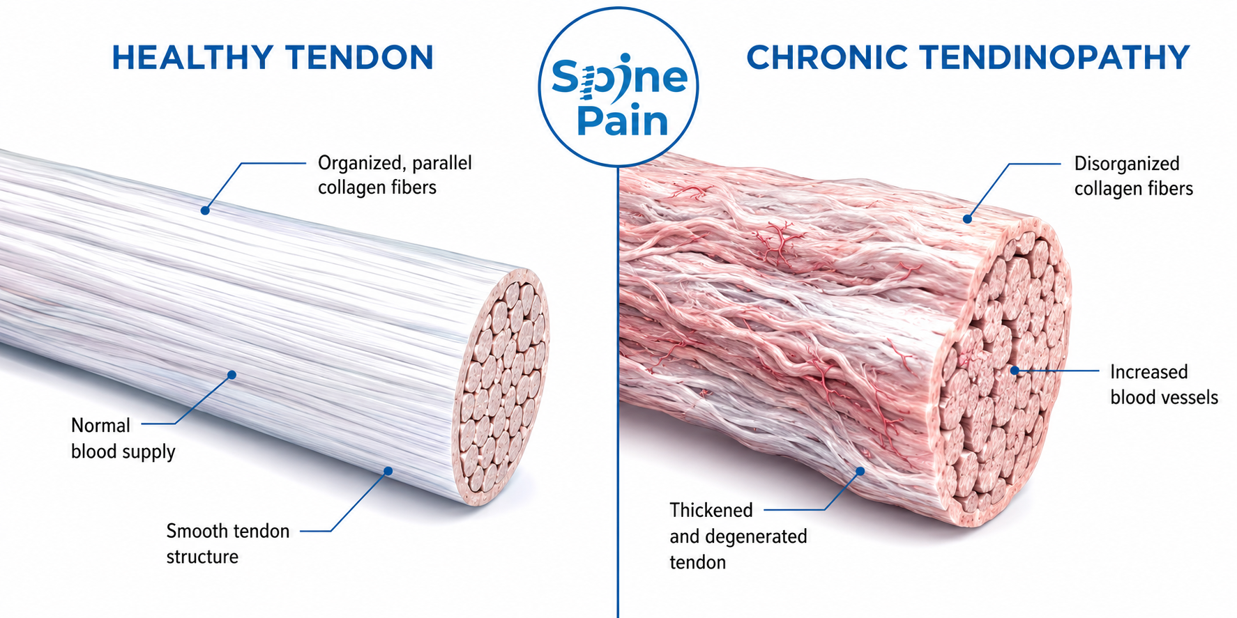
Chronic tendon pain can be frustrating because the problem is not always ongoing inflammation. In many patients, a painful tendon has undergone structural changes collectively known as tendinopathy, including collagen disorganization, tendon thickening, altered blood vessel growth, and an incomplete healing response.
This distinction matters. Treatments designed primarily to suppress inflammation may reduce symptoms, but they do not necessarily address the underlying abnormal tendon tissue.
Percutaneous needle tenotomy (PNT) is a minimally invasive procedure used in selected patients with persistent tendinopathy. Under ultrasound guidance, a needle or specialized instrument is advanced through the skin and directed precisely into the abnormal portion of the tendon. Depending on the technique, the tendon may be repeatedly fenestrated to stimulate a healing response, or abnormal tissue may be mechanically fragmented and removed.
PNT is not one specific device or brand. It describes a broader family of percutaneous tendon procedures. Conventional needle tenotomy, ultrasonic tenotomy systems such as Tenex, and hydroresection systems such as TenJet use different technologies but share the concept of treating chronically abnormal tendon tissue through a small percutaneous approach.
Quick Answer
Percutaneous needle tenotomy is an ultrasound-guided treatment for selected chronic tendon problems that have not improved adequately with conservative care. A needle or small specialized instrument is directed into the abnormal portion of the tendon to create controlled mechanical stimulation. Some techniques use repeated needle fenestration, while others fragment or remove abnormal tendon tissue. The goal is not simply to numb the painful area, but to create an environment in which the tendon can undergo further healing and remodeling. Recovery is gradual and usually includes progressive rehabilitation.
Clinical Perspective
An abnormal tendon on an MRI or ultrasound does not automatically need a procedure. Tendon degeneration is common and can sometimes be present without causing pain. The most important step is determining whether the abnormal tendon actually corresponds to the patient’s symptoms and examination. Percutaneous tenotomy is most useful when the diagnosis, imaging findings, functional limitations, and response to previous treatment all point toward the tendon as the primary pain generator.
Understanding Chronic Tendinopathy
Tendons are strong bands of connective tissue that transfer force from muscle to bone. Healthy tendon fibers are arranged in an organized pattern that allows the tendon to tolerate substantial mechanical loads.
Tendons are also living tissue. They continually respond to loading, activity, injury, and recovery. Problems can develop when repetitive stress or an acute increase in load exceeds the tendon’s ability to adapt and repair itself.
Tendinitis, Tendinosis, and Tendinopathy
The terms tendinitis, tendinosis, and tendinopathy are often used interchangeably, but they do not describe exactly the same process.
Tendinitis traditionally refers to an inflammatory tendon process. Inflammation may play an important role during an acute tendon injury and can also participate in different stages of chronic tendon disease.
Tendinosis describes chronic degenerative changes within the tendon. The normal collagen architecture may become disorganized, the tendon may thicken, and abnormal vascular and cellular changes may develop.
Tendinopathy is the broader clinical term used to describe persistent tendon pain and impaired function associated with these tendon changes.
This is why chronic tendon pain is often more complicated than simply treating “inflammation.”
Why Tendons Can Become Chronically Painful
Tendinopathy often develops when the mechanical demands placed on a tendon repeatedly exceed its ability to recover and adapt. This can occur in competitive athletes, recreational exercisers, people performing repetitive occupational activities, or even after an abrupt increase in ordinary physical activity.
Potential contributors include:
- repetitive tendon loading
- sudden increases in exercise or activity
- biomechanical stress
- previous tendon injury
- incomplete rehabilitation
- age-related tendon changes
- certain metabolic or systemic conditions
- medications or other factors that may affect tendon health
Importantly, imaging abnormalities do not always correlate perfectly with pain. Some people have substantial tendon abnormalities without symptoms, while others experience significant pain with comparatively modest imaging changes.
What Is Percutaneous Needle Tenotomy?
The name describes the basic concept:
- Percutaneous means the procedure is performed through the skin.
- Needle refers to the needle or needle-like instrument used to reach the abnormal tendon.
- Tenotomy refers to controlled mechanical treatment of tendon tissue.
During conventional PNT, a physician uses ultrasound to identify the abnormal portion of the tendon and then performs multiple controlled needle passes through that tissue. This process is also commonly described as tendon fenestration.
The purpose is not to sever the tendon. Rather, the goal is to mechanically stimulate a chronically abnormal region and encourage a renewed healing and remodeling response.
Important: Percutaneous needle tenotomy for tendinopathy should not be confused with a traditional surgical tenotomy in which a tendon may be deliberately released or divided. Modern ultrasound-guided PNT generally targets a focal region of abnormal tendon while preserving the surrounding tendon structure.
How Percutaneous Needle Tenotomy Works
Chronic tendinopathy can represent an unsuccessful or incomplete healing response. One rationale behind PNT is to mechanically disrupt the abnormal region and create a controlled local injury that may restart portions of the body’s healing process.
Mechanical Fenestration
In conventional PNT, the needle is passed repeatedly through the targeted region. These controlled passes mechanically fenestrate abnormal tendon tissue.
Stimulating a Healing Response
The controlled microtrauma created by fenestration may produce localized bleeding and initiate cellular and biochemical processes involved in tissue repair and remodeling. The tendon is then progressively loaded during rehabilitation as healing occurs.
Removing Abnormal Tissue
Not every percutaneous tenotomy technique works by fenestration alone. Specialized technologies can fragment, resect, and evacuate targeted abnormal tissue. These include ultrasonic tenotomy and hydroresection systems.

Different Types of Percutaneous Tenotomy
The terminology surrounding percutaneous tendon procedures can be confusing. PNT, Tenex, and TenJet are sometimes discussed as though they are interchangeable, but their mechanisms are different.
Conventional Needle Tenotomy or Fenestration
Conventional PNT uses a standard needle to make multiple controlled passes through the abnormal tendon under ultrasound guidance. The primary treatment is mechanical fenestration rather than active removal of a substantial amount of tendon tissue.
Percutaneous Ultrasonic Tenotomy
Percutaneous ultrasonic tenotomy uses a specialized instrument that delivers high-frequency mechanical energy to targeted abnormal tissue. Irrigation and aspiration allow fragmented tissue to be removed through the same small treatment site.
Tenex is one technology used to perform this type of ultrasonic tendon treatment.
Hydroresection
Hydroresection uses a pressurized stream of saline to resect targeted abnormal soft tissue while fluid and tissue debris are simultaneously evacuated.
TenJet is a device that uses this approach. Unlike conventional needle fenestration, its primary mechanism includes selective tissue resection rather than repeated needle passes alone.
Key Point: These procedures belong to the same general family of minimally invasive tendon treatments, but they are not identical. A particular device should not be assumed to be superior simply because it uses a more specialized technology.

Why Ultrasound Guidance Matters
Ultrasound provides real-time visualization of both the tendon and the treatment instrument. This is particularly valuable because tendinopathy is often not distributed evenly throughout an entire tendon.
Ultrasound may allow the physician to identify:
- tendon thickening
- changes in tendon fiber organization
- focal areas of tendinosis
- calcification
- partial tearing
- adjacent blood vessels
- nearby nerves
- the relationship between the tendon and bone
The needle or treatment instrument can then be observed continuously as it approaches and treats the targeted region.
Clinical Perspective
The value of ultrasound guidance is not simply that the physician can see a needle. It allows the procedure to be planned around the individual patient’s anatomy. The goal is to identify the clinically relevant abnormal tendon tissue, treat that region deliberately, and avoid unnecessary injury to surrounding structures.
What Conditions Can Be Treated With Percutaneous Needle Tenotomy?
PNT and related percutaneous tenotomy techniques have been studied in several areas of the body. The amount and quality of evidence vary considerably between tendon locations.
Elbow Tendinopathy
The elbow is one of the most commonly studied regions. Potential indications include chronic lateral elbow tendinopathy, commonly called tennis elbow, and medial elbow tendinopathy, commonly called golfer’s elbow.
Shoulder Tendinopathy
Selected patients with chronic rotator cuff tendinopathy may be considered for PNT. Significant partial or complete rotator cuff tears require a different evaluation because tenotomy does not mechanically repair a torn tendon.
Hip and Pelvic Tendinopathy
Gluteus medius and gluteus minimus tendinopathy can contribute to chronic lateral hip pain. Proximal hamstring tendinopathy is another condition in which percutaneous treatment may be considered in appropriately selected patients.
Knee Tendinopathy
Chronic patellar tendinopathy, sometimes called jumper’s knee, has also been treated with percutaneous tenotomy techniques. Selected quadriceps tendon disorders may be considered depending on anatomy and structural integrity.
Achilles Tendinopathy
Percutaneous procedures have been studied for both midportion and insertional Achilles tendinopathy. The Achilles tendon is a major load-bearing structure, so the extent of degeneration and any associated tearing must be carefully evaluated before treatment.
Plantar Fascia
Percutaneous procedures are also used for selected cases of chronic plantar fasciopathy. Technically, however, the plantar fascia is fascia rather than a tendon. It is often included in discussions of percutaneous tenotomy because the treatment principles and technologies can overlap.
Who May Be a Candidate for Percutaneous Needle Tenotomy?
PNT is generally considered after the diagnosis has been established and reasonable conservative treatment has not provided adequate improvement.
A potential candidate may have:
- persistent, localized tendon pain
- symptoms reproduced by loading the affected tendon
- physical examination findings consistent with tendinopathy
- ultrasound or MRI findings that correspond with the clinical problem
- functional limitations despite appropriate rehabilitation
- inadequate improvement with activity modification and other conservative treatments
- tendon anatomy suitable for a percutaneous procedure
There is no single symptom duration or imaging finding that automatically determines candidacy. The decision should be individualized.
When PNT May Not Be the Right Treatment
A minimally invasive procedure is useful only when it addresses the correct problem. PNT may not be appropriate when pain is primarily arising from another structure or when the tendon requires mechanical repair rather than stimulation or debridement.
Examples may include:
- complete tendon rupture
- large or mechanically significant tendon tears
- active infection
- certain bleeding disorders or anticoagulation concerns
- pain arising primarily from a joint rather than the tendon
- nerve-related pain mimicking tendinopathy
- significant structural abnormalities that require surgical repair
Key Point: The correct diagnosis matters more than the procedure. A technically perfect tenotomy directed at the wrong pain generator is unlikely to provide the desired result.
Diagnosing the Actual Pain Generator
The evaluation begins before ultrasound guidance or any procedure is considered.
History
The location of pain, activities that provoke symptoms, duration of the problem, previous injuries, occupational or athletic demands, and response to previous treatments all help determine whether the tendon is likely to be responsible.
Physical Examination
Examination may include palpation of the tendon, resisted muscle testing, tendon-loading maneuvers, range of motion, strength testing, and evaluation of nearby joints and nerves.
Diagnostic Ultrasound
Musculoskeletal ultrasound provides a dynamic view of tendon architecture and can identify focal abnormalities while the patient moves. It can also help evaluate nearby structures that may produce similar symptoms.
MRI
MRI can provide a broader assessment when there is concern for significant tearing, adjacent joint disease, bone abnormalities, or other pathology. Ultrasound and MRI are often complementary rather than competing tests.
What Happens During Percutaneous Needle Tenotomy?
The exact technique varies depending on the tendon and the type of tenotomy being performed, but a typical ultrasound-guided procedure follows several general steps.
- The tendon is examined with ultrasound. The physician identifies the tendon, surrounding anatomy, and the region targeted for treatment.
- The skin is prepared. The treatment area is cleaned using sterile technique.
- Local anesthetic is administered. Anesthetic is used to improve comfort while preserving the ability to treat the intended tendon safely.
- The needle or instrument is positioned. Ultrasound guidance allows its path to be followed in real time.
- The abnormal region is treated. Conventional PNT uses controlled fenestration. Device-assisted procedures may fragment, resect, irrigate, or aspirate targeted tissue.
- The instrument is removed. Because the access site is small, closure may require only a small dressing, although technique varies.
Most percutaneous tendon procedures are performed without the larger incision and tissue exposure associated with open tendon surgery.
Does Percutaneous Needle Tenotomy Hurt?
Local anesthetic is typically used to reduce discomfort from the skin and surrounding tissues. Patients may still notice pressure, movement, or discomfort during treatment depending on the tendon and technique.
Soreness after the procedure is expected. PNT deliberately creates controlled tissue stimulation, so some temporary increase in pain does not necessarily indicate that something has gone wrong.
Severe or progressively worsening pain, significant swelling, drainage, fever, new weakness, or neurological symptoms should be reported to the treating physician.
Percutaneous Needle Tenotomy vs Cortisone Injection
Corticosteroid injection and PNT have fundamentally different treatment goals.
| Feature | Percutaneous Needle Tenotomy | Corticosteroid Injection |
|---|---|---|
| Primary goal | Mechanically treat abnormal tendon tissue and stimulate remodeling | Reduce pain and inflammatory activity |
| Medication required | Not necessarily | Corticosteroid medication |
| Removes abnormal tissue | Only with certain device-assisted techniques | No |
| Rehabilitation | Important part of recovery | Often still important |
Corticosteroid injections can have a role in selected musculoskeletal conditions, but repeated corticosteroid exposure in or around certain tendons requires caution. The appropriate treatment depends on the diagnosis, tendon involved, severity of disease, previous treatment, and individual clinical circumstances.
Percutaneous Needle Tenotomy vs PRP
PNT and platelet-rich plasma (PRP) are often discussed together because both may be used for chronic tendinopathy, but they approach the problem differently.
PNT is primarily a mechanical treatment. The tendon is fenestrated to stimulate a healing response.
PRP is a biologic treatment. A patient’s own blood is processed to create a platelet concentrate, which is then injected into the targeted region with the goal of influencing the local healing environment.
The two approaches can also be combined. In fact, PRP injection into a tendon commonly involves some degree of needle fenestration simply because the needle is passed through the targeted tendon tissue during treatment.
However, adding PRP should not automatically be assumed to make PNT more effective. In a randomized controlled trial involving several types of chronic tendinosis, both PNT alone and PNT combined with leukocyte-rich PRP improved outcomes over time, with no significant overall difference between groups across the study period.
Treatment Tip: The decision between PNT, PRP, or a combined approach should be based on the tendon involved, imaging findings, previous treatment, available evidence, and the goals of the individual patient rather than assuming that more treatment is necessarily better.
Percutaneous Needle Tenotomy vs TenJet
Percutaneous needle tenotomy describes a broad treatment concept. TenJet is a specific device-assisted technique.
Conventional PNT uses repeated needle passes to fenestrate the abnormal portion of the tendon. TenJet uses a high-velocity saline stream within a small treatment window to selectively resect abnormal soft tissue while simultaneously evacuating fluid and tissue debris.
Both are performed percutaneously and can be guided with ultrasound, but their mechanisms are different.
Early clinical studies of TenJet have reported improvements in pain and function in selected patients, including patients with chronic elbow tendinopathy. However, the TenJet evidence base is smaller than the literature for conventional PNT and ultrasonic tenotomy, and current evidence does not establish TenJet as universally superior to other percutaneous approaches.
Learn more: TenJet for Chronic Tendinopathy
Percutaneous Needle Tenotomy vs Tenex
Tenex is another device-assisted percutaneous treatment, but it does not use the same mechanism as TenJet.
Tenex uses ultrasonic mechanical energy to fragment targeted abnormal tissue. Irrigation and aspiration then remove the fragmented material through the small treatment instrument.
A simple way to understand the distinction is:
- Conventional PNT: needle fenestration
- Tenex: ultrasonic fragmentation with irrigation and aspiration
- TenJet: hydroresection using a pressurized saline stream with evacuation
Published studies have reported favorable outcomes after percutaneous ultrasonic tenotomy for several forms of chronic tendinopathy, but systematic reviews have also emphasized the need for larger, higher-quality comparative trials.
At present, there is insufficient evidence to declare one of these technologies the best choice for every tendon or every patient.
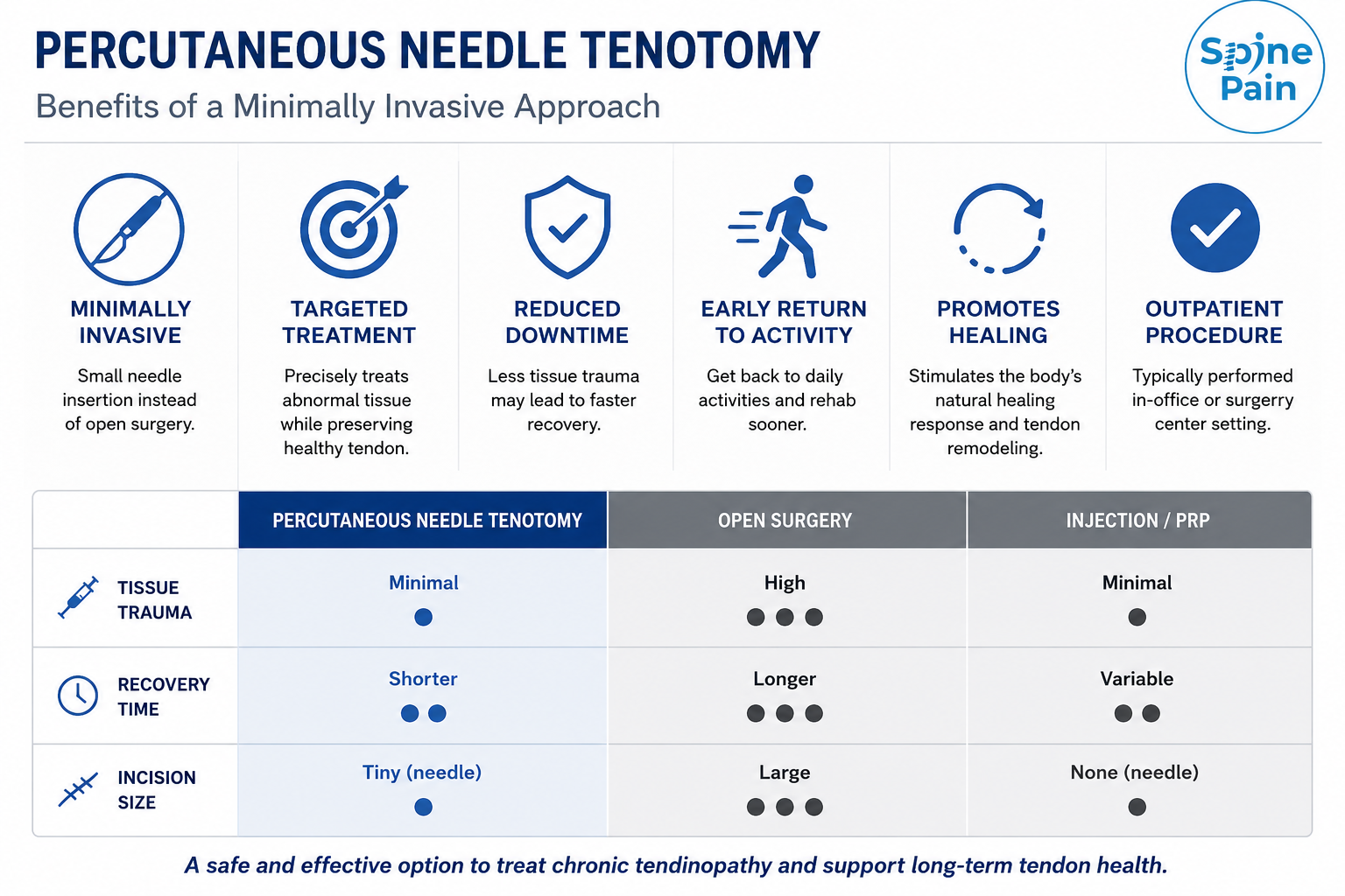
Percutaneous Tenotomy vs Tendon Surgery
Percutaneous tenotomy occupies an important space between conservative treatment and traditional surgery for appropriately selected patients.
| Feature | Percutaneous Tenotomy | Traditional Tendon Surgery |
|---|---|---|
| Access | Small percutaneous entry site | Larger surgical exposure depending on procedure |
| Imaging | Usually real-time ultrasound guidance | Direct surgical visualization and/or imaging |
| Major tendon repair | No | Can repair or reconstruct significant structural tears |
| Recovery | Often shorter, but varies by tendon and treatment | Often longer and procedure-dependent |
The smaller access site of PNT does not make it a substitute for surgery when a tendon has lost mechanical integrity and requires repair. Choosing the least invasive procedure is useful only when that procedure can adequately address the underlying problem.
How Effective Is Percutaneous Needle Tenotomy?
Published research on percutaneous tenotomy is encouraging, but the strength of the evidence varies by technique and tendon location.
A 2024 systematic review evaluated ultrasound-guided PNT and percutaneous ultrasonic tenotomy across multiple tendon locations. The included studies generally reported improvements in pain, function, and patient-reported outcomes, with relatively few adverse events.
However, there are important limitations. Many published studies are relatively small, use different outcome measures, include different tendon locations, and are not randomized controlled trials. These differences make direct comparisons difficult.
The evidence is also not equally developed for every technology. Although the 2024 systematic review searched for studies involving PNT, Tenex, and TenJet, no TenJet studies ultimately met that review’s inclusion criteria. TenJet has separate prospective observational data, including a multicenter study involving chronic elbow tendinopathy, but its overall clinical literature remains more limited.
Clinical Perspective
The available evidence supports percutaneous tenotomy as a reasonable treatment option for selected patients with chronic tendinopathy that has not responded adequately to conservative treatment. It does not establish that every patient will improve, that every tendon responds equally, or that one device-assisted technique is superior to all others.
Why Results Differ Between Patients
Tendinopathy is not one uniform disease. Two patients with pain in the same tendon may have very different structural abnormalities, biomechanics, activity demands, and healing capacity.
Factors that may influence recovery include:
- duration of symptoms
- severity and extent of tendon degeneration
- presence of partial tearing or calcification
- the specific tendon being treated
- age and general health
- smoking and other factors that may impair tissue healing
- biomechanical contributors
- occupational or athletic demands
- adherence to rehabilitation
- returning to heavy loading too quickly
- whether the tendon was actually the primary pain generator
For this reason, published success rates should never be interpreted as a prediction of what will happen to an individual patient.
Recovery After Percutaneous Needle Tenotomy
Recovery after PNT is a biological process rather than an immediate event. The purpose of the procedure is to stimulate or facilitate tissue remodeling, and that process takes time.
The First Several Days
Soreness around the treated tendon is common. Activity is usually modified to protect the area from excessive loading while still allowing appropriate movement.
Early Recovery
Normal daily activity is gradually increased according to the tendon treated, the specific procedure performed, symptoms, and the treating physician’s protocol.
Progressive Rehabilitation
Rehabilitation may progress through range-of-motion exercises, isometric loading, strengthening, and progressively more demanding tendon loading. The exact sequence depends on the tendon and individual clinical situation.
Return to Exercise and Sports
Return to strenuous exercise should be based on healing, strength, symptoms, and functional progression rather than simply reaching a particular date on the calendar.
Key Point: There is no single recovery timeline for every PNT procedure. Treating a common extensor tendon at the elbow and treating a load-bearing Achilles tendon are not biologically or mechanically equivalent.
Why Rehabilitation Is Part of the Treatment
The procedure itself is only one part of tendon recovery.
Tendons adapt to mechanical loading. After treatment, progressive rehabilitation helps expose the healing tendon to increasing levels of stress so that it can regain strength and load tolerance.
The challenge is finding the appropriate balance.
Treatment Tip: Too little loading may leave the tendon inadequately conditioned. Too much loading too early may provoke pain or interfere with recovery. Progressive rehabilitation attempts to stay between those two extremes.
Risks and Potential Complications
Percutaneous tenotomy is minimally invasive, but minimally invasive does not mean risk-free.
Potential risks include:
- temporary post-procedure pain or soreness
- bruising or bleeding
- infection
- injury to surrounding structures
- nerve or blood vessel injury
- persistent or incompletely improved symptoms
- tendon injury or progression of an existing tear
- need for additional treatment
- eventual need for surgery
The risks vary according to the tendon treated, the technique used, the patient’s health, and the surrounding anatomy.
Can PNT Be Combined With Other Treatments?
Yes, but combination treatment should have a clinical rationale rather than simply adding procedures.
Depending on the individual problem, a treatment plan may incorporate:
- structured physical therapy
- progressive tendon-loading exercises
- PRP in selected patients
- correction of biomechanical contributors
- temporary bracing or activity modification
- treatment of another pain generator when more than one structure is involved
The objective is not to perform the greatest number of interventions. It is to identify the relevant pain generator and use the least complex treatment strategy that reasonably addresses the problem.
Frequently Asked Questions
Is percutaneous needle tenotomy surgery?
PNT is generally described as a minimally invasive percutaneous procedure rather than traditional open surgery. It is performed through a small skin entry site, usually under ultrasound guidance. Device-assisted forms of percutaneous tenotomy may require a slightly larger access site than conventional needle fenestration.
Is PNT the same as TenJet?
No. PNT is the broader treatment concept. Conventional PNT uses needle fenestration. TenJet is a specific device that uses hydroresection to selectively resect targeted abnormal tissue.
Is PNT the same as Tenex?
Not exactly. Tenex is a form of percutaneous ultrasonic tenotomy. It uses ultrasonic mechanical energy together with irrigation and aspiration. Conventional PNT uses repeated needle fenestration without an ultrasonic tissue-removal system.
Does PNT remove the tendon?
No. Conventional PNT does not remove the tendon. It creates controlled fenestrations within the targeted abnormal region. Certain device-assisted techniques remove small amounts of abnormal tissue, but they are intended to preserve the surrounding tendon.
Does the tendon grow back after tenotomy?
The term can be misleading because modern PNT for tendinopathy generally does not involve cutting the entire tendon. Instead, a focal abnormal region is mechanically treated. Healing then involves tissue repair and remodeling rather than simply regrowing a completely divided tendon.
How long does recovery take?
Recovery varies considerably. The tendon being treated, severity of tendinopathy, type of procedure, physical demands, and rehabilitation program all matter. Patients should follow a tendon-specific rehabilitation plan rather than relying on a universal timeline.
When can I exercise again?
Light activity may resume relatively early for some procedures, but strenuous tendon loading is generally reintroduced progressively. Return to exercise should be based on symptoms, strength, healing, and the tendon treated.
Can PNT treat a partial tendon tear?
Some patients with tendinopathy and limited partial tearing may be candidates, but this depends on the size, location, depth, and mechanical significance of the tear. Larger or unstable tears may require surgical evaluation because PNT does not mechanically repair a tendon defect.
Is PNT better than PRP?
Current evidence does not support a simple answer that one is universally better. They use different mechanisms and may be appropriate in different circumstances. A randomized trial comparing PNT alone with PNT plus leukocyte-rich PRP did not demonstrate an overall advantage from adding PRP across the study period.
Can PRP and PNT be performed together?
Yes. Needle fenestration can be combined with PRP injection. Whether the combination offers an advantage for a particular patient depends on the clinical circumstances and remains an area of ongoing research.
What happens if PNT does not work?
Persistent symptoms should prompt reassessment rather than automatic repetition of the same procedure. The tendon may require additional rehabilitation, another treatment approach, or surgical evaluation. In some cases, reconsidering the original diagnosis reveals another source of pain.
Can percutaneous needle tenotomy be repeated?
Repeat treatment may be considered in selected circumstances, but persistent symptoms should first be reevaluated. There is little value in repeatedly treating a tendon without understanding why the initial procedure did not produce the expected improvement.
Does insurance cover percutaneous needle tenotomy?
Coverage varies by insurance plan, diagnosis, procedure, and technology used. Some insurers may consider certain percutaneous tendon procedures differently from others. Coverage and authorization should therefore be verified before treatment.
Key Takeaways
- Percutaneous needle tenotomy is a minimally invasive treatment used for selected chronic tendon disorders.
- Chronic tendinopathy is not simply persistent inflammation. Degenerative and failed-healing changes can develop within the tendon.
- Ultrasound guidance allows the abnormal tendon region and surrounding anatomy to be visualized during treatment.
- Conventional PNT, Tenex, and TenJet use different mechanisms. Needle fenestration, ultrasonic fragmentation, and hydroresection should not be treated as interchangeable procedures.
- No percutaneous tenotomy technology has been proven universally superior for every tendon and every patient.
- Rehabilitation is part of the treatment. Tendon remodeling and progressive loading occur over time rather than immediately after the procedure.
- Patient selection matters. Successful treatment begins with identifying the tendon as the actual pain generator.
References
- Maag L, Linder S, Hackett L, et al. Effectiveness of Percutaneous Needle Tenotomy for Tendinopathies: A Systematic Review. Sports Health. 2024;17(4):834-842. doi:10.1177/19417381241275659.
- Kirschner JS, Cheng J, Hurwitz N, et al. Ultrasound-guided percutaneous needle tenotomy alone versus percutaneous needle tenotomy plus platelet-rich plasma injection for the treatment of chronic tendinosis: A randomized controlled trial. PM&R. 2021;13(12):1340-1349. doi:10.1002/pmrj.12583.
- Vajapey S, Ghenbot S, Baria MR, Magnussen RA, Vasileff WK. Utility of Percutaneous Ultrasonic Tenotomy for Tendinopathies: A Systematic Review. Sports Health. 2021;13(3):258-264. doi:10.1177/1941738120951764.
- Strauser-Curtis K, Varacallo CP, Voss TT, Stephens CB, Kapteyn RW. Ultrasound-Guided Tenotomy Via a Hydrosurgery Resection Device Improves Symptoms of Chronic Elbow Tendinopathy: A Multi-Center Prospective Study. Journal of Orthopaedic Experience & Innovation. 2022;3(1). doi:10.60118/001c.34671.
Related Educational Articles
Continue Learning
TenJet for Chronic Tendinopathy explains how hydroresection differs from conventional needle fenestration.
Additional related guides on PRP, ultrasound-guided procedures, and tendon-specific treatment options can help patients understand where percutaneous tenotomy fits within a broader treatment plan.
About This Guide
This resource is provided for educational purposes and should not replace personalized medical advice, diagnosis, or treatment from a qualified healthcare professional.



