
SI Joint Fusion: Candidacy, RFA vs Fusion & Recovery
Interventional Spine and Pain Management Physician, Chief of the Division of Pain Medicine at Good Samaritan University Hospital, and Founder of SpinePain Solutions. Dr. Sharma completed fellowship training in pain medicine at Johns Hopkins and has practiced interventional spine and pain medicine for more than 22 years.
Minimally invasive sacroiliac joint fusion, also called SI joint fusion, is intended to stabilize a painful SI joint after the diagnosis has been carefully evaluated and appropriate nonsurgical care has not provided enough improvement. It is not a first-line treatment for ordinary low-back or buttock pain.
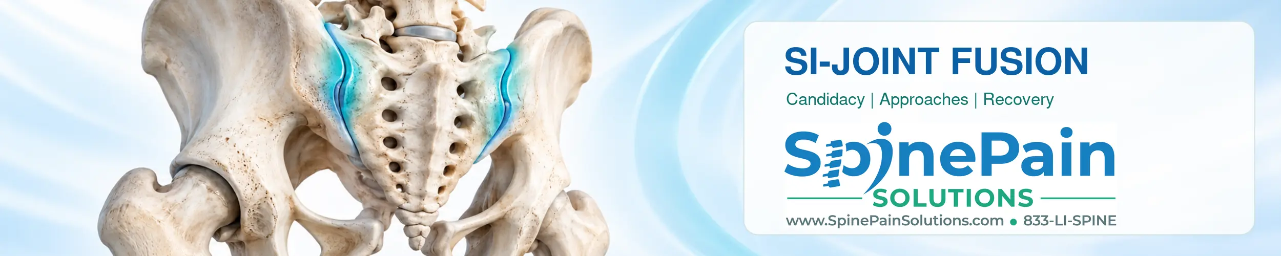
The SI joint connects the sacrum at the base of the spine to the ilium of the pelvis. Symptoms from this region can overlap with lumbar spine, hip, nerve-root and other posterior pelvic conditions. For that reason, successful treatment begins with diagnostic precision rather than an implant choice.
Practice update
At SpinePain Solutions, Dr. Sharma now performs most appropriate SI-joint fusion procedures using SacriX. Posterior LinQ remains an option for selected patients. This reflects the current practice pattern; it does not mean that one device is universally better for every anatomy, diagnosis or clinical situation.
What Is Minimally Invasive SI-Joint Fusion?
SI-joint fusion uses an implant, graft or fixation system to stabilize the joint and encourage bone growth across it. The procedure is performed through a smaller surgical corridor than traditional open fusion, but a small incision does not eliminate the need for bone healing, activity precautions or follow-up.
Different systems use different trajectories and implant concepts. A transfixing system crosses the joint to provide mechanical fixation. A posterior intra-articular system places a graft within the joint through a posterior corridor. Results and recovery from one technique should not automatically be generalized to another.
When Should SI-Joint Fusion Be Considered?
Quick answer
SI-joint fusion may be considered when persistent, function-limiting pain has been reasonably localized to the SI joint, appropriate nonsurgical treatment has not provided adequate improvement, competing lumbar and hip conditions have been evaluated, and an image-guided diagnostic injection provides convincing pain and functional improvement. No single symptom, test, scan or injection percentage establishes candidacy by itself.
Fusion is a later treatment option. The decision should combine the pain history, examination, imaging, response to treatment, diagnostic injection findings, patient goals, procedural risk and the evidence for the proposed approach.
| Findings that may support consideration | Reasons to evaluate or treat further first |
|---|---|
| Persistent pain below L5 consistent with an SI-joint pattern | Acute, mild or steadily improving pain |
| Meaningful limitation of walking, standing, sleep, work or daily activity | Symptoms without substantial functional limitation |
| Several provocation maneuvers reproduce the familiar pain | Examination does not reproduce the usual pain or points elsewhere |
| Lumbar, hip, neurological and inflammatory causes have been evaluated | Untreated radiculopathy, hip disease, fracture, infection or another likely source |
| Appropriate rehabilitation and nonsurgical care did not provide enough improvement | Adequate conservative treatment has not been attempted |
| Strong, activity-correlated response to an image-guided diagnostic injection | Weak, inconsistent or poorly documented injection response |
| Realistic goals centered on function and meaningful improvement | Expectation of guaranteed or complete pain elimination |
| Acceptable infection, bleeding, anesthesia and bone-healing risk | Active infection or unacceptable procedural or healing risk |
How Candidacy Is Confirmed
A typical evaluation includes the location and behavior of the pain, a neurological and musculoskeletal examination, several SI-joint provocation maneuvers and assessment of the lumbar spine and hips. Imaging can identify arthritis, fracture, inflammatory disease, tumor, infection or a competing diagnosis, but routine degenerative findings do not prove that the SI joint is the pain source.
When the result will change treatment, an image-guided local-anesthetic injection may add diagnostic evidence. The most useful response occurs during the expected anesthetic period and includes improvement while repeating an activity that normally reproduces the patient’s usual pain. Read the dedicated guide to interpreting an SI-joint injection.
How Much Injection Relief Is Required?
There is no single cutoff that applies to every insurer, jurisdiction, proposed approach or clinical decision. Some policies and professional groups favor two image-guided injections with at least 75% relief. Other clinical guidance uses lower thresholds in selected diagnostic or prognostic settings. The number should not be separated from the timing, technical accuracy, activity tested and purpose of the block.
A positive injection supports the diagnosis but does not guarantee fusion success. Anesthetic spread, sedation, expectation effects, reduced activity and other pain generators can affect the result. The applicable coverage policy should be verified before scheduling because requirements change and do not always match clinical guideline thresholds.
SacriX Versus Posterior LinQ SI-Joint Fusion
SacriX and LinQ are different minimally invasive approaches rather than interchangeable versions of one procedure. SacriX is an FDA-cleared implant system that uses a percutaneous transfixing corridor across the SI joint. LinQ uses a posterior intra-articular corridor to place a structural cortical allograft within the joint.
| Decision factor | SacriX transfixing approach | Posterior LinQ approach |
|---|---|---|
| General corridor | Percutaneous lateral-oblique transfixing trajectory across the joint | Posterior intra-articular trajectory into the joint |
| Implant concept | Metal implants are designed to stabilize the ilium and sacrum across the joint | A structural cortical allograft is placed within the joint to support stabilization and fusion |
| Weight bearing | Temporary protected or partial weight bearing may be prescribed, depending on the plan | Some protocols permit earlier weight bearing, but instructions remain individualized |
| Evidence | Transfixing/lateral fusion has more comparative evidence overall, but results are conflicting and device-specific | Posterior evidence is growing but remains lower certainty and should not be equated with transfixing evidence |
| Technical concerns | Trajectory, implant position, nerve irritation, bleeding, fracture, loosening or nonunion | Graft position or migration, inadequate stabilization, persistent pain or nonunion |
| Current practice use | Used for most appropriate SI-joint fusion procedures in Dr. Sharma’s current practice | Retained as an option for selected patients |
No approach should be presented as universally safer, less invasive or more effective. The choice depends on the pain diagnosis, anatomy, bone quality, previous surgery, implant goals, procedural risk, physician experience, payer requirements and the quality of evidence for that exact system. Learn more about the SacriX approach.
How Minimally Invasive SI-Joint Fusion Is Performed
The procedure is generally performed with the patient positioned face down. Imaging guidance is used to identify the SI joint and guide the planned surgical corridor. After sterile preparation and anesthesia, the physician creates a small incision, prepares the pathway and places the selected implant or graft according to the system’s technique. Imaging is used to assess position before the incision is closed.
Many procedures are completed in an outpatient setting, although the location of care and discharge plan depend on the approach, anesthesia, medical conditions, pain control and safe mobility. The patient should receive written wound-care, medication, weight-bearing and follow-up instructions before leaving.
Alternatives Before SI-Joint Fusion
Treatment should match the most likely pain generator. Depending on the diagnosis, alternatives may include:
- Targeted physical therapy, pelvic stabilization and progressive conditioning
- Activity and ergonomic modification
- Appropriate medication management
- Image-guided therapeutic SI-joint injection
- Sacral lateral-branch radiofrequency ablation when anatomically appropriate and covered
- Treatment of lumbar radiculopathy, facet pain, hip disease or another competing source
- Evaluation and treatment of inflammatory sacroiliitis
- Continued nonsurgical management when expected benefit does not justify fusion risk
Fusion should not be used to bypass evaluation of a correctable hip, lumbar, neurological, infectious or inflammatory condition. Review the broader guide to SI-joint dysfunction and diagnosis.
SI-Joint Radiofrequency Ablation Versus Fusion: When Is Each Used?
Different targets, different treatment goals
SI-joint RFA reduces pain transmission from selected nerves supplying the posterior SI-joint complex. It does not stabilize or fuse the joint. Fusion aims to stabilize a carefully selected painful joint and requires surgical recovery and bone healing. Failed RFA does not automatically mean fusion is needed, and a positive joint injection is not universal clearance for either procedure.
The SI-joint complex includes the joint cavity and posterior ligaments and surrounding structures. Sacral lateral-branch RFA treats selected posterior nerve supply, often including the L5 dorsal ramus and sacral lateral branches. An intra-articular injection primarily tests pain arising inside the joint. The diagnostic pathway for fusion and the prognostic nerve-block pathway for RFA are therefore not interchangeable.
| Decision factor | SI-joint RFA | SI-joint fusion |
|---|---|---|
| Goal | Reduce pain transmission from selected posterior SI structures | Stabilize the joint and promote fusion |
| Selection | Clinical assessment and appropriate lateral-branch prognostic blocks | Combined history, examination, competing-cause assessment, imaging and diagnostic joint-injection findings |
| Implant | No permanent implant or intended bone fusion | Implant or graft remains; biological fusion takes time |
| Durability | Benefit may diminish; repeat treatment may be considered after reassessment | Intended permanent stabilization, but pain relief and successful fusion are not guaranteed |
| Recovery | Usually outpatient; temporary soreness or neuritis may occur; no bone-healing period | Wound care, activity limits and possibly protected weight bearing; healing continues over months |
| Evidence | Randomized evidence supports selected anatomically appropriate techniques | Evidence varies by system and approach and includes conflicting randomized findings |
How the Choice Is Made
RFA may be considered for persistent posterior SI-joint complex pain when conservative care has not provided adequate improvement and appropriate prognostic blocks support the targeted nerves. Fusion may be considered for persistent, disabling joint pain when the separate candidacy assessment above is convincing and the expected benefit justifies implant, surgical and bone-healing risks.
Neither procedure should bypass evaluation of hip disease, lumbar radiculopathy, fracture, infection or inflammatory sacroiliitis. Rehabilitation, pelvic stabilization, medication management, therapeutic injection and treatment of another pain generator remain alternatives. RFA is not mandatory before every fusion, and fusion is not an automatic next step when RFA wears off.
Durability, Risks and Evidence Limits
A multicenter randomized trial found cooled RFA improved pain and function more than standard medical management at three months in selected patients. Its later observational crossover follow-up reported benefit through 12 months in some patients. These studies did not compare RFA directly with fusion, do not establish one treatment as universally better, and should not be generalized to every RFA technique. Read the randomized trial and 12-month follow-up.
RFA risks include temporary pain flare, neuritis, numbness, bleeding, infection, burns, unintended nerve injury and inadequate relief. Fusion has the additional implant, nonunion and surgical risks discussed below. Neither a positive block nor temporary relief guarantees a durable outcome.
Coverage varies by insurer, jurisdiction, treatment target and proposed fusion approach. Block requirements for one procedure should not be applied automatically to another. Review the applicable policy with the treating team. Learn more about radiofrequency ablation and SI-joint injection interpretation.
What the Evidence Shows and What It Cannot Prove
Positive pragmatic randomized studies have reported improvement after minimally invasive SI-joint fusion compared with nonsurgical management in carefully selected patients. However, a 2024 double-blind randomized trial did not demonstrate that minimally invasive fusion was superior to sham surgery at six months. These findings make balanced counseling essential.
The 2025 multispecialty consensus guidelines characterize the evidence for lateral minimally invasive fusion as conflicting and the evidence for posterior fusion as lower certainty. Study populations, diagnostic criteria, implants, surgical corridors and follow-up differ. Results from one device or approach should not be transferred automatically to another.
Important limitation
A diagnostic injection and careful selection reduce uncertainty; they do not predict an individual result with certainty. SI-joint fusion may reduce pain from the treated joint without eliminating pain from the lumbar spine, hips, nerves or other structures.
How Long Is Recovery After Minimally Invasive SI-Joint Fusion?
Quick answer
Recovery varies with the approach, implant system, bone quality, whether one or both sides are treated, other spine or hip conditions and work demands. Many patients go home the same day and begin short walks early, sometimes with crutches or a walker. Light daily activity may resume over several weeks, while unrestricted activity and biological fusion can take several months.
The operating physician’s instructions take priority over a generic timeline. SacriX and posterior LinQ patients should not assume that their weight-bearing limits, rehabilitation schedule or return-to-work date will be identical.
| Recovery period | Typical focus | Questions to clarify |
|---|---|---|
| First 24-72 hours | Incisional pain control, wound protection, short walks, fall prevention and prescribed assistive-device use | May I bear full weight? How do I use crutches or a walker? |
| First 1-2 weeks | Gradual household activity, incision monitoring and compliance with weight-bearing and movement limits | When can I shower, drive and return for wound review? |
| Weeks 2-6 | Progressive walking and activity based on the procedure and follow-up findings | Do weight-bearing limits continue? Is modified work appropriate? |
| Weeks 6-12 | Strengthening or formal rehabilitation when cleared; gradual return to more demanding activity | When can lifting, exercise and full work duties resume? |
| Three months and beyond | Continued bone healing, conditioning and assessment of persistent or recurrent symptoms | Is activity unrestricted? Is imaging or further evaluation needed? |
This is a general framework, not a promise. Incisional improvement can occur before the internal stabilization and bone-healing process is complete.
Weight Bearing, Walking and Assistive Devices
Walking commonly begins early when medically safe, but the amount of weight placed on the treated side must follow the specific postoperative plan. Some posterior protocols permit early weight bearing. Some transfixing procedures use temporary protected or partial weight bearing. Bone quality, balance, pain, implant position and other conditions may change the plan.
If crutches or a walker are prescribed, patients should understand how long to use them and how to manage stairs safely. A small incision or rapidly improving pain should not be used as permission to advance activity ahead of schedule.
Wound Care and Early Activity
Keep the incision and dressing protected according to the written instructions. Showering depends on the closure and dressing. Baths, swimming and hot tubs generally wait until the incision is fully healed and the physician provides clearance. Short, frequent walks are usually preferable to prolonged bed rest, but lifting, forceful twisting and higher-impact activity may be restricted.
Driving
Driving generally requires that the patient is no longer taking medication that impairs alertness, can enter and exit the vehicle safely, can sit comfortably and has adequate strength and reaction time for emergency braking. The side treated and any weight-bearing restriction also matter. Resume driving only after the operating team provides clearance.
Returning to Work
Sedentary work may resume earlier than physical employment, but commuting, prolonged sitting and the need to change position should be considered. Jobs involving lifting, climbing, prolonged driving, patient handling, machinery or unpredictable physical demands require later individualized clearance. Modified duty may be useful. Avoid promising a universal one- or two-week return.
Physical Therapy and Exercise
The timing of rehabilitation depends on the approach and physician protocol. Early aggressive rotation, loading or hip strengthening may be inappropriate even when walking is encouraged. When cleared, therapy generally progresses from safe mobility and walking toward hip, gluteal and trunk conditioning.
Recovery Limitations and Expectations
- Early pain improvement does not prove that biological fusion is complete.
- Postoperative soreness does not automatically mean the procedure failed.
- Recovery may be slower with bilateral treatment, osteoporosis, smoking, diabetes, deconditioning or other spine and hip disorders.
- Posterior and transfixing procedures should not share an identical recovery promise.
- SI-joint fusion may reduce pain from the treated joint without eliminating every low-back, buttock, hip or leg symptom.
- Therapy can temporarily increase soreness; severe or new symptoms require reassessment.
- Driving, return-to-work and unrestricted-activity dates are individualized.
Risks and Warning Signs
Potential risks include:
- Infection or wound complications
- Bleeding or hematoma
- Nerve irritation or neurological injury
- Implant or graft malposition, migration or loosening
- Fracture
- Persistent, recurrent or worsening pain
- Nonunion or pseudarthrosis
- Adjacent pelvic, hip or lumbar pain
- Anesthesia complications
- Revision, additional surgery or implant removal
Risk varies with the technique and system. A transfixing trajectory can create concerns about implant position, nerve irritation, bleeding or fracture. Posterior approaches can create concerns about graft position or migration, insufficient stabilization and nonunion. Neither list predicts one patient’s outcome.
Contact the treating team promptly
Report fever, drainage, spreading redness, worsening swelling, wound opening, uncontrolled or rapidly increasing pain, new weakness or numbness, or loss of bowel or bladder control. Chest pain, shortness of breath or major progressive neurological symptoms require urgent medical evaluation.
Questions to Ask Before SI-Joint Fusion
- What findings make the SI joint the most likely pain source?
- Which competing lumbar, hip, neurological or inflammatory causes have been evaluated?
- How should my diagnostic injection response be interpreted?
- What does my insurer require before authorizing fusion?
- Why are you recommending SacriX, posterior LinQ or another approach for me?
- What evidence supports this exact procedure and implant system?
- What improvement is realistic, and which symptoms may remain?
- Will I need crutches, a walker or temporary protected weight bearing?
- When can I shower, drive, work, exercise and begin physical therapy?
- Which symptoms require an urgent call?
- What happens if the joint does not fuse or pain persists?
SI-Joint Fusion Frequently Asked Questions
When should SI-joint fusion be considered?
SI-joint fusion may be considered when persistent, function-limiting pain has been reasonably localized to the SI joint, appropriate nonsurgical care has not provided adequate improvement, competing lumbar and hip conditions have been evaluated, and image-guided diagnostic injection findings support the diagnosis. No single symptom, examination maneuver, image or injection percentage establishes candidacy by itself.
How is candidacy for SI-joint fusion confirmed?
Candidacy is based on the combined findings from the history, pain location, several examination maneuvers that reproduce the usual pain, evaluation for lumbar and hip causes, appropriate imaging, response to nonsurgical care and an image-guided diagnostic injection when needed. The expected benefit must also justify the procedural and recovery risks.
How much injection relief is required before SI-joint fusion?
There is no universal percentage for every patient, insurer or fusion approach. The response should occur during the expected local-anesthetic window and should include improvement while repeating a normally painful activity. Some policies and societies favor two injections with at least 75% relief, while other clinical guidance uses lower thresholds in selected settings. The applicable policy and clinical purpose must be checked.
What is the difference between SacriX and posterior LinQ SI-joint fusion?
SacriX uses a percutaneous transfixing approach designed to place implants across the SI joint. LinQ uses a posterior intra-articular approach that places a structural allograft into the joint. The surgical corridor, implant concept, postoperative instructions and evidence base differ. Neither approach should be described as best for every patient.
Which SI-joint fusion approach does Dr. Sharma use?
Dr. Sharma currently performs most appropriate SI-joint fusion procedures using SacriX, while posterior LinQ remains an option for selected patients. This practice pattern does not prove that one system is universally superior. The choice depends on diagnosis, anatomy, bone quality, previous surgery, risk, coverage and the technical goals of treatment.
How long is recovery after minimally invasive SI-joint fusion?
Recovery varies with the surgical approach, implant system, bone quality, weight-bearing instructions, other health conditions and work demands. Many patients go home the same day and begin short walks early, sometimes with crutches or a walker. Light activity may resume over several weeks, but unrestricted activity and biological fusion can take several months.
Will I need crutches or a walker after SI-joint fusion?
Possibly. Assistive-device and weight-bearing instructions depend on the fusion approach, implant system, bone quality, balance and physician protocol. Some patients may bear weight early, while others require temporary partial or protected weight bearing. Follow the written instructions for the specific procedure rather than a generic online timeline.
When can I drive after SI-joint fusion?
Driving usually waits until the patient is no longer taking impairing medication, can enter and exit the vehicle safely, can sit comfortably and has enough strength and reaction time for emergency braking. Weight-bearing restrictions and the side treated may also matter. The operating physician should provide clearance.
When can I return to work after SI-joint fusion?
Return to work depends on healing, commuting and job demands. Sedentary work may resume earlier than work involving lifting, climbing, prolonged driving, patient handling or heavy equipment. Modified duty may be appropriate. There is no reliable one- or two-week promise that applies to every patient or approach.
When is SI-joint radiofrequency ablation used instead of fusion?
SI-joint radiofrequency ablation may be considered for selected posterior SI-joint complex pain when appropriate prognostic nerve blocks support the target and nonsurgical care has not provided enough improvement. It reduces pain transmission without stabilizing or fusing the joint. Fusion is a more invasive option for carefully selected persistent joint pain after a separate diagnostic and risk assessment. Neither treatment is automatically the next step after the other.
Does failed SI-joint RFA mean I need fusion?
No. A failed or short-lived RFA should prompt reassessment of the diagnosis, targeted nerves, block response, technique and competing lumbar or hip pain sources. It does not prove joint instability or establish fusion candidacy. A positive intra-articular SI-joint injection also does not automatically predict lateral-branch RFA success because the anatomical targets differ.
Does SI-joint fusion guarantee pain relief?
No. Carefully selected patients may improve, but fusion does not guarantee complete or permanent pain relief. It may reduce SI-joint pain without treating pain from the lumbar spine, hip, nerves or other structures. Diagnostic injections reduce uncertainty but cannot predict an individual outcome with certainty.
Selected References
- McCormick ZL, et al. Consensus practice guidelines on sacroiliac joint complex pain. 2025.
- Cohen SP, et al. Cooled radiofrequency ablation versus standard medical management for chronic sacroiliac joint pain: a multicenter, randomized comparative effectiveness study. 2024.
- Cohen SP, et al. Cooled radiofrequency ablation provides extended clinical utility in chronic sacroiliac joint pain: 12-month observational crossover follow-up. 2025.
- Randers EM, et al. The effect of minimally invasive sacroiliac joint fusion compared with sham operation: a double-blind randomized placebo-controlled trial. 2024.
- Cleveland Clinic. Sacroiliac Joint Fusion. Reviewed July 23, 2025.
- U.S. Food and Drug Administration. SacriX Sacroiliac Joint Fusion System 510(k) summary.
- SacriX. Sacroiliac joint fusion system and procedural information.
- PainTEQ. LinQ posterior SI-joint stabilization system information for clinicians.
Dr. Amit Sharma & our minimally invasive pain & spine team.



