

Mid-Back Pain: Causes, Diagnosis & Treatment
Mid-back pain, also called thoracic back pain, is pain arising between the base of the neck and the bottom of the rib cage. It may feel like an ache between the shoulder blades, a focal painful spot beside the spine, pain that wraps around the ribs, or occasionally pain accompanied by numbness, weakness, or problems with balance.
The thoracic spine is different from both the cervical spine above it and the lumbar spine below it. Every thoracic vertebra is connected to the rib cage. The spinal cord also travels through the thoracic spinal canal. These anatomical differences create pain generators and neurologic problems that simply do not exist in quite the same way in the lower back.
Most mid-back pain is ultimately musculoskeletal. Muscles, thoracic facet joints, ribs, posture, and mechanical strain account for a large share of cases. But the thoracic region also requires us to think about compression fractures, nerve pain, spinal cord compression, inflammatory disease, infection, malignancy, and even conditions originating in the chest or abdomen.
That is why diagnosing thoracic pain should not begin with an MRI report. It begins by answering a much more useful question:
What structure is actually producing the pain?
The Short Version
Mid-back pain can be divided into three useful patterns: axial pain centered around the thoracic spine, radicular pain traveling around the rib cage along a thoracic nerve, and myelopathic pain or symptoms caused by involvement of the spinal cord. The history, examination, and imaging are then matched to determine whether the source is muscular, joint-related, disc-related, neurologic, fracture-related, or coming from somewhere outside the spine.
When mid-back pain needs prompt medical evaluation
Seek urgent evaluation when thoracic pain is accompanied by:
- New or progressive weakness or numbness in the legs
- Difficulty walking, unusual falls, or loss of balance
- Loss of bowel or bladder control
- Numbness involving the trunk, legs, or genital region
- A new band-like feeling of tightness, numbness, or altered sensation around the chest or abdomen
Prompt assessment is also appropriate for severe pain following trauma, new pain in a person with osteoporosis or long-term corticosteroid use, fever, unexplained weight loss, significant immunosuppression, known cancer, or persistent severe pain at rest or at night.
Chest pressure, severe shortness of breath, fainting, or sudden severe chest or upper-back pain should be treated as a medical emergency rather than assumed to be a spine problem.
Why the Thoracic Spine Is Different
The thoracic spine consists of twelve vertebrae, labeled T1 through T12. It begins below the cervical spine and extends to the thoracolumbar junction, where the relatively rigid chest-bearing portion of the spine transitions into the much more mobile lumbar region.
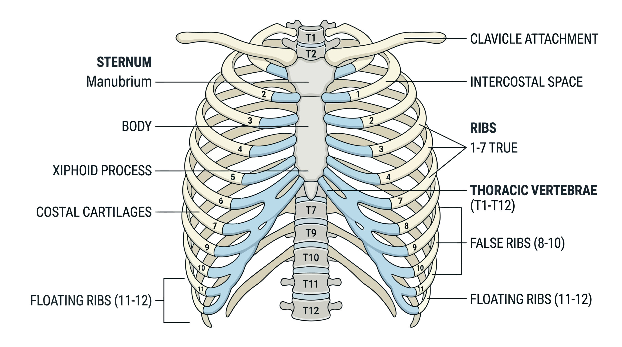
The thoracic spine consists of 12 vertebrae and is mechanically linked to the rib cage, making it more stable and less mobile than the cervical or lumbar spine.
There are several important anatomical differences.
- Every thoracic vertebral level participates in supporting the rib cage.
- The rib cage makes the thoracic spine more mechanically stable than the neck or lower back.
- The thoracic facet joints have a different orientation from lumbar facets and contribute to rotation while limiting excessive flexion and extension.
- Ribs form joints with the spine at the costovertebral and costotransverse articulations.
- Thoracic nerve roots travel around the torso along the ribs rather than down an arm or leg.
- The spinal cord remains present throughout the thoracic canal, making spinal cord symptoms especially important.
The result is a region where pain from muscles, joints, ribs, nerves, discs, vertebral bodies, and the spinal cord can overlap substantially.
The Three Important Patterns of Mid-Back Pain
One of the most useful first steps is determining whether the pain behaves primarily as axial pain, radicular pain, or a spinal cord problem.
1. Axial Thoracic Pain
Axial pain remains centered around the thoracic spine, shoulder-blade region, or adjacent ribs rather than following a nerve around the chest.
Patients may describe:
- A dull ache between the shoulder blades
- A focal painful spot next to the spine
- Stiffness after prolonged sitting
- Pain when twisting or extending the trunk
- Tenderness over a muscle, rib, facet joint, or spinous process
- Pain that changes with posture or activity
Possible generators include muscle and fascial pain, thoracic facet joints, costovertebral or costotransverse joints, the vertebral bodies, discs, ligaments, and less commonly a compression fracture.
2. Thoracic Radicular Pain
Thoracic radicular pain occurs when a thoracic spinal nerve becomes irritated or compressed. Instead of running down an arm or leg, the thoracic nerves travel around the trunk along the ribs.
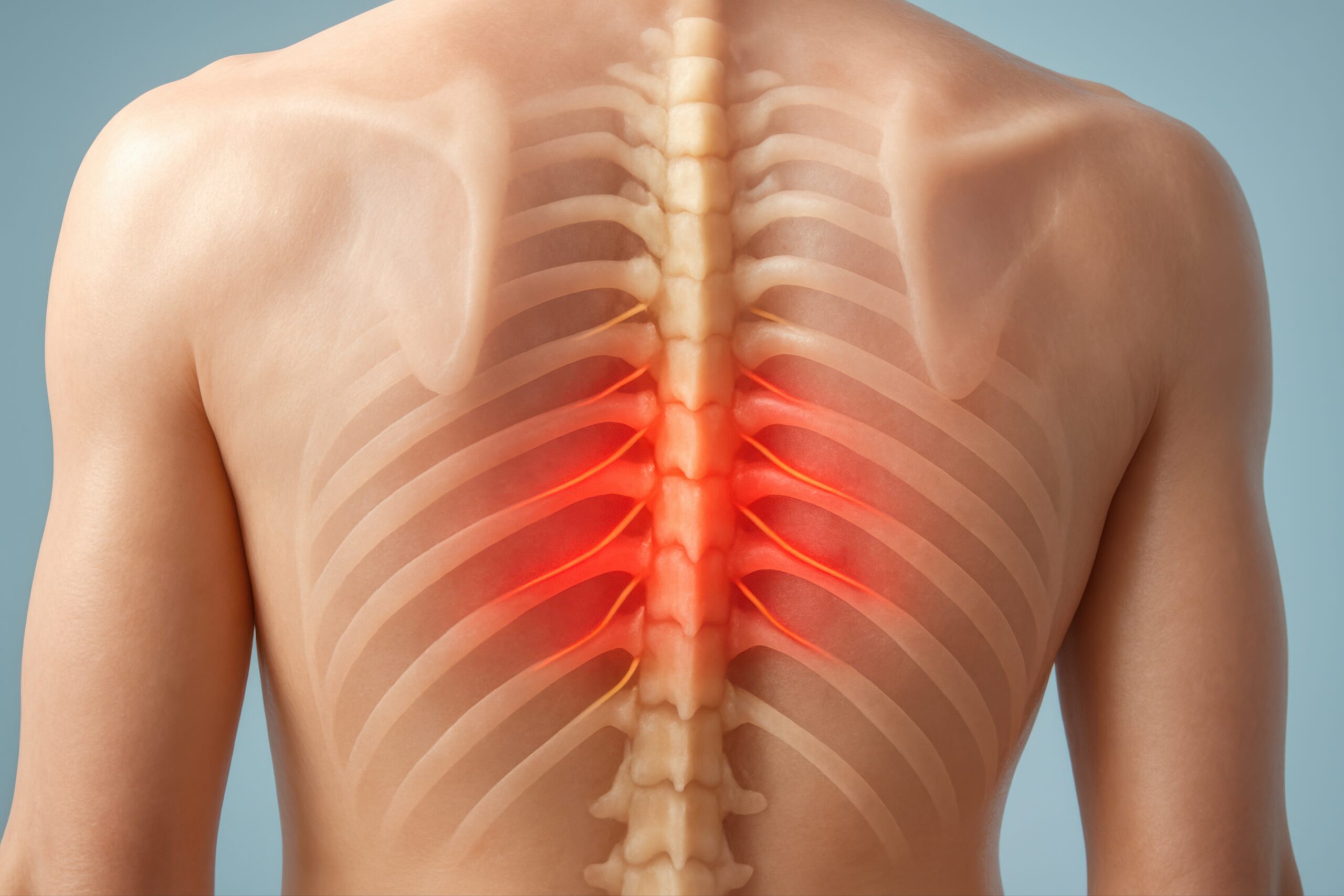
Thoracic nerve roots travel around the trunk along the ribs. Irritation of a thoracic nerve can therefore cause pain, burning, tingling, or numbness that wraps around the chest or abdomen.
The result can be a strip, band, or semicircle of pain traveling from the spine toward the side of the chest or abdomen.
Patients may describe the pain as:
- Burning
- Sharp
- Electric
- Stabbing
- Tingling
- Numb
- A tight band around one side of the chest or abdomen
A thoracic disc herniation or foraminal narrowing can produce this pattern, although intercostal neuralgia, shingles, abdominal-wall conditions, and other disorders can mimic it.
This distinction matters. A patient saying, “My mid-back hurts” and a patient saying, “The pain begins next to my spine and wraps around my left ribs toward my abdomen” may have completely different pain generators.
3. Thoracic Myelopathy: When the Spinal Cord Is Involved
The third pattern is the one that requires particular attention. Myelopathy means dysfunction of the spinal cord itself.
Unlike an irritated thoracic nerve root, spinal cord compression can affect everything below the involved level. Symptoms may include:
- Leg heaviness or weakness
- Unusual stiffness in the legs
- Difficulty walking
- Loss of balance
- Changes in leg coordination
- Numbness or altered sensation below a particular level on the trunk
- Changes in bowel or bladder function
Thoracic spinal cord compression may result from severe stenosis, a large disc herniation, tumor, infection, fracture, or other structural abnormality. These symptoms require timely neurologic evaluation and usually MRI.
Pain Between the Shoulder Blades Does Not Always Come From the Mid-Back
This is one of the most important diagnostic traps.
A patient points directly between the shoulder blade and the spine. It is tempting to conclude that a rhomboid muscle or thoracic joint must be responsible. Sometimes that is correct. Sometimes the actual source sits several inches higher.
The lower cervical spine frequently refers pain into the upper thoracic and scapular region. Cervical facet joints, discs, and nerve roots can all produce pain felt around the shoulder blade.
That means persistent pain between the shoulder blades should often trigger examination of the neck as well as the thoracic spine.
The shoulder itself can also refer discomfort into this region. Problems such as rotator cuff tendinopathy may coexist with or mimic spine pain.
Location alone does not identify the source. Where pain is felt and where pain originates are not always the same place.
Common Causes of Mid-Back Pain
Muscle and Myofascial Pain
Muscular and myofascial pain is among the most frequent explanations for uncomplicated mid-back discomfort.
The trapezius, rhomboids, paraspinal muscles, latissimus, intercostal muscles, and other stabilizers of the shoulder blade and thoracic spine work continuously to maintain posture and control movement.
Common triggers include:
- Prolonged computer or desk work
- Repetitive lifting
- Sudden increases in exercise
- Poor conditioning
- Carrying loads asymmetrically
- Prolonged driving
- Muscle strain after coughing or illness
Myofascial pain frequently produces palpable tender areas or trigger points. The pain generally changes with movement or posture and may improve with heat, stretching, massage, physical therapy, strengthening, and activity modification.
Persistent focal muscle pain may occasionally be treated with trigger point injections.
Thoracic Facet Joint Pain
Facet joints are the small paired joints at the back of each spinal segment. Just as they can become painful in the cervical and lumbar spine, thoracic facet joints can develop arthritis, inflammation, injury, or mechanical overload.
Facet-mediated thoracic pain is usually axial. It may be felt:
- Beside the spine
- Between the shoulder blades
- Across part of the mid-back
- Occasionally toward the posterior ribs
Extension, twisting, prolonged standing, or rotation may aggravate symptoms, but no single movement conclusively diagnoses a painful facet joint.
Imaging can demonstrate arthritis but cannot prove that a facet joint hurts. When the diagnosis remains important and uncertain, appropriately performed medial branch blocks may be used diagnostically.
If controlled diagnostic blocks confirm facet-mediated pain and symptoms remain significant despite conservative treatment, radiofrequency ablation may be considered in appropriately selected patients.
Costovertebral and Costotransverse Joint Pain
The thoracic spine has a category of joints that the neck and lower back simply do not have.
Each rib articulates with the spine through the costovertebral and costotransverse joints. Together they allow the ribs to move during breathing while keeping the chest wall mechanically stable.
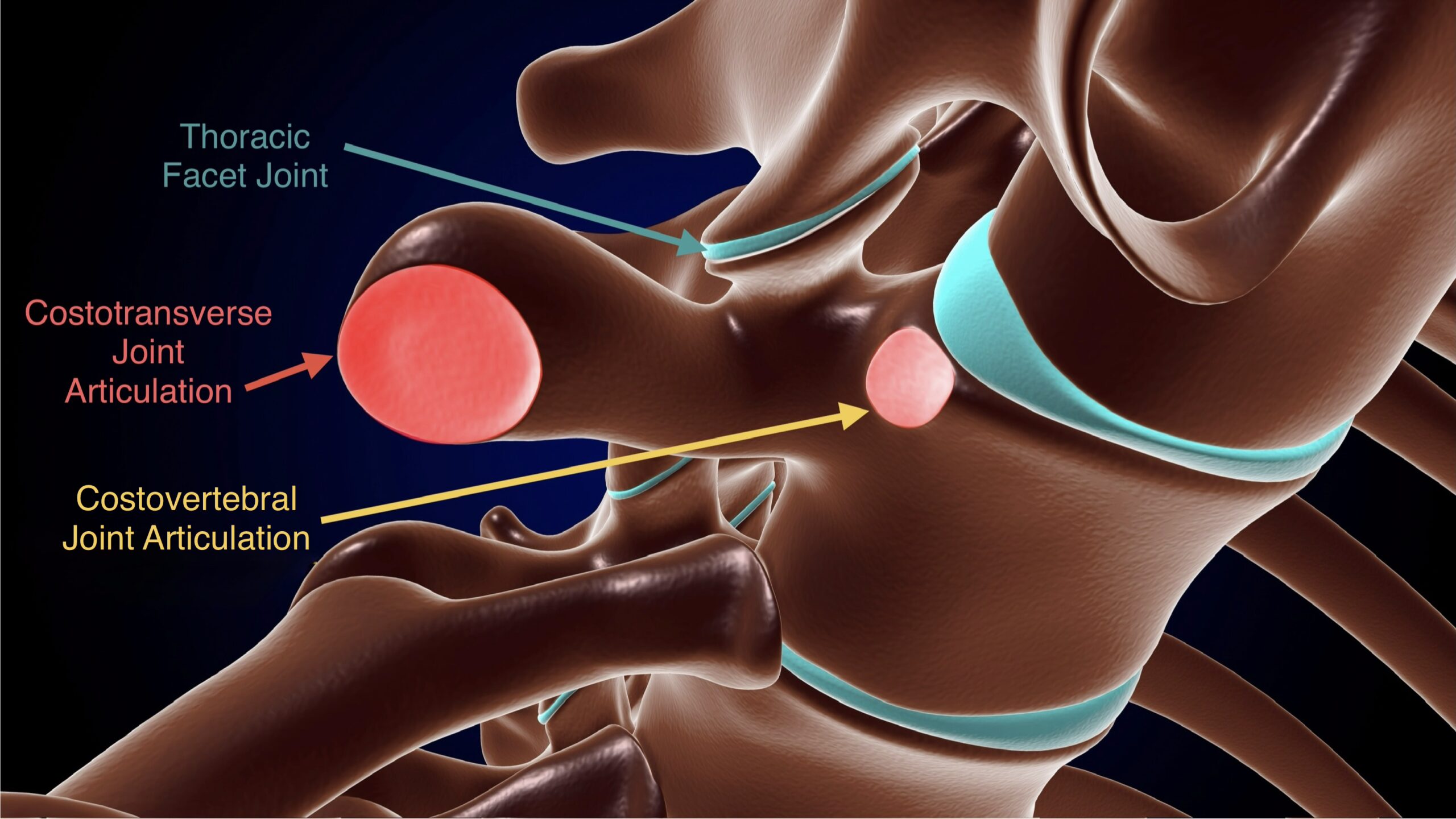
The thoracic spine has unique rib-spine articulations. The costovertebral and costotransverse joints move with breathing and trunk rotation and can become sources of mid-back and rib pain.
These joints can become irritated by trauma, repetitive movement, arthritis, coughing, altered thoracic mechanics, or other stresses.
Pain is often felt several centimeters lateral to the center of the spine and can sometimes travel along the rib. Deep breathing, coughing, rotation, or pressure over the involved rib articulation may reproduce symptoms.
Because rib-joint pain may extend toward the side or front of the chest, cardiac and pulmonary causes may need to be excluded before labeling chest-wall pain as musculoskeletal.
This rib-spine interface is one of the reasons mid-back pain deserves to be treated as its own anatomical category rather than simply as “upper back pain.”
Thoracic Disc Bulge or Herniated Disc
Thoracic disc abnormalities are considerably less familiar to patients than cervical or lumbar disc problems.
A thoracic disc bulge or herniated disc may cause no symptoms at all. When symptomatic, the presentation depends on what the disc affects.
- Axial pain: discomfort centered in the thoracic spine.
- Radicular pain: pain or altered sensation wrapping around a rib toward the chest or abdomen.
- Spinal cord compression: leg weakness, balance difficulty, altered sensation, or other signs of myelopathy.
This is another example of why the MRI cannot be interpreted in isolation. A disc abnormality becomes clinically meaningful when its location and anatomy match the patient’s symptoms and examination.
Thoracic Spinal Stenosis
Spinal stenosis means narrowing of the space available for neurologic structures.
In the lumbar spine, stenosis commonly affects the nerve roots and produces leg pain or neurogenic claudication. In the thoracic spine, significant central narrowing can affect the spinal cord.
For that reason, thoracic stenosis accompanied by progressive walking difficulty, leg weakness, abnormal reflexes, or bowel or bladder changes is a very different clinical problem from uncomplicated mechanical back pain.
Vertebral Compression Fracture
A vertebral compression fracture is an important cause of sudden thoracic and thoracolumbar pain, particularly in people with osteoporosis.
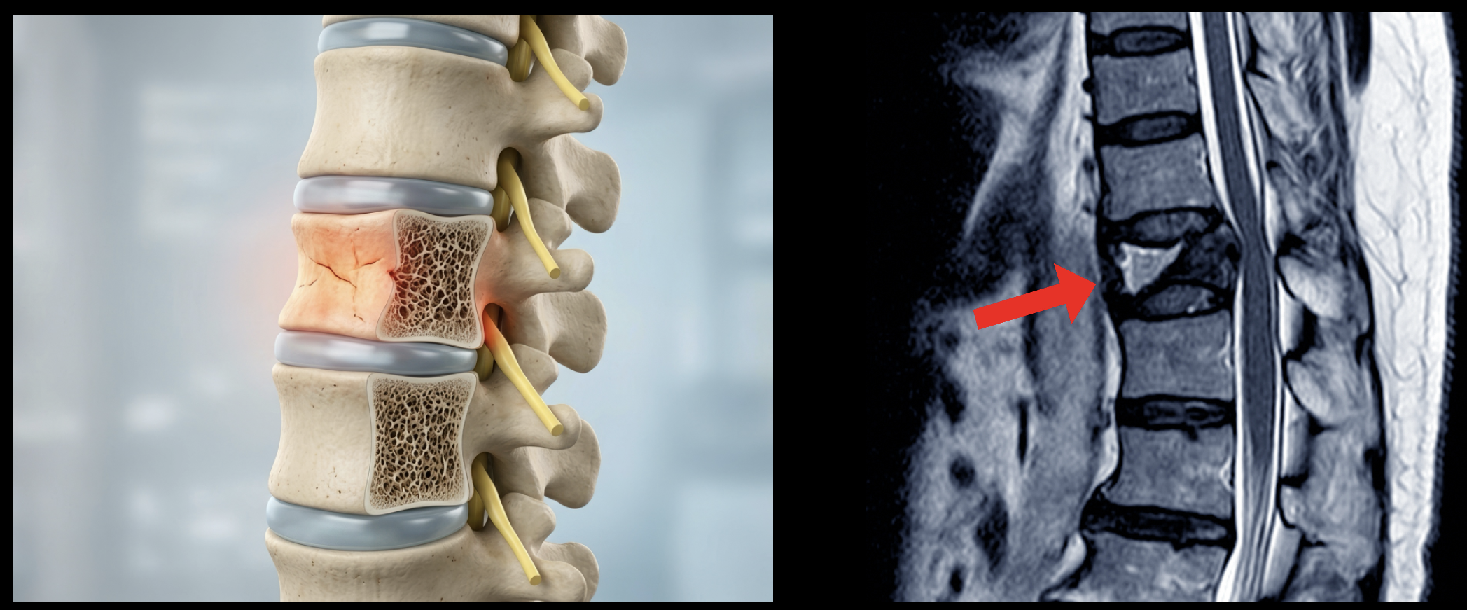
A vertebral compression fracture causes loss of height and collapse of the vertebral body. Osteoporosis is an important risk factor, and some fractures occur after surprisingly minor stress or trauma.
Fractures do not always follow dramatic trauma. In weakened bone, an apparently minor event such as lifting, bending, coughing, or a low-energy fall may be enough.
Typical clues include:
- Sudden focal mid-back pain
- Pain worse when standing or changing position
- Marked focal tenderness
- Older age
- Known osteoporosis or osteopenia
- Previous fragility fracture
- Long-term corticosteroid exposure
When a compression fracture is identified, the evaluation should not stop with the painful vertebra. The underlying bone health matters because one fragility fracture increases concern for future fractures.
Many fractures are managed conservatively. In appropriately selected patients with persistent severe pain from a confirmed acute or subacute fracture, vertebral augmentation, including kyphoplasty, may be considered.
Thoracic Spondylosis
Spondylosis is the broad term for degenerative changes in the spine, including disc-space narrowing, osteophytes, and facet-joint degeneration.
These changes become increasingly common with age and are not automatically painful. The challenge, as in every other part of the spine, is distinguishing an incidental imaging finding from the structure actually producing symptoms.
Intercostal Neuralgia
The intercostal nerves travel beneath the ribs around the chest wall. Injury or irritation can produce intercostal neuralgia.
Patients frequently describe a burning, stabbing, hypersensitive, or electric pain following the course of a rib.
Potential causes include:
- Thoracic or chest surgery
- Rib trauma
- Nerve entrapment
- Shingles
- Postherpetic neuralgia
- Occasionally unexplained nerve irritation
When shingles-related nerve pain continues after the acute rash resolves, it may represent postherpetic neuralgia.
Depending on the diagnosis and severity, intercostal nerve blocks may sometimes help confirm and treat the involved nerve.
Spinous Process and Posterior Element Pain
Very focal pain directly over the midline may arise from the posterior elements of the spine rather than the disc or nerve roots.
If one particular bony prominence is exquisitely tender, the pattern may point toward spinous process pain, local ligamentous irritation, trauma, or another focal posterior source.
This is another situation in which careful palpation and examination can reveal information an MRI report alone cannot provide.
Kyphosis, Scoliosis, and Structural Alignment
The normal thoracic spine has a gentle kyphotic curve. Excessive kyphosis, scoliosis, prior compression fractures, and other structural changes can alter load distribution across the discs, muscles, facet joints, and ribs.
The curvature itself is not necessarily the pain generator. Many people with scoliosis or increased kyphosis have little or no pain. The clinical question is whether the altered mechanics are overloading a particular structure.
In adolescents and young adults, structural thoracic kyphosis may occasionally be related to Scheuermann disease.
Inflammatory Back Pain
Not all spinal pain is caused by mechanical wear or injury.
Inflammatory conditions such as axial spondyloarthritis can cause back pain and stiffness, often beginning at a younger age. Clues may include prolonged morning stiffness, improvement with movement, pain during the second half of the night, alternating buttock pain, or associated inflammatory conditions.
A pattern suggestive of inflammatory disease warrants a different workup from ordinary mechanical thoracic pain.
Mid-Back Pain That Comes From Somewhere Else
The thoracic spine sits behind the chest and upper abdomen. Consequently, pain felt in the mid-back is not automatically a spine problem.
Potential non-spinal sources include conditions involving the:
- Heart and major blood vessels
- Lungs and pleura
- Esophagus
- Gallbladder
- Pancreas
- Kidneys
- Stomach and upper gastrointestinal tract
This does not mean that every episode of mid-back pain requires an extensive medical investigation. It means the clinical history matters.
Pain associated with exertional chest pressure, difficulty breathing, fever, significant abdominal symptoms, fainting, or an abrupt severe onset should not simply be attributed to arthritis or muscle strain.
For patients whose chest-wall pain remains after cardiac, pulmonary, and gastrointestinal causes have been appropriately evaluated, see our guide to non-cardiac, non-pulmonary chest pain.
Pain near the front of the chest may also arise from costochondritis, while pain traveling along the rib can arise from intercostal neuralgia.
Dr. Amit Sharma & our minimally invasive pain & spine team.
How We Diagnose the Source of Mid-Back Pain
A useful diagnosis comes from putting together several pieces rather than treating the MRI as the answer key.
Step 1: Understand the Pain Pattern
The history often narrows the possibilities considerably before imaging is reviewed.
Important questions include:
- Is the pain in the center, beside the spine, or underneath a shoulder blade?
- Does it wrap around a rib?
- Is it burning, aching, electric, sharp, or pressure-like?
- Did it begin suddenly or gradually?
- Does twisting reproduce it?
- Does breathing change it?
- Is it related to meals or exertion?
- Is it worse with sitting, standing, lying down, or changing position?
- Does neck movement reproduce the shoulder-blade pain?
- Is there numbness, tingling, weakness, or difficulty walking?
- Was there a fall, accident, coughing episode, or lifting injury?
- Is osteoporosis present?
Step 2: Examine More Than the Painful Spot
A thoracic evaluation should not consist solely of pressing where it hurts.
Depending on the presentation, the examination may include:
- Thoracic range of motion
- Facet-loading maneuvers
- Palpation of thoracic paraspinal and scapular muscles
- Examination of the ribs and costovertebral region
- Assessment of the cervical spine
- Shoulder examination
- Sensory examination of the trunk
- Strength testing of the legs
- Reflex testing
- Gait and balance assessment
- Signs suggesting spinal cord dysfunction
The surrounding regions matter because cervical disease, shoulder pathology, chest-wall disorders, and visceral conditions can all masquerade as thoracic spine pain.
Step 3: Decide Whether Imaging Is Necessary
Not every patient with mid-back pain needs an immediate MRI.
When pain is acute, appears mechanical, the neurologic examination is normal, and no concerning features are present, an initial period of conservative treatment is often appropriate.
Imaging becomes more important when there is:
- Significant trauma
- Concern for compression fracture
- Known osteoporosis or major fracture risk
- Progressive neurologic symptoms
- Clinical evidence of myelopathy
- Concern for malignancy or infection
- Persistent symptoms that fail appropriate conservative treatment
- A need to evaluate a suspected structural lesion before intervention
X-Ray
X-rays are useful for evaluating vertebral height, compression fractures, major degenerative changes, kyphosis, scoliosis, and overall alignment.
They cannot adequately evaluate the spinal cord, nerve roots, or most disc pathology.
MRI
MRI is particularly valuable when the spinal cord, discs, nerve roots, infection, malignancy, or an acute/subacute compression fracture needs to be assessed.
An MRI may identify:
- Thoracic disc herniation
- Spinal cord compression
- Spinal stenosis
- Neural foraminal narrowing
- Compression fracture
- Bone-marrow abnormalities
- Inflammatory or infectious abnormalities
- Other lesions requiring further investigation
But an abnormal MRI does not automatically reveal the pain generator. Structural abnormalities can exist without causing symptoms.
CT
CT provides excellent detail of bone and may be useful when evaluating fracture anatomy, complex osseous abnormalities, or situations where MRI cannot answer the relevant question.
Step 4: Match the Story, Examination, and Imaging
This is the same principle that guides our approach to neck pain and low-back pain.
A patient may have several abnormalities on MRI. One level may show a disc bulge. Several facet joints may look arthritic. There may be mild scoliosis and degenerative changes at multiple levels.
The goal is not to treat every abnormal word in the radiology report.
The goal is to identify which finding, if any, matches:
- Where and how the patient hurts
- What the physical examination demonstrates
- What the imaging shows anatomically
Pain + examination + imaging. When all three point to the same structure, confidence in the diagnosis rises substantially.
Step 5: Diagnostic Injections When the Answer Remains Unclear
Imaging sometimes narrows the possibilities without providing a definitive answer.
For example, several thoracic facet joints may appear degenerative, but the MRI cannot tell us whether any one of them is actually producing the pain.
In selected cases, image-guided diagnostic injections can temporarily anesthetize a suspected structure. A carefully interpreted response can provide another piece of evidence about the source of pain.
Examples include diagnostic medial branch blocks for suspected facet pain and selected nerve blocks when a peripheral or intercostal nerve is suspected.
These procedures should answer a specific clinical question. They should not be used simply because an MRI contains multiple abnormalities.
Treatment of Mid-Back Pain
There is no single “mid-back pain treatment” because mid-back pain is a symptom rather than a diagnosis.
Treatment should follow the identified or most likely pain generator.
Conservative Treatment
For uncomplicated mechanical and muscular thoracic pain, conservative treatment is usually the appropriate starting point.
This may include:
- Remaining appropriately active
- Temporary modification of aggravating activities
- Thoracic mobility exercises
- Scapular stabilization
- Postural retraining
- Physical therapy
- Core and posterior-chain strengthening
- Heat or ice when helpful
- Appropriate nonprescription or prescription medication
Prolonged bed rest is rarely useful for ordinary mechanical spine pain. The aim is generally to control symptoms while progressively restoring comfortable movement and function.
Trigger Point Injections
When a persistent component of pain is clearly myofascial and focal, trigger point injections may sometimes be added to an exercise and rehabilitation program.
The injection is not a substitute for addressing the mechanical or conditioning factors that allowed the muscle pain to persist.
Thoracic Medial Branch Blocks and Radiofrequency Ablation
When the thoracic facet joints are suspected as the source of persistent axial pain, medial branch blocks can help test that diagnosis.
If the diagnostic response supports facet-mediated pain and appropriate criteria are met, radiofrequency ablation may provide longer-lasting relief by interrupting the small sensory nerves supplying the painful facet joints.
The key word is confirmed. Facet arthritis on imaging alone is not enough.
Intercostal Nerve Blocks
For selected patients with pain following an intercostal nerve distribution, an intercostal nerve block may be used diagnostically and therapeutically.
The first job is determining whether the problem truly arises from the intercostal nerve rather than a thoracic nerve root, rib joint, chest-wall structure, or internal organ.
Thoracic Epidural Steroid Injection
Thoracic epidural procedures are not routine treatment for nonspecific mid-back pain.
In carefully selected cases in which clinical findings and imaging identify thoracic nerve-root inflammation or another appropriate epidural pain generator, an epidural steroid injection may sometimes be considered.
Because the thoracic anatomy includes the spinal cord, procedure selection and technique require particular attention to anatomy and risk.
Treatment of Compression Fractures
A painful vertebral compression fracture follows a different pathway from ordinary muscular or facet-mediated pain.
Treatment may include analgesic management, bracing in selected cases, activity modification, osteoporosis evaluation and treatment, and time.
When severe fracture pain persists and imaging confirms that the fracture remains the likely active pain generator, minimally invasive vertebral augmentation may be considered.
Kyphoplasty is one form of vertebral augmentation in which the fractured vertebral body is stabilized from within.
The decision depends on fracture age, imaging characteristics, symptoms, overall health, and whether the fracture actually explains the pain.
When Surgery or Urgent Specialist Evaluation Is Needed
Most thoracic pain does not require surgery.
Referral for surgical or urgent specialist assessment becomes more important when there is:
- Progressive spinal cord dysfunction
- Significant thoracic cord compression
- Progressive weakness
- Unstable fracture
- Severe structural compression associated with neurologic deterioration
- Tumor or infection requiring surgical management
The objective is not to escalate treatment because pain has lasted a certain number of days. It is to escalate when the diagnosis and clinical situation justify it.
What Your Pain Pattern Can Tell Us
| Pain Pattern | Possible Sources | Important Clues |
|---|---|---|
| Between shoulder blade and spine | Muscle, thoracic facet, cervical referred pain | Check whether neck motion reproduces symptoms |
| Focal pain beside the spine | Facet, rib joint, myofascial pain | Tenderness and pain with rotation may help localize it |
| Pain wrapping around a rib | Thoracic radiculopathy, intercostal neuralgia, shingles | Burning, electric, numb, or band-like quality |
| Pain with deep breathing or coughing | Rib/chest-wall structures, costovertebral joint, pulmonary causes | Chest and lung causes may need exclusion |
| Sudden focal pain after minor trauma | Compression fracture | Especially important with osteoporosis or older age |
| Mid-back pain plus gait or leg changes | Possible spinal cord involvement | Requires prompt neurologic assessment |
These patterns help organize the differential diagnosis but are not diagnostic by themselves.
When Should You See a Spine Specialist?
A mild episode after an obvious strain that steadily improves may never require specialist treatment.
An evaluation becomes more useful when:
- Pain persists despite reasonable conservative treatment
- Pain repeatedly returns
- The cause remains uncertain
- Pain follows a rib or appears neurologic
- Imaging shows several abnormalities and it is unclear which one matters
- There is concern for a compression fracture
- Pain significantly interferes with sleep, work, or normal activity
- A targeted diagnostic or interventional procedure is being considered
Urgent evaluation is appropriate when neurologic deficits, spinal cord symptoms, significant trauma, infection risk, cancer-related concerns, or other red flags are present.
The Goal Is Not to Treat the MRI
Thoracic MRI reports can contain intimidating language: disc protrusion, degeneration, osteophytes, facet arthropathy, foraminal narrowing, kyphosis, hemangioma, endplate change, and other abnormalities.
Some findings matter. Some do not.
The essential question is whether the abnormality explains the patient’s actual clinical presentation.
That is why our approach to mid-back pain is the same diagnostic principle used throughout spine care:
Match the pain pattern to the examination and the imaging.
Then treat the confirmed source.
For conditions affecting the areas immediately above and below the thoracic spine, see our guides to neck pain and low-back pain.
You can also explore the complete spine conditions guide.
Dr. Amit Sharma & our minimally invasive pain & spine team.
Frequently Asked Questions
What is the most common cause of mid-back pain?
Muscular and mechanical problems are common causes of uncomplicated mid-back pain. Postural strain, overuse, myofascial pain, thoracic facet joints, and rib-spine joints can all produce symptoms. The exact source cannot reliably be determined from pain location alone.
What causes pain between the shoulder blades?
Pain between the shoulder blades may arise from muscles, thoracic facet joints, rib joints, or the thoracic spine. Importantly, the lower cervical spine can also refer pain into the shoulder-blade region, so persistent parascapular pain often requires examination of the neck as well as the mid-back.
Can a pinched nerve in the mid-back cause pain around the ribs?
Yes. Thoracic nerve roots travel from the spine around the trunk. Irritation of one of these nerves can produce thoracic radicular pain that feels like a burning, electric, sharp, numb, or band-like sensation extending around the rib cage toward the chest or abdomen.
Why does my mid-back hurt when I take a deep breath?
Pain that changes with breathing may come from muscles, ribs, costovertebral or costotransverse joints, intercostal nerves, or other chest-wall structures. However, lung, pleural, cardiac, and other internal causes can also produce breathing-related chest or back pain. New or significant symptoms should be medically evaluated rather than assumed to be musculoskeletal.
Does all mid-back pain need an MRI?
No. Uncomplicated acute thoracic back pain without neurologic abnormalities or other red flags does not necessarily require immediate imaging. MRI becomes much more important when symptoms suggest spinal cord or nerve involvement, cancer, infection, significant trauma, fracture, or when persistent symptoms remain unexplained despite appropriate conservative treatment.
Can a compression fracture happen without a major injury?
Yes. When osteoporosis weakens a vertebra, a compression fracture may occur after relatively minor stress, including a small fall, lifting, bending, or sometimes no memorable traumatic event. Sudden focal mid-back pain in someone with osteoporosis or substantial fracture risk deserves medical evaluation.
Can neck problems cause pain in the mid-back?
Yes. Cervical joints, discs, and nerve roots can refer pain into the upper thoracic and shoulder-blade region. Pain that changes with neck position or is accompanied by arm pain, tingling, numbness, or weakness raises the possibility that the cervical spine is contributing.
What are the warning signs of spinal cord compression in the thoracic spine?
Possible warning signs include progressive leg weakness, leg stiffness, difficulty walking, loss of balance, unusual numbness below a level on the trunk, and changes in bowel or bladder function. These symptoms warrant prompt medical evaluation.
Can thoracic facet arthritis cause mid-back pain?
Yes. Thoracic facet joints can become painful from degeneration, injury, or mechanical stress. Imaging may show arthritis, but imaging alone cannot prove that a facet joint is the source. In selected patients, diagnostic medial branch blocks can help determine whether the facet joints are actually generating the pain.
What are costovertebral and costotransverse joints?
They are small joints connecting each rib to the thoracic spine. Because these joints participate in rib movement during breathing and trunk rotation, they can occasionally become painful. Symptoms may be felt next to the spine and sometimes extend along the adjacent rib.
How do you determine which structure is causing mid-back pain?
The diagnosis is based on matching the pain pattern, physical examination, and imaging rather than relying on any one of them alone. When several potential pain generators remain possible, carefully selected image-guided diagnostic injections can sometimes provide additional information.



