
L5 vs S1 Radiculopathy: How to Tell the Difference
- Posted on: Aug 21 2026
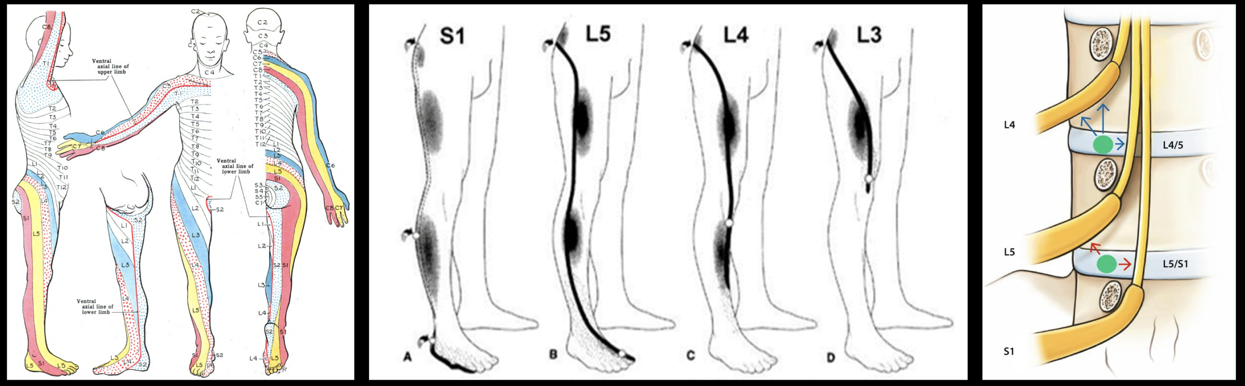
Pain starts in your lower back or buttock, travels down the leg, and reaches the foot. Your MRI shows abnormalities at L4-5, L5-S1, or perhaps both. Is the problem the L5 nerve root or the S1 nerve root?
This is one of the most common localization problems in lumbar radiculopathy. L5 and S1 sit next to each other, their pain distributions overlap, and both can produce what patients commonly call sciatica.
There are useful differences, however. L5 more commonly affects the top of the foot and big toe and may weaken muscles that lift the foot or big toe. S1 more commonly affects the back of the calf and outer or bottom of the foot and may weaken the muscles used to push off the ground or stand on the toes.
Even those patterns are not absolute. The best distinction comes from combining the pain pattern with numbness, muscle weakness, reflex changes, physical examination, and the exact location of nerve compression on MRI.
Quick Summary: L5 radiculopathy commonly causes symptoms along the outer leg and top of the foot toward the big toe, with possible weakness lifting the big toe or foot. S1 radiculopathy more often travels down the back of the leg toward the outer or bottom of the foot, with possible weakness pushing the foot downward and a reduced Achilles reflex. These patterns overlap, so pain location alone cannot reliably distinguish L5 from S1.
L5 vs S1 Radiculopathy at a Glance
| Finding | L5 Radiculopathy | S1 Radiculopathy |
|---|---|---|
| Typical pain | Outer leg toward top of foot | Back of leg toward lateral foot |
| Sensory clue | Top of foot, big toe, first web space | Lateral foot and sometimes sole |
| Motor clue | Big-toe extension, dorsiflexion, foot eversion | Plantar flexion |
| Walking clue | Difficulty heel-walking | Difficulty toe-walking |
| Reflex clue | No consistently useful routine deep-tendon reflex | Achilles reflex may be reduced |
| Common disc lesion | L4-5 paracentral disc affecting traversing L5 | L5-S1 paracentral disc affecting traversing S1 |
| Another important location | L5-S1 foraminal or far-lateral disease affecting exiting L5 | L5-S1 lateral recess affecting traversing S1 |
These are typical patterns, not rigid rules. Individual nerve distributions overlap, and not every patient develops every sensory, motor, or reflex finding.
What Does L5 Radiculopathy Feel Like?
The L5 nerve root commonly produces symptoms extending from the buttock or outer portion of the leg toward the top of the foot.
Patients may describe:
- Pain along the outer thigh or leg
- Burning or electric pain extending below the knee
- Tingling or numbness over the top of the foot
- Numbness toward the big toe or first web space
- Weakness lifting the big toe
- Weakness lifting the foot toward the shin
- Difficulty walking on the heel
- Toe drag or foot drop when weakness becomes more severe
The big toe deserves particular attention. The extensor hallucis longus, which lifts the big toe upward, receives important L5 innervation. Weakness of great-toe extension can therefore be a useful clue when L5 radiculopathy is suspected.
Our L5 nerve root pain guide covers the anatomy, symptoms, causes, and treatment of L5 involvement in much greater detail.
What Does S1 Radiculopathy Feel Like?
S1 symptoms tend to shift toward the posterior leg and lateral foot.
Patients may experience:
- Buttock pain
- Pain traveling down the back of the thigh
- Burning or shooting pain through the posterior calf
- Numbness or tingling along the outer edge of the foot
- Sensory changes involving the sole of the foot
- Weakness pushing the foot downward
- Difficulty repeatedly standing on the toes
- A reduced Achilles reflex
The functional clue is often push-off strength. The gastrocnemius and soleus muscles used for plantar flexion receive substantial S1 contribution. Significant S1 dysfunction can therefore make repeated single-leg toe raises noticeably weaker on the affected side.
Top of the Foot vs Outside of the Foot
One of the simplest distinctions patients can understand is where sensory symptoms reach the foot.
L5: think top of the foot and toward the big toe.
S1: think outer edge of the foot and toward the sole.
This distinction is useful, particularly when there is actual numbness rather than pain alone.
But it is not a diagnostic law. Sensory territories overlap between people, and radicular pain often extends beyond the neat boundaries shown on dermatome illustrations.
Heel Walking vs Toe Walking
The difference between L5 and S1 becomes more useful when we stop asking only where does it hurt? and start asking what has become weak?
L5: Heel Walking
Walking on the heels requires the ankle dorsiflexors to keep the front of the foot elevated.
L5 contributes substantially to this movement. A patient with meaningful L5 motor involvement may have difficulty keeping the forefoot elevated during heel walking.
Testing great-toe extension separately can add another useful L5 clue.
S1: Toe Walking
Standing on the toes requires plantar flexion.
Because S1 contributes strongly to the gastrocnemius and soleus muscles, S1 dysfunction can make repeated toe raises difficult.
A single toe raise may still be possible despite weakness. Repeated single-leg heel raises can make subtle asymmetry easier to detect during a clinical examination.
These maneuvers provide clues, but new weakness should be professionally evaluated rather than diagnosed from a home test.
Dr. Amit Sharma & our minimally invasive pain & spine team.
The Achilles Reflex Gives S1 an Advantage
S1 has another useful localization clue that L5 largely lacks.
The Achilles tendon reflex primarily evaluates the S1 nerve root, with contribution from S2. When S1 is significantly affected, the ankle jerk may be diminished compared with the opposite side.
L5 does not have an equally dependable routine deep-tendon reflex.
That does not mean a normal Achilles reflex excludes S1 radiculopathy, or that a reduced reflex automatically proves it. Reflexes vary with age and other neurologic conditions.
But when posterior leg pain, lateral-foot numbness, plantar-flexion weakness, and a diminished Achilles reflex all occur on the same side, the pieces begin to tell a much more coherent story.
Why L5 and S1 Pain Can Look So Similar
Dermatome diagrams make the nervous system look more orderly than it really is.
They typically show each nerve root occupying a sharply defined stripe of skin. Actual lumbar radicular pain frequently does not respect those boundaries.
Neighboring nerve roots overlap. Individual anatomy varies. Pain itself may spread beyond an area of objective sensory loss.
That is why two patients with confirmed L5 radiculopathy may draw noticeably different pain maps, and why an S1 patient may describe symptoms extending into territory that appears “L5” on a textbook diagram.
Our broader article on L4 vs L5 vs S1 nerve root pain explains why pain location should be treated as a clue rather than a stand-alone diagnostic test.
Which Disc Causes L5 Radiculopathy?
This is where the numbering becomes counterintuitive.
The L5 nerve root does not have to be compressed by the L5-S1 disc.
In fact, one of the classic causes of L5 radiculopathy is a posterolateral L4-5 disc herniation.
Why?
The L5 nerve root is already traveling downward through the spinal canal behind the L4-5 disc before it eventually exits the spine through the L5-S1 neural foramen.
At L4-5, it is therefore the traversing L5 nerve root.
L4-5 paracentral disc herniation → commonly affects the traversing L5 nerve root.
This is why an MRI saying “L4-5 disc herniation” can perfectly explain symptoms diagnosed clinically as L5 radiculopathy.
Which Disc Causes S1 Radiculopathy?
A typical posterolateral or paracentral L5-S1 disc herniation can compress the nerve traveling behind that disc.
That nerve is the traversing S1 nerve root.
L5-S1 paracentral disc herniation → commonly affects the traversing S1 nerve root.
So the classic relationship is:
- L4-5 disc → L5 radiculopathy
- L5-S1 disc → S1 radiculopathy
But there is an important exception, and it explains why simply reading the disc level from an MRI report can be misleading.
How Can L5 and S1 Both Be Affected at L5-S1?
This is one of the most useful pieces of anatomy for understanding an MRI.
At the L5-S1 level, two different nerve roots are nearby:
- The L5 nerve root is exiting through the L5-S1 neural foramen.
- The S1 nerve root is traveling downward through the spinal canal toward its exit below.
Therefore, the location of the abnormality at L5-S1 matters as much as the level itself.
Foraminal or Far-Lateral L5-S1 Disease
Narrowing or disc material within or lateral to the L5-S1 foramen can compress the exiting L5 nerve root.
This can produce L5 symptoms even though the abnormal disc is L5-S1.
Paracentral or Lateral-Recess L5-S1 Disease
Disc material or stenosis closer to the spinal canal can compress the traversing S1 nerve root.
This produces the more familiar S1 radiculopathy associated with L5-S1 disease.
Same spinal level. Different nerve roots.
At L5-S1, foraminal disease can affect L5, while lateral-recess or paracentral disease can affect S1. The MRI location matters, not simply the words “L5-S1.”
What If the MRI Shows Both L4-5 and L5-S1 Problems?
This is extremely common.
An MRI may show an L4-5 disc protrusion capable of affecting L5 and, on the same scan, L5-S1 stenosis capable of affecting L5 or S1.
The MRI then gives us several anatomically possible explanations.
It does not automatically tell us which one hurts.
The useful question becomes:
Which MRI abnormality matches the patient’s symptoms, sensory findings, weakness, reflexes, and examination?
This distinction matters because treating every abnormality visible on an MRI is neither necessary nor desirable.
Degenerative changes become increasingly common with age, including in people who have no corresponding symptoms.
What If Pain, Examination, and MRI Do Not Agree?
Sometimes they do not.
A patient may have symptoms compatible with either L5 or S1 while the MRI shows abnormalities capable of affecting both.
Depending on the clinical situation, additional evaluation may include electrodiagnostic testing or a carefully targeted selective nerve root block.
A selective nerve root block places a small amount of medication around a specific spinal nerve under image guidance. When appropriately selected and interpreted, the response can provide additional information about whether that particular root is contributing substantially to the patient’s pain.
It is not a perfect test. Diagnostic blocks should supplement the history, examination, and imaging rather than replace them.
Could L5 Symptoms Actually Come From the Peroneal Nerve?
Yes, and this is an important mimic.
The common peroneal nerve travels around the outside of the knee near the fibular head. Compression or injury there can cause:
- Weakness lifting the foot
- Foot drop
- Numbness over portions of the top of the foot
Those findings can resemble L5 radiculopathy.
But L5 and peroneal nerve problems do not affect every muscle in the same way. Examination of foot inversion, eversion, great-toe movement, hip abduction, sensory distribution, and other findings can help determine whether the problem originates at the lumbar nerve root or farther down the leg.
This distinction is important enough to deserve its own discussion rather than being reduced to a few lines here.
Does L5 or S1 Radiculopathy Always Cause Back Pain?
No.
Some patients have prominent lower back pain followed by leg symptoms. Others have relatively little back pain and predominantly experience pain, numbness, tingling, or weakness in the leg or foot.
A lumbar nerve root can be irritated even when the patient’s main complaint is nowhere near the lumbar spine.
This is one reason a patient with unexplained foot symptoms may eventually discover that the source is in the lower back.
Does L5 or S1 Radiculopathy Always Mean Surgery?
No.
Many patients with lumbar radicular pain improve without surgery, particularly when there is no progressive motor deficit or other urgent neurologic finding.
Treatment depends on the cause, severity, duration, neurologic findings, and impact on function.
Options may include:
- Activity modification while maintaining appropriate movement
- Physical therapy and rehabilitation
- Anti-inflammatory or other appropriate medications
- Epidural steroid injection for selected patients
- Selective nerve root injection when more precise targeting or diagnostic information is needed
- Surgical decompression when appropriate for severe or progressive neurologic compression or persistent disabling symptoms
The goal is not simply to treat “sciatica.” It is to understand which nerve is affected, where it is affected, and why.
When L5 or S1 Symptoms Need Urgent Evaluation
Most L5 and S1 radiculopathy is painful but not an emergency.
Prompt medical evaluation is important for:
- New or rapidly worsening leg weakness
- New significant foot drop
- Increasing difficulty walking
- Severe weakness affecting ankle or foot function
- New numbness involving the saddle or genital region
- Loss of bowel or bladder control
- Difficulty initiating urination or urinary retention
- Severe bilateral neurologic symptoms
- Fever or systemic illness accompanying severe spinal pain
- Symptoms following significant trauma
Bowel or bladder dysfunction and saddle numbness may indicate compression of multiple cauda equina nerve roots and require emergency evaluation.
So Is It L5 or S1?
If we had to reduce the distinction to its most useful clinical clues:
- Top of foot or big toe + weakness lifting the big toe or foot → think L5.
- Back of calf or lateral foot + weak push-off + reduced Achilles reflex → think S1.
But “think” is the important word.
Neither pattern is sufficiently reliable to diagnose a nerve root from symptoms alone.
The strongest diagnosis occurs when several independent pieces of evidence agree:
- The pain distribution is compatible with the nerve.
- Sensory changes support the same root.
- Muscle testing identifies matching weakness.
- Reflex findings provide additional support where applicable.
- The MRI demonstrates compression in the correct anatomical location.
When those findings point in different directions, the correct response is not to force the patient into a dermatome diagram. It is to investigate why they do not match.
For a detailed discussion of L5 anatomy, including how the same L5 nerve can be compressed at both L4-5 and L5-S1, see our L5 nerve root pain guide.
Dr. Amit Sharma & our minimally invasive pain & spine team.
Frequently Asked Questions
What is the main difference between L5 and S1 radiculopathy?
L5 more commonly affects the outer leg, top of the foot, and big toe and may weaken great-toe extension or ankle dorsiflexion. S1 more commonly affects the back of the calf and lateral or plantar foot and may weaken plantar flexion. S1 can also reduce the Achilles reflex.
Is big toe numbness L5 or S1?
Numbness toward the big toe or first web space is more characteristic of L5 involvement. S1 sensory symptoms are more commonly felt toward the lateral or plantar foot. These sensory distributions overlap, so big-toe numbness alone cannot prove L5 radiculopathy.
Is outside-of-the-foot numbness L5 or S1?
Numbness along the lateral edge of the foot is more characteristic of S1. L5 symptoms more commonly involve the top of the foot and region toward the big toe.
Does L5 radiculopathy affect the Achilles reflex?
Not typically. The Achilles reflex primarily evaluates S1, with contribution from S2. L5 does not have an equally reliable routine deep-tendon reflex, so strength and sensory testing become particularly important when evaluating suspected L5 radiculopathy.
Why does an L4-5 disc herniation cause L5 radiculopathy?
The L5 nerve root travels downward behind the L4-5 disc before exiting the spine at L5-S1. A typical paracentral L4-5 disc herniation can therefore compress the traversing L5 nerve root.
Can L5-S1 cause either L5 or S1 radiculopathy?
Yes. At L5-S1, foraminal or far-lateral disease can affect the exiting L5 nerve root, while paracentral or lateral-recess disease can affect the traversing S1 nerve root. The location of the abnormality is therefore as important as the spinal level.
Which nerve root causes difficulty walking on the heels?
L5 dysfunction can weaken ankle dorsiflexion and may make heel walking difficult. Severe weakness can contribute to toe drag or foot drop, although other neurologic conditions can produce similar findings.
Which nerve root causes difficulty walking on the toes?
S1 dysfunction can weaken plantar flexion and make repeated toe raises or toe walking difficult. A reduced Achilles reflex may provide another clue to S1 involvement.
Can L5 radiculopathy cause foot drop?
Yes. Significant L5 motor dysfunction can contribute to weakness lifting the foot and produce foot drop. However, peroneal nerve injury and other neurologic disorders can also cause foot drop, so new weakness requires proper evaluation.
Can you have L5 or S1 radiculopathy without back pain?
Yes. Some patients have predominantly leg or foot pain, numbness, tingling, or weakness with little accompanying low back pain. The location of the symptoms does not necessarily identify where along the nerve pathway the problem originates.
References
- Lumbar radicular pain. Review of lumbar nerve-root anatomy, clinical patterns, diagnosis and management. PMC
- Allegri M, et al. Mechanisms of low back pain: a guide for diagnosis and therapy. Discussion of radicular pain, dermatomal sensory loss and myotomal weakness. PMC
- Murphy DR, et al. Pain patterns and descriptions in patients with radicular pain: Does the pain necessarily follow a specific dermatome? PMC
- Iversen T, et al. Accuracy of physical examination for chronic lumbar radiculopathy. PMC
- Radicular Back Pain. StatPearls. National Center for Biotechnology Information. NCBI Bookshelf
Tagged with: L5 Radiculopathy, Leg Pain, Lumbar Radiculopathy, S1 Radiculopathy, Sciatica
Posted in: News, Special Report



