
L4 vs L5 vs S1 Nerve Root Pain: How to Tell
- Posted on: Aug 21 2026
You have pain running down your leg. Your MRI mentions several abnormal discs. One doctor says L4, another says L5, and the report mentions narrowing near S1.
So which nerve is actually causing the pain?
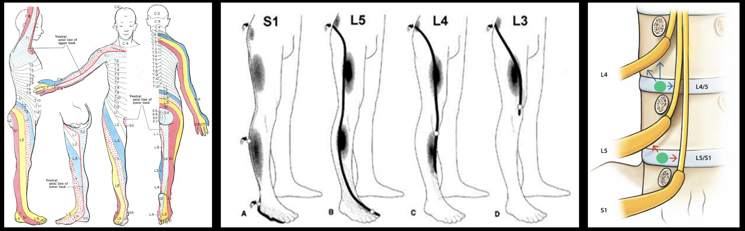
L4 vs L5 vs S1 nerve root pain can produce different patterns of numbness, weakness, and radiating leg symptoms. Textbook diagrams make those patterns look remarkably precise: L4 goes here, L5 goes there, S1 follows another stripe.
Real patients are less cooperative.
Pain from neighboring lumbar nerve roots frequently overlaps, and pain location alone often cannot reliably distinguish an L4 problem from L5 or S1. The more useful clues are usually the combination of where sensation changes, which muscles become weak, which reflexes change, what the examination shows, and whether the MRI abnormality fits the same anatomy.
Quick Summary: L4 problems more often affect the front of the thigh, knee and inner lower leg and may weaken knee extension. L5 problems commonly involve the outer leg, top of the foot and big toe and may weaken great-toe extension or foot movement. S1 problems more often affect the back of the leg and outside or bottom of the foot and may weaken push-off or toe-walking. These are useful patterns, but they overlap. Pain location alone should not be used to diagnose the affected nerve root.
First: What Do L4, L5, and S1 Actually Mean?
L4, L5, and S1 are spinal nerve roots arising from the lower lumbar and upper sacral regions.
After leaving the spine, fibers from these roots combine with other nerves within the lumbosacral plexus and ultimately contribute to the major nerves supplying the leg.
When a lumbar nerve root becomes inflamed or compressed, the result can include:
- Radiating leg pain
- Burning or electric sensations
- Numbness or tingling
- Muscle weakness
- Changes in reflexes
Pain generated by irritation of a spinal nerve root is called radicular pain. When nerve-root dysfunction produces objective sensory or motor abnormalities, the term radiculopathy becomes particularly appropriate.
For a broader explanation, see our lumbar radiculopathy guide.
L4 vs L5 vs S1: The Quick Comparison
| Nerve Root | Common Sensory Area | Weakness That May Occur | Reflex Clue | Functional Clue |
|---|---|---|---|---|
| L4 | Front of thigh, knee, medial lower leg | Knee extension; sometimes ankle dorsiflexion | Patellar reflex may decrease | Knee may buckle; difficulty rising or climbing stairs |
| L5 | Outer leg, top of foot, big toe/first web space | Great-toe extension, foot dorsiflexion or eversion, hip abduction | No consistently useful routine deep-tendon reflex | Difficulty heel-walking; toe drag or foot weakness |
| S1 | Back of calf, lateral foot, sometimes sole | Ankle plantar flexion | Achilles reflex may decrease | Difficulty toe-walking or pushing off |
These are clinical tendencies rather than rigid boundaries. Individual nerve distributions overlap, and not every patient develops every finding.
What Does L4 Nerve Root Pain Feel Like?
L4 nerve-root involvement tends to produce symptoms higher in the leg than L5 or S1.
Possible symptoms include:
- Pain in the lower back or front of the thigh
- Pain around the knee
- Numbness or tingling toward the inner lower leg
- Weakness straightening the knee
- A feeling that the knee is unstable or buckles
- A reduced patellar or knee-jerk reflex
L4 motor fibers contribute importantly to the quadriceps, which straighten the knee. That is why significant L4 dysfunction can show up not simply as pain, but as difficulty climbing stairs, rising from a low chair, or trusting the knee while walking.
What Does L5 Nerve Root Pain Feel Like?
L5 is one of the most frequently involved lumbar nerve roots.
The classic pattern includes symptoms traveling through the outer portion of the leg toward the top of the foot and big toe.
Possible L5 findings include:
- Buttock or outer-leg pain
- Burning or tingling extending below the knee
- Numbness over the top of the foot
- Big-toe or first-web-space numbness
- Weakness lifting the big toe
- Weakness lifting or turning the foot
- Difficulty heel-walking
- More severe weakness producing toe drag or foot drop
L5 is particularly interesting because there is no routinely dependable deep-tendon reflex equivalent to the L4 patellar reflex or S1 Achilles reflex. Diagnosis therefore depends heavily on the sensory and motor pattern.
We cover this nerve in much greater detail on our cornerstone page: L5 Nerve Root Pain: Symptoms, Causes & Treatment.
What Does S1 Nerve Root Pain Feel Like?
S1 symptoms generally shift toward the posterior and lateral portion of the leg.
Possible findings include:
- Pain traveling from the buttock down the back of the thigh or calf
- Numbness or tingling toward the lateral foot
- Symptoms involving the sole or outer edge of the foot
- Weakness pushing the foot downward
- Difficulty standing or walking on the toes
- A diminished or absent Achilles reflex
Functionally, S1 weakness often appears during the push-off phase of walking. A patient may notice difficulty repeatedly rising onto the toes of the affected leg.
Dr. Amit Sharma & our minimally invasive pain & spine team.
Can You Tell the Nerve Root Just From Where the Pain Travels?
Not reliably.
This is one of the most important points on this page.
Dermatome diagrams are useful teaching tools, but they can create the false impression that nerve-root pain follows sharply defined colored stripes on the leg.
Real nerve pain is much less orderly.
Studies of patients with confirmed radicular pain have found that lumbar pain frequently does not follow a classic dermatomal pattern. L5 pain in particular may vary substantially from the textbook distribution.
That means statements such as:
“The pain reaches my big toe, therefore it must be L5.”
or:
“My pain goes down the back of the calf, therefore it must be S1.”
are useful clues, but not diagnoses.
Pain tells us where to look. Numbness, weakness, reflexes, examination and matching imaging tell us whether we are probably right.
Numbness Can Be More Helpful Than Pain Location
When a nerve root is sufficiently affected to cause sensory loss, the distribution of numbness or altered sensation can sometimes provide better localization than the distribution of pain.
For example:
- L4: altered sensation around the knee or medial lower leg
- L5: altered sensation across the dorsal foot toward the big toe
- S1: altered sensation toward the posterior calf and lateral foot
Even these distributions overlap, but they become much more useful when they agree with the motor examination.
Muscle Weakness May Be the Strongest Clue
Pain is subjective and variable. True motor weakness is a different kind of finding.
If a particular group of muscles is weaker on one side and the weakness corresponds to a nerve-root pattern, the diagnosis becomes more convincing.
L4: Knee Extension
The quadriceps receive important L3-L4 input. Significant L4 involvement may make it difficult to straighten the knee against resistance.
Patients may report knee buckling, difficulty climbing stairs, or difficulty rising from a seated position.
L5: Big Toe and Foot Movement
L5 contributes substantially to:
- Great-toe extension
- Ankle dorsiflexion
- Foot eversion
- Hip abduction
Weakness lifting the big toe can be a particularly useful L5 clue.
Severe weakness may contribute to foot drop, although L5 radiculopathy is only one of several possible causes of foot drop.
S1: Push-Off Strength
S1 contributes strongly to plantar flexion, the movement used to push the foot downward.
One practical test is repeated single-leg toe raises. Significant asymmetry can suggest S1 motor dysfunction when the rest of the presentation agrees.
Reflexes Give Us Another Piece of the Puzzle
Reflexes are particularly useful because they are less dependent on how a patient describes pain.
- L4: the patellar reflex may be diminished
- L5: there is no consistently reliable routine deep-tendon reflex
- S1: the Achilles reflex may be diminished
A reduced reflex does not prove a particular radiculopathy, and reflexes can change with age or other neurologic conditions.
But when a reflex change appears on the same side as matching weakness, numbness and radiating pain, it becomes much more meaningful.
Which Disc Level Causes L4, L5, or S1 Symptoms?
This is where lumbar anatomy confuses many patients.
The disc level and the affected nerve root often have different numbers.
In a typical posterolateral disc herniation:
- L3-4 disc herniation may compress the traversing L4 nerve root
- L4-5 disc herniation may compress the traversing L5 nerve root
- L5-S1 disc herniation may compress the traversing S1 nerve root
This happens because the nerve destined to exit at the next level is already traveling downward through the spinal canal behind the disc.
But this rule is not universal.
A foraminal or far-lateral disc herniation can affect the nerve that is exiting at that same level. For example, foraminal disease at L5-S1 can compress the exiting L5 nerve root rather than S1.
This distinction between traversing and exiting nerve roots is particularly important when interpreting an MRI.
What If the MRI Shows Problems at L4-5 and L5-S1?
That is not unusual at all.
As we get older, MRI frequently reveals abnormalities at multiple lumbar levels. A scan might show:
- An L4-5 disc protrusion
- L4-5 lateral-recess stenosis
- L5-S1 disc-height loss
- L5-S1 foraminal narrowing
- Facet arthritis at both levels
If the patient has leg pain, which abnormality deserves treatment?
The answer should not be: all of them.
Instead, we try to match three things:
Symptoms + neurologic examination + matching anatomy on the MRI
When those three pieces converge on the same nerve root, confidence in the diagnosis rises considerably.
What If the Exam and MRI Still Do Not Agree?
This is where spine diagnosis becomes more interesting.
Imagine that a patient has:
- Leg pain compatible with L5 or S1
- Numbness that does not perfectly follow either dermatome
- An L4-5 abnormality that could affect L5
- An L5-S1 abnormality that could affect S1 or the exiting L5 nerve
Simply reading the MRI report may not answer the question.
Depending on the situation, additional evaluation may include more detailed neurologic testing, electrodiagnostic studies, or an image-guided selective nerve root block.
A selective block can temporarily anesthetize a particular nerve root and may provide additional diagnostic evidence when multiple levels could plausibly explain the symptoms.
It is not a perfect test, and its result still needs to be interpreted in context.
L5 vs S1: The Comparison Patients Ask About Most
L5 and S1 are probably the two nerve roots patients have the most difficulty distinguishing.
A useful shorthand is:
L5 tends to move toward the top of the foot and big toe.
S1 tends to move toward the back of the calf and outside of the foot.
But the motor examination often tells us more:
- L5: difficulty lifting the big toe or foot
- S1: difficulty pushing downward or standing repeatedly on the toes
And the reflex examination adds another distinction:
- L5: no dependable standard reflex
- S1: Achilles reflex may be reduced
Because this comparison deserves more detail, we will address L5 vs S1 radiculopathy separately rather than trying to squeeze every diagnostic nuance into one article.
Could It Be Something Other Than a Lumbar Nerve Root?
Absolutely.
Symptoms resembling L4, L5, or S1 radiculopathy can also arise from problems elsewhere.
Potential mimics include:
- Hip arthritis
- Greater trochanteric pain
- Peripheral neuropathy
- Peroneal nerve compression
- Sciatic nerve injury
- Peripheral vascular disease
- Sacroiliac joint pain
- Piriformis/deep gluteal syndromes
- Knee or foot disorders
This is particularly important for L5-like symptoms, because peroneal nerve disorders can also produce dorsal-foot numbness and foot weakness.
A dermatome diagram cannot distinguish those problems. A neurologic examination often can.
When Does Nerve Root Pain Need Urgent Evaluation?
Most lumbar radicular pain is uncomfortable rather than dangerous.
However, prompt evaluation is important for:
- New or rapidly progressive leg weakness
- New significant foot drop
- Severe weakness affecting walking
- Loss of bowel or bladder control
- Difficulty initiating urination or urinary retention
- New numbness in the saddle or genital region
- Severe bilateral leg neurologic symptoms
- Fever or systemic illness with severe back pain
- Major trauma
Bowel or bladder dysfunction and saddle numbness may indicate compression of multiple cauda equina nerve roots and require emergency evaluation.
So How Do We Really Tell L4, L5, and S1 Apart?
Not by memorizing three colored lines on a dermatome chart.
Instead, diagnosis becomes progressively stronger as several independent clues point toward the same nerve:
- Pain pattern suggests which roots should be considered.
- Sensory examination looks for a matching area of numbness or altered sensation.
- Strength testing looks for a matching myotomal weakness.
- Reflexes can provide additional evidence for L4 or S1.
- MRI determines whether there is an anatomical lesion capable of affecting that nerve.
- Diagnostic testing may occasionally help when several levels remain plausible.
The diagnosis should explain the patient, not merely the MRI.
If you are specifically dealing with pain, numbness, or weakness involving the top of the foot or big toe, our detailed L5 nerve root pain guide explains that nerve in greater depth.
Dr. Amit Sharma & our minimally invasive pain & spine team.
Frequently Asked Questions
Where does L4 nerve root pain travel?
L4 symptoms more commonly involve the front of the thigh, knee, and medial lower leg. L4 dysfunction may also weaken knee extension and reduce the patellar reflex. Pain patterns overlap, so location alone cannot confirm an L4 radiculopathy.
Where does L5 nerve root pain travel?
L5 symptoms commonly involve the outer leg, top of the foot, and big toe or first web space. L5 dysfunction can weaken great-toe extension, ankle dorsiflexion, foot eversion, or hip abduction. There is no consistently reliable routine deep-tendon reflex for L5.
Where does S1 nerve root pain travel?
S1 symptoms more commonly travel down the back of the thigh and calf toward the lateral or plantar foot. S1 dysfunction may weaken ankle plantar flexion, make toe-walking difficult, and reduce the Achilles reflex.
What is the difference between L5 and S1 radiculopathy?
L5 commonly affects sensation toward the top of the foot and big toe and may weaken big-toe extension or dorsiflexion. S1 more commonly affects the posterior calf and lateral foot and may weaken plantar flexion. The Achilles reflex may decrease with S1 involvement, while L5 does not have an equally reliable routine reflex.
Can you identify a pinched nerve from pain location alone?
Not reliably. Lumbar radicular pain frequently does not follow a perfect textbook dermatome. Pain location is useful for narrowing the possibilities, but sensory loss, motor weakness, reflex changes, examination findings, and matching imaging provide more reliable localization.
Which nerve root causes big toe numbness?
The L5 nerve root is commonly associated with altered sensation over the top of the foot and toward the big toe or first web space. Peripheral nerve problems can create similar symptoms, so big-toe numbness does not automatically prove L5 radiculopathy.
Which nerve root causes difficulty walking on the toes?
S1 dysfunction can weaken ankle plantar flexion, which may make repeated toe raises or toe-walking difficult. Significant weakness should be medically evaluated rather than diagnosed from a home test alone.
Why can an L4-5 disc herniation cause L5 symptoms?
The L5 nerve root travels downward behind the L4-5 disc before exiting the spine at L5-S1. A typical posterolateral L4-5 disc herniation can therefore compress the traversing L5 nerve root.
What if my MRI shows both L4-5 and L5-S1 abnormalities?
Multilevel abnormalities are common. The important question is which finding matches the patient’s symptoms and neurologic examination. When more than one level remains plausible, additional diagnostic testing, including a carefully selected nerve-root block in some patients, may provide more information.
References
- Lumbar radicular pain. Review of lumbar nerve-root anatomy, clinical patterns and assessment. PMC
- Allegri M, et al. Mechanisms of low back pain: a guide for diagnosis and therapy. Discussion of radicular pain, radiculopathy, dermatomal sensory loss and myotomal weakness. PMC
- Murphy DR, et al. Pain patterns and descriptions in patients with radicular pain: Does the pain necessarily follow a specific dermatome? PMC
- Iversen T, et al. Accuracy of physical examination for chronic lumbar radiculopathy. PMC
- Radicular Back Pain. StatPearls. National Center for Biotechnology Information. NCBI Bookshelf
Tagged with: L5 Nerve Root, Leg Pain, Lumbar Radiculopathy, Nerve Pain, Sciatica
Posted in: News, Special Report