

Lumbar Spinal Stenosis: Pathology-Driven Treatment Selection Before Surgical Referral
Lumbar spinal stenosis is not one disease — it is four or five distinct structural problems that require different treatments. This clinical guide covers precise imaging workup, ligamentum flavum measurement thresholds, the evidence behind MILD and InSpan, and a stepwise decision framework for non-surgical providers managing this diagnosis before and after surgical referral.
The Spine Doesn’t Lie — But the Options Aren’t Always Explained
A comprehensive guide to lumbar spinal stenosis: what it is, why it develops, and why the path from diagnosis to relief is not a straight line to the operating room.
One in ten people over the age of 60 has symptomatic lumbar spinal stenosis. Most of them spent months, sometimes years, being told their back pain was “just aging,” until the day they couldn’t walk through a grocery store without stopping to sit down. The diagnosis is common; the explanation of what is actually happening in the spine, and what the realistic options are, is far less common. This article is for patients navigating that gap, and for the primary care physicians and physician assistants who send them forward.
Part One: What Is Lumbar Spinal Stenosis?
The word “stenosis” comes from the Greek for narrowing. Lumbar spinal stenosis, or LSS, describes the gradual reduction in the space available for the nerves of the lower spine — the spinal cord’s terminal structure, called the cauda equina, and the individual nerve roots that branch from it to supply the legs, bladder, and bowel.
The spine is not a static structure. It is a dynamic, load-bearing column that absorbs and distributes the forces of every step, every seated hour, and every decade of wear. The spaces within it — the central canal, the lateral recesses flanking it, and the foraminal tunnels through which individual nerves exit — are maintained by the health of the discs, the facet joints, and the posterior ligaments. When any of these structures degenerate, those spaces narrow. When they narrow enough to compress nerve tissue, stenosis becomes symptomatic.
Lumbar spinal stenosis is currently estimated to affect over 103 million people worldwide. In the United States alone, more than 200,000 adults carry this diagnosis annually, a number projected to grow substantially as the population ages. It is the most common indication for spinal surgery in patients over the age of 65.
Part Two: The Causes — Understanding What Is Narrowing the Canal
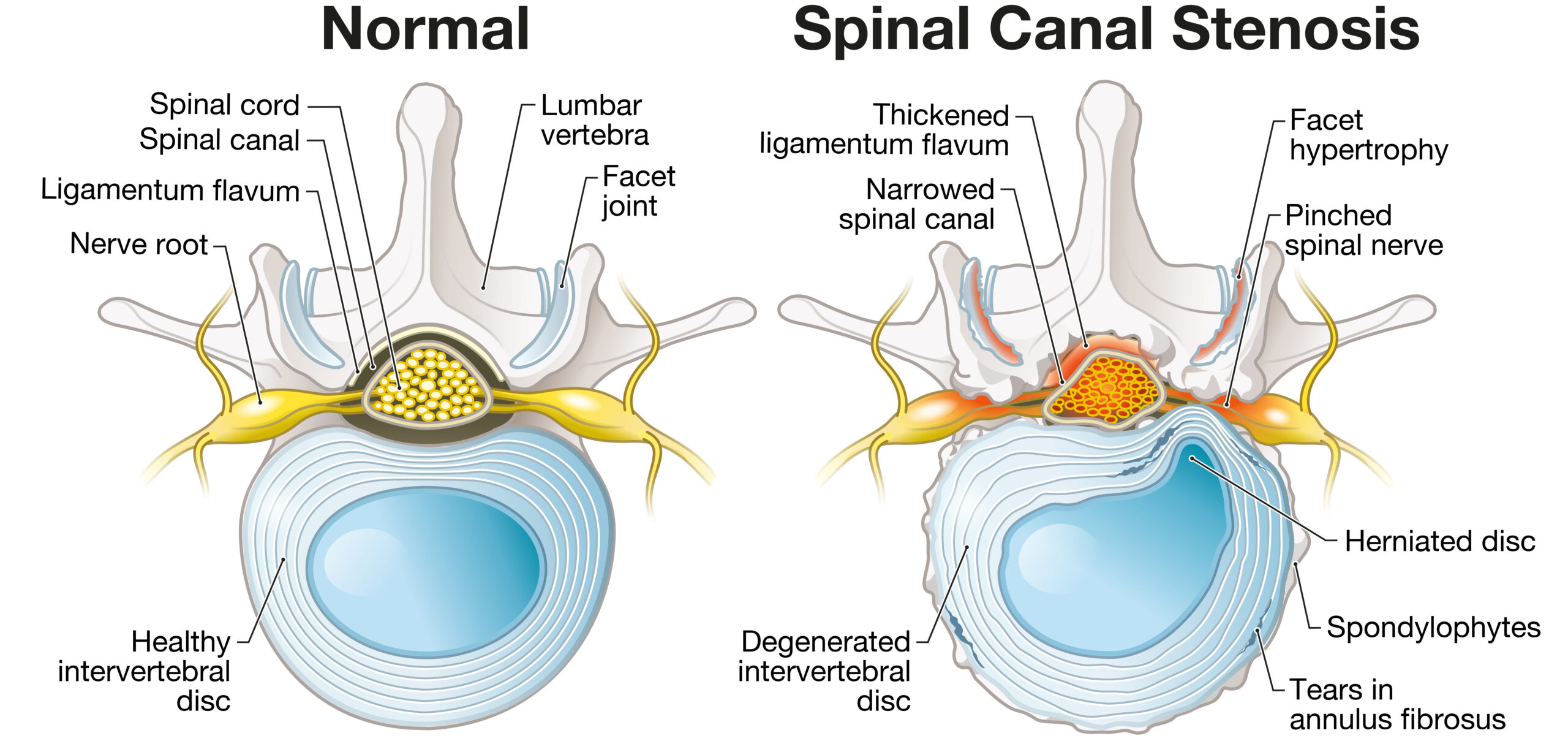
There is no single cause of lumbar spinal stenosis. It is the end product of the degenerative cascade — a cumulative process in which multiple structures fail gradually, often compounding one another. Understanding the source of narrowing in a specific patient determines which treatments are appropriate.
Ligamentum Flavum Hypertrophy
The ligamentum flavum is a paired elastic ligament running along the posterior wall of the spinal canal, connecting the laminae of adjacent vertebrae. Its normal function is to limit excessive spinal flexion and return the spine to neutral posture. With age, chronic mechanical stress, and inflammatory changes, this ligament thickens — a process called hypertrophy.
A recent decade-long retrospective study found that ligamentum flavum hypertrophy prevalence among LSS surgical patients rose from 24.5% to 44.7% between 2013 and 2023, with patients over 60 showing significantly higher rates. The L4-L5 level is most commonly affected. When the ligamentum flavum exceeds 2.5 mm in thickness — measured on axial MRI — it begins to encroach meaningfully on the posterior spinal canal, and the clinical threshold for consideration of targeted intervention is crossed.
Facet Joint Hypertrophy and Osteophyte Formation
The facet joints are the paired bony articulations at the posterior of each vertebral level, providing rotational stability to the spine. Degenerative arthritis of these joints produces bony overgrowth, called osteophytes, that can encroach on the lateral recesses and neural foramina — the tunnels through which individual nerve roots exit the central canal. Facet-driven stenosis is fundamentally a bony problem, which distinguishes it clinically and therapeutically from soft-tissue ligamentous stenosis.
Intervertebral Disc Degeneration and Herniation
Disc degeneration reduces the height of the intervertebral space, shifting load to the posterior elements and accelerating facet arthritis. A herniated or bulging disc can compress nerve roots directly, contributing to both central and foraminal stenosis. Disc-driven stenosis and facet-driven bony stenosis together represent the cases least suited to soft-tissue decompression procedures, and most likely to require surgical evaluation.
Spondylolisthesis
Degenerative spondylolisthesis refers to the forward slippage of one vertebral body relative to the one below it, caused by failure of the posterior stabilizing structures. Even low-grade spondylolisthesis, Grade I, defined as less than 25% forward translation, narrows the canal dynamically — the slip worsens in extension and reduces in flexion. It also introduces a component of segmental instability that influences treatment selection fundamentally: a decompression that does not address instability risks accelerating slip progression.
Congenital Stenosis
A small subset of patients — estimated at under 5% — develop stenosis on the basis of constitutionally narrow canals, a developmental finding present from birth. These patients may become symptomatic at a younger age than their degenerative counterparts and with less structural change required to produce symptoms.
Part Three: What Does Stenosis Feel Like? The Symptom Landscape
The hallmark symptom of lumbar spinal stenosis is neurogenic claudication. This is a reproducible pattern of pain, heaviness, numbness, or weakness in the buttocks and legs — bilateral more often than unilateral — that is brought on by standing and walking, and relieved by sitting, bending forward, or lying down. The relief with forward flexion is not coincidental: spinal flexion increases the cross-sectional area of the spinal canal, momentarily reducing nerve compression.
Distinguishing neurogenic claudication from vascular claudication is one of the first and most consequential clinical decisions in this workup. Vascular claudication — leg pain from arterial insufficiency — is relieved by rest alone, without the need to sit or bend forward. Walking uphill worsens vascular claudication because of greater exertion and oxygen demand, but improves neurogenic claudication because uphill posture induces slight flexion. The bicycle test formalizes this distinction: cycling, which keeps the spine in relative flexion, does not provoke neurogenic claudication but will provoke vascular claudication through muscle exertion. Patients with risk factors for peripheral arterial disease, including smoking history, diabetes, and hypertension, require vascular evaluation before any spinal workup proceeds.
Radiculopathy — sharp, electric, dermatomal pain radiating down one leg — can coexist with neurogenic claudication but has a different anatomical source: focal nerve root compression at a specific level, most often from a disc herniation or severe foraminal stenosis. Its management follows a different track.
Part Four: The Diagnostic Workup — Matching Imaging to Symptoms
A diagnosis of lumbar spinal stenosis requires both radiographic evidence of canal narrowing and correlating clinical symptoms. Imaging alone is insufficient — studies consistently show that many individuals with anatomical evidence of stenosis on MRI are entirely asymptomatic. The MRI confirms the anatomy; the history and physical examination confirm the diagnosis.
MRI with T2-weighted axial and sagittal sequences is the gold standard. It allows grading of central canal stenosis, measurement of ligamentum flavum thickness bilaterally, evaluation of disc morphology, and grading of foraminal stenosis on each side at each level. CT scanning adds bony detail — particularly relevant for facet assessment and surgical planning — but does not replace MRI for soft tissue evaluation.
Flexion-extension radiographs are essential when spondylolisthesis is identified on MRI. Static imaging captures a single position; dynamic films reveal whether the slip translates meaningfully with movement, which defines instability. A slip that increases by more than 3–4 mm between flexion and extension views signals dynamic instability, a finding that materially changes the treatment conversation.
Grading stenosis precisely — central versus lateral recess versus foraminal, and at which levels — is not a formality. It is the map that determines the appropriate treatment route. A patient with severe central stenosis from ligamentum flavum hypertrophy at L4-L5 requires a fundamentally different intervention than a patient with foraminal stenosis at L3-L4 from disc height loss and facet overgrowth, even if both present with similar walking limitations.
Part Five: Conservative Care — Goals, Methods, and Honest Outcomes
Conservative care is not a placeholder before surgery. For a meaningful subset of patients, it is the destination. A 2022 systematic review in BMC Musculoskeletal Disorders found that supervised physical therapy produced outcomes comparable to surgical decompression in mild-to-moderate lumbar spinal stenosis. Long-term follow-up data from conservative management cohorts shows that approximately 57% of patients maintain their clinical improvement at five years without procedural intervention — though those with severe intermittent claudication, especially walking tolerance under 100 meters, show significantly higher rates of deterioration and represent a group requiring closer monitoring and earlier escalation.
Physical Therapy
Flexion-based exercise programs reduce lumbar lordosis, temporarily widening the central canal. Core stabilization training redistributes spinal loading away from the posterior elements. The goal is not to reverse the anatomical stenosis — it is to improve functional tolerance, build compensatory muscular support, and extend the walking distance before symptom onset.
Activity Modification
Avoiding prolonged lumbar extension, such as standing at a counter, walking downhill, or carrying loads in upright posture, reduces dynamic narrowing of the stenotic canal during symptom-provoking activities. This is not rest — it is strategic posture management. A structured activity modification plan, clearly communicated, meaningfully reduces symptom frequency without further deconditioning patients.
Medications
NSAIDs address the inflammatory component of nerve root irritation and facet arthropathy. Neuropathic agents, including gabapentin and pregabalin, can reduce the quality of leg pain — its burning, electric, or numb character — though they do not address the mechanical compression driving it. Opioid analgesics have no place in the long-term management of spinal stenosis; they provide no structural relief and carry well-documented risks in the older population most affected by this disease.
Epidural Steroid Injections
The North American Spine Society holds a Grade B evidence recommendation supporting interlaminar epidural steroid injections for short- to medium-term relief of neurogenic claudication from lumbar spinal stenosis. The mechanism is reduction of local inflammatory mediators around compressed nerve roots and epidural venous congestion — not structural decompression.
ESI response carries diagnostic and prognostic value beyond the relief it provides: patients who respond meaningfully to a targeted epidural injection have confirmed the inflammatory-vascular component of their symptoms, which predicts better outcomes from subsequent targeted decompression procedures. Non-responders with severe anatomical stenosis represent a group for whom structural intervention — minimally invasive or surgical — deserves earlier consideration.
The honest limits of conservative care: it does not stop the underlying degenerative process. Ligamentum flavum does not thin with physical therapy. Facets do not remodel. Disc height does not restore. Conservative care manages symptoms and preserves function; it does not modify the disease.
Part Six: When Conservative Care Has Done Its Work — The Minimally Invasive Tier
When a patient has genuinely completed a structured conservative care program — not a brief trial, but a committed effort including physical therapy, activity modification, and at minimum one well-targeted epidural injection — and symptoms persist or progress, two minimally invasive procedural options occupy the space between injections and open surgery. Both are performed without general anesthesia. Neither requires hospitalization.
The MILD Procedure
MILD, or minimally invasive lumbar decompression, targets ligamentum flavum hypertrophy as the primary driver of central stenosis. Through a small percutaneous incision and under fluoroscopic guidance, a specialized instrument removes and debulks hypertrophied ligamentum flavum tissue from its laminar attachments posteriorly. The posterior ligamentous tension across the stenotic segment is reduced, decompressing the neural elements without implantation.
Patient selection for MILD is specific. The procedure requires MRI-confirmed ligamentum flavum hypertrophy of at least 2.5 mm, central canal stenosis with a dural sac area below 100 mm², the absence of spondylolisthesis, and the absence of significant instability. It is best suited to the patient whose primary structural problem is posterior soft tissue — not bony — compression.
What the evidence shows is instructive. The MOTION trial, a prospective multicenter randomized controlled trial, demonstrated that MILD plus conventional medical management significantly outperformed medical management alone at one year, with objective improvements in walking capacity and no device- or procedure-related adverse events. Clinical improvement is real and documented.
However, a separate multicenter RCT published in World Neurosurgery in 2020, using pre- and post-procedure MRI in 665 patients, found no measurable change in ligamentum flavum thickness or dural sac dimensions on imaging after MILD. The most defensible interpretation is that MILD produces relief through tension redistribution and posterior ligamentous disruption rather than gross anatomical decompression — a mechanism that explains both its responders and its finite durability.
For the right patient — particularly those with significant surgical risk, no instability, and ligamentum flavum-dominant stenosis — a procedure that provides meaningful functional improvement without general anesthesia or implants represents a legitimate clinical gain.
The InSpan Interlaminar Fixation System
InSpan represents a distinct therapeutic strategy: not debulking tissue, but mechanically restoring the geometry of the posterior spinal segment. The device is placed between adjacent laminae through a small posterior incision, anchoring there and performing two functions simultaneously — limiting pathological extension, the position that maximally narrows the stenotic canal, and providing a scaffold for progressive interlaminar fusion.
The biomechanical effect is quantified in the literature. In extension specifically, this device class increases canal area, subarticular diameter, foraminal area, and foraminal width — findings from cadaveric studies corroborated by in vivo CT morphometrics demonstrating statistically significant foraminal diameter enlargement at the implanted level.
InSpan’s strongest indications are the cases where MILD is least appropriate: single-level stenosis with low-grade spondylolisthesis or mild instability, foraminal stenosis where restoration of foraminal height is the primary therapeutic need, and MILD non-responders with persistent neurogenic claudication. It provides structural commitment that MILD does not. It also preserves posterior anatomy and avoids pedicle violation, maintaining the surgical access planes if escalation eventually becomes necessary.
Part Seven: A Pathology-Driven Decision Framework
The following framework is not a protocol — it is a clinical map. Every step is a decision point, not a mandatory passage.
Step 1 — Confirm the Diagnosis is Spinal, Not Vascular
Neurogenic claudication and vascular claudication overlap in symptoms and in patient demographics. Vascular evaluation is not optional in patients with relevant risk factors. Sending a patient for spinal intervention before ruling out arterial insufficiency is a diagnostic error.
Step 2 — Define the Anatomy Precisely
The MRI report must characterize which structures are causing narrowing at which levels: ligamentum flavum, disc, facet, spondylolisthesis, or a combination. Ligamentum flavum thickness must be specifically measured. Dynamic films are required if spondylolisthesis is identified. This step determines which interventions apply.
Step 3 — Red Flags Exit the Stepwise Pathway Immediately
Cauda equina syndrome, including bladder or bowel dysfunction from nerve compression, requires emergency surgical evaluation. Rapid progressive neurological deficit, significant spinal deformity, high-grade spondylolisthesis, and stenosis from bony lateral recess encroachment from facet overgrowth or disc herniation are all surgical referral criteria. These patients do not belong in a stepwise minimally invasive pathway.
Step 4 — A Genuine Trial of Conservative Care
The operative word is genuine. A six-week course of directed physical therapy, structured activity modification, and a well-targeted epidural steroid injection at the symptomatic level or levels constitutes a minimum. ESI response is diagnostic — it informs the next decision.
Step 5 — MILD if the Anatomy Fits
Ligamentum flavum thickness of 2.5 mm or greater, central stenosis dominance, no instability, no spondylolisthesis, and adequate surgical risk to benefit from avoidance of implants all support consideration of MILD. Set the expectation explicitly: MILD is a durable functional improvement, not a structural cure.
Step 6 — InSpan if MILD Is Not Indicated, or After MILD
Spondylolisthesis with mild instability, foraminal stenosis requiring height restoration, and MILD non-responders with ongoing neurogenic claudication and appropriate anatomy may be candidates for InSpan. InSpan in single-level stable disease without severe deformity represents an alternative to proceeding directly to open laminectomy or fusion — with a smaller footprint, preserved future surgical options, and no requirement for general anesthesia.
Step 7 — Surgical Referral When Warranted, Without Hesitation
Multilevel severe disease, progressive neurological deficit despite minimally invasive intervention, patient values prioritizing definitive anatomical correction, or anatomy that is clearly beyond what minimally invasive strategies can address all warrant surgical referral. A referral is not a failure of the stepwise approach — it is the stepwise approach working correctly.
Part Eight: A Note on Informed Decision-Making
Patients navigating a new diagnosis of lumbar spinal stenosis are frequently overwhelmed by the volume of information, the urgency they may feel after years of progressive symptoms, and the varying perspectives of the specialists they consult. Some important principles apply regardless of where a patient is in their care.
Not all stenosis progresses. Approximately 57% of patients managed conservatively maintain their condition at five years. Stenosis visible on MRI is not an automatic mandate for intervention — the clinical picture drives the decision, not the image alone.
The cause of stenosis determines the treatment. A patient whose stenosis is entirely from ligamentum flavum hypertrophy with no instability has different options than a patient with equivalent symptoms from facet overgrowth and a grade I slip. These are not interchangeable clinical situations, even when the presenting complaint is identical.
Sequence matters. Irreversible procedures — fusion in particular — eliminate anatomical options and create adjacent segment loading. They have a clear role in the right patient. They are most defensible when less invasive options have been genuinely offered and have genuinely failed.
Second opinions are not disloyalty. Every major spine society, including the North American Spine Society and the American Academy of Orthopaedic Surgeons, endorses a second opinion for elective spinal surgery. A patient who asks “is there something less invasive worth trying first?” is asking a medically appropriate question, and one that deserves a thorough answer.
Conclusion: The Spine Doesn’t Need to Be Rushed
Lumbar spinal stenosis is a degenerative disease with a variable natural history, a range of contributing anatomical causes, and a treatment landscape that spans from stretching exercises to multi-level spinal fusion. The patients who do best are those who receive a diagnosis precise enough to match their treatment to their anatomy, and clinicians patient enough to sequence that treatment logically.
The minimally invasive options available today — MILD, InSpan, and endoscopic decompression — did not exist a generation ago. They exist now because the gap between injections and open surgery is real, it is large, and it contains a meaningful population of patients who deserve the options that gap holds.
For patients reading this: bring the anatomy conversation to your next appointment. Ask what is specifically causing your stenosis. Ask what the least invasive option is for your specific anatomy. Understand that “wait and try conservative care” is often the right answer — and that it is not the same as being told nothing can be done.
For referring physicians and PAs: the precision of the referral letter changes the patient’s outcome. When you specify which structure is causing stenosis, at which level, with which ligamentum flavum measurement, you are not doing the specialist’s job — you are giving the specialist the roadmap to do theirs.
Dr. Amit Sharma, MD, is a Johns Hopkins fellowship-trained interventional pain management and spine care physician with clinical faculty affiliation at Columbia University. He practices at AmitSharmaMD.com across four locations in Long Island, New York: Bay Shore, Bethpage, Commack, and Huntington. This article is for educational purposes and does not constitute individualized medical advice. If you are experiencing symptoms consistent with lumbar spinal stenosis, please consult a qualified physician for a complete evaluation.



