
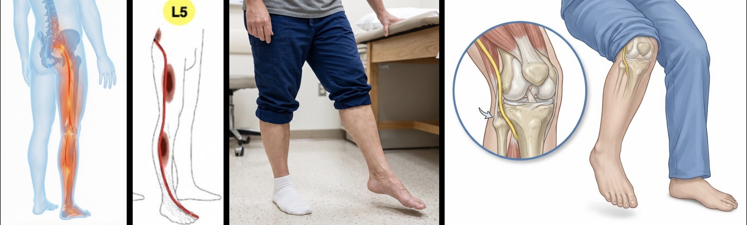
L5 Radiculopathy vs Peroneal Neuropathy: Foot Drop
- Posted on: Aug 23 2026
Your foot has become weak. The toes catch on the floor, the foot may slap when you walk, and lifting the front of the foot toward the shin has become difficult.
This is commonly called foot drop.
Two important causes can look remarkably similar: L5 radiculopathy, where a nerve root is compressed in the lower spine, and peroneal neuropathy, where a peripheral nerve is compressed or injured farther down the leg, most commonly near the outside of the knee.
L5 radiculopathy vs peroneal neuropathy distinction matters because the treatment, prognosis, and urgency may be very different.
A peroneal nerve compressed after prolonged leg crossing, weight loss, positioning, or pressure near the fibular head may improve substantially with time and conservative treatment. A progressively weakening L5 nerve compressed by a large lumbar disc herniation may require a much faster decision about surgical decompression.
So the useful question is not simply:
“Do I have foot drop?”
It is:
“Where along the nervous system is the weakness coming from, and is the nerve still recovering or continuing to deteriorate?”
Quick Summary: Both L5 radiculopathy and peroneal neuropathy can cause foot drop and numbness over the top of the foot. L5 becomes more likely when weakness also affects big-toe extension, foot inversion, or hip abduction and when symptoms extend upward into the leg or buttock. Peroneal neuropathy becomes more likely when weakness mainly affects dorsiflexion and eversion, symptoms begin around the outside of the knee, and foot inversion remains strong. Recovery is possible with either condition, but progressive or severe weakness from lumbar nerve-root compression should not simply be watched indefinitely because duration and severity of weakness influence the chance of motor recovery.
What Is Foot Drop?
Foot drop describes weakness of the muscles that lift the foot and toes upward.
Instead of smoothly clearing the ground during each step, the front of the foot may hang downward.
Patients may notice:
- The toes catching on carpet or uneven ground
- Tripping more frequently
- The foot slapping onto the floor
- Needing to lift the knee higher while walking
- Difficulty walking on the heel
- Weakness lifting the big toe
Foot drop is a sign of neurologic dysfunction, not a diagnosis by itself.
The problem can arise anywhere along the pathway controlling ankle dorsiflexion, including the brain, spinal cord, lumbar nerve roots, sciatic nerve, peroneal nerve, or the muscles themselves.
In patients with lower-back or leg symptoms, two common diagnostic considerations are L5 radiculopathy and peroneal neuropathy.
Why L5 Radiculopathy and Peroneal Neuropathy Look Similar
The overlap is anatomical.
The L5 nerve root leaves the lower spine and contributes nerve fibers that eventually travel through the sciatic nerve and into portions of the peroneal nerve.
Therefore, an injury near the beginning of that pathway at the lumbar spine and an injury farther downstream near the knee can affect some of the same muscles.
Both conditions can produce:
- Weak ankle dorsiflexion
- Weak toe extension
- Difficulty heel-walking
- Numbness over portions of the top of the foot
- Foot drop
If we examine only dorsiflexion strength, the two can therefore look nearly identical.
The distinction comes from testing movements and sensory territories that lie outside the peroneal nerve but still depend on L5.
L5 Radiculopathy vs Peroneal Neuropathy at a Glance
| Finding | L5 Radiculopathy | Peroneal Neuropathy |
|---|---|---|
| Low-back or buttock pain | May be present | Usually absent |
| Radiating leg pain | Common | Usually more localized around lateral leg/foot |
| Ankle dorsiflexion | May be weak | May be weak |
| Foot eversion | May be weak | Often weak |
| Foot inversion | May be weak | Usually preserved |
| Hip abduction | May be weak | Usually preserved |
| Big-toe extension | Often useful L5 clue | Can also be weak |
| Numbness | Outer leg/top of foot/big toe pattern possible | Lateral leg and/or dorsum of foot depending on lesion |
| Fibular-head tenderness | Not typical | May be present |
| MRI lumbar abnormality | May show L5 compression | May be incidental or normal |
The Most Useful Exam Clue: Foot Inversion
This is one of the most useful clinical distinctions.
The muscles that invert the foot receive important L5 input but are supplied through the tibial nerve, not the common peroneal nerve.
Therefore:
Foot drop + weak inversion → think more strongly about L5 radiculopathy or a more proximal nerve lesion.
Foot drop + preserved inversion → isolated peroneal neuropathy becomes more likely.
No single maneuver is perfect, but this distinction is much more informative than simply asking whether the patient can dorsiflex the ankle.
Hip Abduction Gives Us Another L5 Clue
L5 also contributes to the gluteus medius, an important muscle used to move the leg outward and stabilize the pelvis during walking.
This muscle is supplied through the superior gluteal nerve, far upstream from the peroneal nerve.
Therefore, weakness of hip abduction accompanying foot drop provides evidence that the problem may be proximal, such as an L5 radiculopathy, rather than an isolated peroneal neuropathy at the knee.
This is why a neurologic examination for foot drop should not stop at the ankle.
What Causes L5 Foot Drop?
L5 weakness most commonly results from compression or inflammation of the L5 nerve root.
Important causes include:
- L4-5 disc herniation compressing the traversing L5 root
- L4-5 lateral-recess stenosis
- L5-S1 foraminal stenosis compressing the exiting L5 root
- L5-S1 far-lateral disc herniation
- Spondylolisthesis
- Severe degenerative foraminal narrowing
- Less commonly, tumor, infection, trauma, or postoperative nerve-root injury
The same L5 nerve can therefore be compressed at more than one spinal level.
Our L5 nerve root pain guide explains this traversing-versus-exiting anatomy in greater detail.
Can L4 Radiculopathy Also Cause Foot Drop?
Yes. Foot drop is most commonly associated with L5 dysfunction, but ankle dorsiflexion is not controlled by L5 alone.
The tibialis anterior muscle, which lifts the foot toward the shin, receives important input from both the L4 and L5 nerve roots. Significant L4 radiculopathy can therefore produce dorsiflexion weakness and, in some patients, a foot-drop pattern.
The surrounding examination helps separate them.
- L4 involvement is more likely to include weakness of knee extension, sensory changes around the knee or medial lower leg, and a reduced patellar reflex.
- L5 involvement more commonly produces weakness of great-toe extension, foot eversion, and sometimes hip abduction, with sensory symptoms toward the top of the foot and big toe.
In practice, foot drop should not automatically be labeled “L5 radiculopathy.” The entire motor, sensory, and reflex pattern needs to be examined.
What Causes Peroneal Neuropathy?
The common peroneal nerve, also called the common fibular nerve, wraps around the fibular head near the outside of the knee.
It is relatively superficial there, making it vulnerable to compression.
Potential triggers include:
- Habitual leg crossing
- Prolonged kneeling or squatting
- Rapid or major weight loss
- Prolonged bed rest
- Compression during surgery or anesthesia positioning
- Tight casts, braces, or splints
- Knee trauma
- Fibular-head fracture
- Ganglion or other cystic lesions
- Masses or tumors
Some patients can identify a clear event. Others cannot.
Dr. Amit Sharma & our minimally invasive pain & spine team.
Can Foot Drop Recover Without Surgery?
Yes, sometimes.
But the answer depends heavily on what caused the foot drop.
This is where lumping every foot drop into a single category becomes dangerous.
Peroneal Neuropathy Can Recover Spontaneously
When the peroneal nerve is compressed but not severely or permanently damaged, recovery can occur with time.
Conservative management may include:
- Removing pressure from the fibular head
- Avoiding leg crossing or prolonged compression
- Physical therapy
- Range-of-motion exercises
- Strengthening as nerve function returns
- An ankle-foot orthosis when necessary to prevent falls
- Monitoring motor recovery
A 2026 multicenter randomized study attempted to compare maximal conservative treatment with surgical neurolysis in patients who still had peroneal-entrapment foot drop 6 to 14 weeks after onset.
The study had to stop early because only 26 patients could be enrolled. That makes its conclusions necessarily limited.
Nevertheless, both groups improved in strength, walking, and quality of life, and time to recovery was actually faster in the conservatively managed group. The investigators noted that recruitment itself was difficult partly because spontaneous recovery was common.
That does not mean surgery has no role in peroneal entrapment. It means that selected patients with stable peroneal neuropathy can have a meaningful chance of recovery without immediate surgery.
L5 Foot Drop Can Also Recover Without Surgery, But the Evidence Is Different
Spontaneous recovery from L5-related foot drop has been reported.
There are published cases of even substantial foot drop from lumbar disc herniation recovering rapidly with conservative treatment.
But this evidence consists largely of individual case reports rather than strong comparative trials.
That matters.
A rare dramatic recovery proves that recovery can happen. It does not tell us that waiting is the safest strategy for the average patient with significant motor weakness.
For lumbar foot drop, the stronger body of evidence involves patients undergoing decompression, and that literature consistently suggests that the severity and duration of weakness influence the likelihood of motor recovery.
Pain and weakness should not be treated as the same clock.
A patient with severe leg pain but normal strength can often undergo a reasonable period of conservative treatment.
A patient whose foot is becoming progressively weaker deserves a different conversation. Once meaningful motor loss is present, waiting indefinitely for pain treatment to work may reduce the window in which the nerve has the best chance to recover.
What Does the Evidence Say About Recovery From Lumbar Foot Drop?
Most of the stronger outcome data involve patients who underwent lumbar decompression for foot drop caused by disc herniation or degenerative compression.
A 2025 systematic review and individual-patient-data meta-analysis found several factors associated with better motor recovery after surgery:
- Shorter duration of weakness
- Less severe weakness before surgery
- Younger age
- Early improvement after decompression
Patients treated surgically within approximately six weeks of the onset of foot drop were substantially more likely to experience greater final motor recovery than patients with longer-standing weakness.
A separate meta-analysis comparing early with delayed surgery also found better recovery when decompression occurred within approximately one month rather than later.
Another systematic review reported improvement in motor strength after decompression in approximately 84.5% of pooled surgical patients, although outcomes varied substantially between studies.
These studies do not prove that every patient with foot drop needs immediate surgery.
They do tell us something clinically important:
When foot drop is caused by lumbar nerve-root compression, time and remaining motor strength appear to matter.
Does Complete Foot Drop Recover?
It can, but prognosis generally becomes less favorable as weakness becomes more severe.
Doctors often grade muscle strength from 0 to 5:
- 5/5: normal strength
- 4/5: movement against resistance but weaker than normal
- 3/5: movement against gravity but not meaningful resistance
- 2/5: movement only when gravity is eliminated
- 1/5: visible or palpable contraction without useful movement
- 0/5: no detectable muscle contraction
The 2025 meta-analysis found that patients who still had at least some useful preoperative motor function had better recovery than those presenting with complete paralysis.
This is intuitive biologically: a nerve producing 3/5 strength is dysfunctional, but it may not be injured to the same degree as a nerve producing no motor response at all.
Again, none of these numbers should be used to predict an individual patient’s outcome with certainty.
How Long Does Nerve Recovery Take?
Nerves recover more slowly than pain often improves.
A disc-related inflammatory component may settle over days or weeks, while meaningful motor recovery can continue over months.
The exact timeline depends on:
- How severely the nerve was injured
- How long compression was present
- Whether compression has been relieved
- Whether axons were temporarily blocked or structurally damaged
- Age and general health
- Diabetes or other conditions affecting nerve recovery
Improvement may therefore occur gradually rather than suddenly.
In postoperative lumbar foot drop, early recovery during the first several months is encouraging. In the 2025 individual-patient analysis, improvement by three months strongly predicted additional eventual recovery.
When Is Conservative Treatment Reasonable?
There is no universal rule, because the cause and severity matter.
Conservative treatment may be reasonable in selected patients when:
- The weakness is mild
- Strength is stable or already improving
- The suspected cause is transient peroneal compression
- There is no severe structural nerve compression requiring decompression
- Electrodiagnostic findings and clinical examination support observation
- The patient can walk safely with appropriate support
Even then, “conservative treatment” should not mean ignoring the weakness.
Motor strength should be reassessed. A patient going from 4/5 to 3/5 is not following the same pathway as someone improving from 3/5 to 4/5.
When Does Waiting Become Risky?
The threshold for urgent specialist evaluation becomes much lower when there is:
- Rapidly progressive weakness
- New complete or near-complete foot drop
- A large disc herniation clearly compressing the matching L5 root
- Severe foraminal or lateral-recess compression with matching motor loss
- Weakness involving multiple muscle groups
- Bilateral foot drop
- Bowel or bladder dysfunction
- Saddle numbness
- Other signs of cauda equina compression
The concern is not merely current disability.
Persistent severe nerve compression can eventually produce injury that does not fully reverse even after the mechanical pressure is removed.
That is why progressive foot drop is different from persistent sciatica with normal strength.
Can an Epidural Injection Treat Foot Drop?
An epidural steroid injection can reduce inflammation around an irritated lumbar nerve root and may substantially improve radicular pain.
But an injection does not mechanically remove a large disc fragment or enlarge a severely narrowed foramen.
That distinction becomes particularly important when the dominant problem is progressive motor weakness rather than pain.
An epidural procedure may be appropriate in selected patients with pain and stable mild weakness, depending on the anatomy and clinical circumstances.
It should not delay appropriate surgical evaluation when strength is substantially or progressively deteriorating.
What Role Does EMG Play?
Electrodiagnostic testing, usually involving nerve-conduction studies and electromyography, can be particularly helpful when the examination cannot clearly distinguish L5 radiculopathy from peroneal neuropathy.
EMG may help determine:
- Whether the lesion is at the nerve root or peripheral nerve
- Which muscles demonstrate denervation
- Whether the injury is acute or chronic
- Whether signs of reinnervation are developing
- Whether more than one nerve is involved
Timing matters because electrodiagnostic abnormalities evolve after nerve injury. A very early study can occasionally be less informative than a study performed after sufficient time has passed for denervation changes to develop.
EMG does not replace the examination or MRI. It answers a different question.
What Does MRI Tell Us?
Lumbar MRI is useful when the clinical picture suggests L5 radiculopathy.
Important locations include:
- L4-5: disc herniation or lateral-recess narrowing affecting the traversing L5 nerve
- L5-S1: foraminal or far-lateral disease affecting the exiting L5 nerve
But finding an L4-5 disc bulge does not automatically prove that it caused the foot drop.
Degenerative MRI abnormalities are common.
If the examination looks much more like a peroneal neuropathy and the lumbar MRI shows only mild incidental degeneration, assuming that the spine caused the weakness can lead treatment in the wrong direction.
Why the Exact Diagnosis Changes Treatment
Consider two patients with apparently identical difficulty lifting the foot.
Patient A lost substantial weight, routinely crosses the legs, has focal symptoms around the fibular head, preserved inversion and hip abduction, and no back or proximal leg symptoms.
That pattern points toward peroneal neuropathy, where pressure avoidance, bracing, rehabilitation, electrodiagnostic assessment, and observation may be entirely reasonable depending on severity and progression.
Patient B developed severe radiating leg pain followed by progressive dorsiflexion and big-toe weakness, weak inversion, and MRI evidence of a large L4-5 disc extrusion compressing the L5 nerve.
That is a very different problem.
Giving both patients the same generic diagnosis of “foot drop” misses the part of the diagnosis that actually determines treatment.
Dr. Amit Sharma & our minimally invasive pain & spine team.
Frequently Asked Questions
How can you tell L5 radiculopathy from peroneal neuropathy?
Both can cause dorsiflexion weakness and foot drop. Weak foot inversion or hip abduction favors a more proximal L5 nerve-root problem because those muscles receive L5 input but are not supplied by the peroneal nerve. Preserved inversion with weakness mainly in dorsiflexion and eversion makes isolated peroneal neuropathy more likely. The complete neurologic examination, imaging, and sometimes EMG are used together.
Can foot drop recover without surgery?
Yes, depending on the cause. Peroneal compression can recover spontaneously in selected patients, and conservative treatment is often appropriate when weakness is stable or improving. Spontaneous recovery from lumbar disc-related foot drop has also been reported, but stronger evidence shows that duration and severity of lumbar motor weakness affect recovery, so significant or progressive foot drop warrants prompt specialist evaluation.
How long does foot drop take to recover?
Recovery varies considerably. Mild compression may improve over weeks, while motor recovery after more significant nerve injury may continue for months. Cause, severity, duration of compression, age, general health, and whether the nerve is showing early recovery all influence the timeline.
Does earlier surgery improve recovery from lumbar foot drop?
Evidence from surgical studies suggests that shorter duration of weakness is associated with better motor recovery. Meta-analyses have found better outcomes when decompression is performed earlier rather than after prolonged motor weakness. The appropriate timing for an individual patient depends on the severity, progression, imaging findings, and overall clinical picture.
Can peroneal neuropathy heal on its own?
Yes. Peroneal neuropathy caused by transient compression can improve when pressure is removed and the nerve is allowed to recover. A 2026 randomized study, although small and stopped early, found improvement with both conservative treatment and surgical neurolysis. Persistent, severe, or worsening weakness still requires reassessment.
Does an epidural steroid injection fix foot drop?
An epidural steroid injection may reduce inflammation and radicular pain around an L5 nerve root, but it does not physically remove a large disc herniation or open severe foraminal narrowing. Progressive motor weakness should prompt reassessment rather than relying on pain relief alone.
Can L5 radiculopathy cause foot drop without back pain?
Yes. Some patients with L5 radiculopathy have predominantly leg or foot symptoms and very little low-back pain. The absence of back pain therefore does not exclude a lumbar nerve-root problem.
Where is the peroneal nerve commonly compressed?
The common peroneal nerve is especially vulnerable near the fibular head on the outside of the knee, where it travels close to the skin. Leg crossing, prolonged pressure, weight loss, positioning, trauma, braces, or local lesions can contribute to compression.
Can EMG tell whether foot drop comes from the back or the peroneal nerve?
EMG and nerve-conduction studies can provide important localization information by examining the pattern of nerve and muscle abnormalities. They are particularly useful when examination and imaging do not clearly distinguish L5 radiculopathy from peroneal neuropathy. Results still need to be interpreted with the clinical examination and imaging.
References
- Mirza AB, et al. Prognostic factors and surgical outcomes of foot drop secondary to lumbar degenerative disease: a systematic review and individual patient data meta-analysis. European Spine Journal. 2025. PubMed
- Song SY, et al. Surgical decompression timing for patients with foot drop from lumbar degenerative diseases: a meta-analysis. European Spine Journal. 2022. PubMed
- Oosterbos C, et al. A comparison of conservative versus surgical treatment in patients with foot drop due to peroneal nerve entrapment: results of a prematurely terminated randomized controlled trial. Brain & Spine. 2026. PubMed
- Saeed F, et al. Spontaneous Recovery in Complete Foot Drop in a Case of Lumbar Disc Herniation: A Neurological Surprise. Cureus. 2022. PMC
- Berger A, et al. Prognostic indicators of surgical outcome in painful foot drop: a systematic review and meta-analysis. European Spine Journal. 2021. PubMed
- Fortier LM, et al. An Update on Peroneal Nerve Entrapment and Neuropathy. Review of peroneal nerve anatomy, diagnosis, and treatment. PMC
- Nori SL, Stretanski MF. Foot Drop. StatPearls. National Center for Biotechnology Information. NCBI Bookshelf
Tagged with: Foot Drop, L5 Radiculopathy, Lumbar Radiculopathy, Nerve Pain, Peroneal Neuropathy
Posted in: News, Special Report